WO2023170240A1 - Anti-ceacam5 antibodies and conjugates and uses thereof - Google Patents
Anti-ceacam5 antibodies and conjugates and uses thereof Download PDFInfo
- Publication number
- WO2023170240A1 WO2023170240A1 PCT/EP2023/056081 EP2023056081W WO2023170240A1 WO 2023170240 A1 WO2023170240 A1 WO 2023170240A1 EP 2023056081 W EP2023056081 W EP 2023056081W WO 2023170240 A1 WO2023170240 A1 WO 2023170240A1
- Authority
- WO
- WIPO (PCT)
- Prior art keywords
- seq
- antibody
- amino acid
- acid sequence
- sequence
- Prior art date
Links
- 101000914324 Homo sapiens Carcinoembryonic antigen-related cell adhesion molecule 5 Proteins 0.000 claims abstract description 169
- 206010028980 Neoplasm Diseases 0.000 claims abstract description 159
- 229940127121 immunoconjugate Drugs 0.000 claims abstract description 114
- 201000011510 cancer Diseases 0.000 claims abstract description 84
- 238000011282 treatment Methods 0.000 claims abstract description 52
- 102000047627 human CEACAM5 Human genes 0.000 claims abstract description 46
- 239000003795 chemical substances by application Substances 0.000 claims abstract description 35
- 239000008194 pharmaceutical composition Substances 0.000 claims abstract description 32
- 150000007523 nucleic acids Chemical class 0.000 claims abstract description 19
- 108020004707 nucleic acids Proteins 0.000 claims abstract description 14
- 102000039446 nucleic acids Human genes 0.000 claims abstract description 14
- 210000004027 cell Anatomy 0.000 claims description 260
- 125000003275 alpha amino acid group Chemical group 0.000 claims description 178
- 102100025475 Carcinoembryonic antigen-related cell adhesion molecule 5 Human genes 0.000 claims description 128
- 238000000034 method Methods 0.000 claims description 126
- 230000027455 binding Effects 0.000 claims description 78
- 239000003814 drug Substances 0.000 claims description 67
- 235000001014 amino acid Nutrition 0.000 claims description 57
- 238000006467 substitution reaction Methods 0.000 claims description 45
- 230000014509 gene expression Effects 0.000 claims description 44
- 230000035772 mutation Effects 0.000 claims description 40
- 241000282567 Macaca fascicularis Species 0.000 claims description 38
- ZVYVPGLRVWUPMP-FYSMJZIKSA-N exatecan Chemical group C1C[C@H](N)C2=C(CN3C4=CC5=C(C3=O)COC(=O)[C@]5(O)CC)C4=NC3=CC(F)=C(C)C1=C32 ZVYVPGLRVWUPMP-FYSMJZIKSA-N 0.000 claims description 38
- 229950009429 exatecan Drugs 0.000 claims description 36
- 230000012010 growth Effects 0.000 claims description 33
- 230000002401 inhibitory effect Effects 0.000 claims description 33
- 208000001333 Colorectal Neoplasms Diseases 0.000 claims description 31
- 239000012472 biological sample Substances 0.000 claims description 31
- 206010009944 Colon cancer Diseases 0.000 claims description 29
- 108091028043 Nucleic acid sequence Proteins 0.000 claims description 27
- 102000004190 Enzymes Human genes 0.000 claims description 20
- 108090000790 Enzymes Proteins 0.000 claims description 20
- 229940088598 enzyme Drugs 0.000 claims description 20
- 229940127089 cytotoxic agent Drugs 0.000 claims description 19
- 101000914326 Homo sapiens Carcinoembryonic antigen-related cell adhesion molecule 6 Proteins 0.000 claims description 16
- 208000005718 Stomach Neoplasms Diseases 0.000 claims description 16
- 239000002254 cytotoxic agent Substances 0.000 claims description 16
- 206010017758 gastric cancer Diseases 0.000 claims description 15
- 201000011549 stomach cancer Diseases 0.000 claims description 15
- 108010021625 Immunoglobulin Fragments Proteins 0.000 claims description 14
- 102000008394 Immunoglobulin Fragments Human genes 0.000 claims description 14
- 102100025473 Carcinoembryonic antigen-related cell adhesion molecule 6 Human genes 0.000 claims description 13
- 206010061902 Pancreatic neoplasm Diseases 0.000 claims description 13
- 201000002528 pancreatic cancer Diseases 0.000 claims description 13
- 229910052717 sulfur Inorganic materials 0.000 claims description 13
- 235000018417 cysteine Nutrition 0.000 claims description 12
- 125000004434 sulfur atom Chemical group 0.000 claims description 12
- 208000000461 Esophageal Neoplasms Diseases 0.000 claims description 11
- 206010030155 Oesophageal carcinoma Diseases 0.000 claims description 11
- 201000004101 esophageal cancer Diseases 0.000 claims description 11
- 208000015486 malignant pancreatic neoplasm Diseases 0.000 claims description 11
- 208000008443 pancreatic carcinoma Diseases 0.000 claims description 11
- 229940002612 prodrug Drugs 0.000 claims description 11
- 239000000651 prodrug Substances 0.000 claims description 11
- 230000002285 radioactive effect Effects 0.000 claims description 11
- 101000914320 Homo sapiens Carcinoembryonic antigen-related cell adhesion molecule 8 Proteins 0.000 claims description 10
- XUJNEKJLAYXESH-UHFFFAOYSA-N cysteine Natural products SCC(N)C(O)=O XUJNEKJLAYXESH-UHFFFAOYSA-N 0.000 claims description 10
- 206010060862 Prostate cancer Diseases 0.000 claims description 9
- 208000000236 Prostatic Neoplasms Diseases 0.000 claims description 9
- 101000914321 Homo sapiens Carcinoembryonic antigen-related cell adhesion molecule 7 Proteins 0.000 claims description 8
- 239000003085 diluting agent Substances 0.000 claims description 8
- 108010062802 CD66 antigens Proteins 0.000 claims description 6
- 108010017739 LAGA Proteins 0.000 claims description 6
- 239000003937 drug carrier Substances 0.000 claims description 6
- YJAGIIHSFUDVBG-OOEBKATBSA-N laga peptide Chemical compound C([C@H](NC(=O)[C@H](CCC(O)=O)NC(=O)[C@H](C)NC(=O)[C@@H](NC(=O)[C@H](C)N)CC(C)C)C(=O)N[C@@H](CCC(=O)OC(=O)[C@H](CC(C)C)NC(=O)[C@H](C)NC(=O)[C@H](CCC(=O)OC(=O)[C@H](CC(C)C)NC(=O)[C@H](C)NC(=O)[C@H](C)NC(=O)[C@H](CCC(O)=O)NC(=O)[C@@H](N)CC=1C2=CC=CC=C2NC=1)NC(=O)[C@H](C)NC(=O)[C@H](CCC(O)=O)NC(=O)[C@H](C)N)C(=O)OC(=O)CC[C@H](NC(=O)[C@H](C)NC(=O)[C@H](CC(C)C)NC(=O)[C@H](C)NC(=O)[C@H](CC(C)C)NC(=O)[C@H](C)N)C(=O)N[C@@H](C)C(=O)OC(=O)[C@H](C)NC(=O)[C@H](C)NC(=O)[C@H](CC(C)C)NC(=O)[C@H](C)NC(=O)[C@H](CC(C)C)NC(=O)[C@@H](N)CCC(O)=O)C1C=NC=N1 YJAGIIHSFUDVBG-OOEBKATBSA-N 0.000 claims description 6
- 210000004962 mammalian cell Anatomy 0.000 claims description 6
- 239000003053 toxin Substances 0.000 claims description 6
- 231100000765 toxin Toxicity 0.000 claims description 6
- 108700012359 toxins Proteins 0.000 claims description 6
- 229940123780 DNA topoisomerase I inhibitor Drugs 0.000 claims description 5
- 239000000365 Topoisomerase I Inhibitor Substances 0.000 claims description 5
- 210000001163 endosome Anatomy 0.000 claims description 5
- 239000000546 pharmaceutical excipient Substances 0.000 claims description 5
- 102000053187 Glucuronidase Human genes 0.000 claims description 4
- 108010060309 Glucuronidase Proteins 0.000 claims description 4
- 239000003242 anti bacterial agent Substances 0.000 claims description 4
- 239000002246 antineoplastic agent Substances 0.000 claims description 4
- 208000020816 lung neoplasm Diseases 0.000 claims description 4
- UOWVMDUEMSNCAV-WYENRQIDSA-N rachelmycin Chemical compound C1([C@]23C[C@@H]2CN1C(=O)C=1NC=2C(OC)=C(O)C4=C(C=2C=1)CCN4C(=O)C1=CC=2C=4CCN(C=4C(O)=C(C=2N1)OC)C(N)=O)=CC(=O)C1=C3C(C)=CN1 UOWVMDUEMSNCAV-WYENRQIDSA-N 0.000 claims description 4
- 102000046810 human CEACAM6 Human genes 0.000 claims description 3
- UQVNRKBFAXNOGA-LWTNMJDUSA-N (E)-tomaymycin Chemical compound CO[C@H]1NC2=CC(O)=C(OC)C=C2C(=O)N2C\C(=C\C)C[C@@H]12 UQVNRKBFAXNOGA-LWTNMJDUSA-N 0.000 claims description 2
- 239000012624 DNA alkylating agent Substances 0.000 claims description 2
- 229940124087 DNA topoisomerase II inhibitor Drugs 0.000 claims description 2
- 229940123237 Taxane Drugs 0.000 claims description 2
- UQVNRKBFAXNOGA-IUODEOHRSA-N Tomaymycin Natural products CO[C@H]1Nc2cc(O)c(OC)cc2C(=O)N3CC(=CC)C[C@H]13 UQVNRKBFAXNOGA-IUODEOHRSA-N 0.000 claims description 2
- 239000000317 Topoisomerase II Inhibitor Substances 0.000 claims description 2
- 241000863480 Vinca Species 0.000 claims description 2
- 229940088710 antibiotic agent Drugs 0.000 claims description 2
- 108010044540 auristatin Proteins 0.000 claims description 2
- PSNOPSMXOBPNNV-VVCTWANISA-N cryptophycin 1 Chemical class C1=C(Cl)C(OC)=CC=C1C[C@@H]1C(=O)NC[C@@H](C)C(=O)O[C@@H](CC(C)C)C(=O)O[C@H]([C@H](C)[C@@H]2[C@H](O2)C=2C=CC=CC=2)C/C=C/C(=O)N1 PSNOPSMXOBPNNV-VVCTWANISA-N 0.000 claims description 2
- AMRJKAQTDDKMCE-UHFFFAOYSA-N dolastatin Chemical class CC(C)C(N(C)C)C(=O)NC(C(C)C)C(=O)N(C)C(C(C)C)C(OC)CC(=O)N1CCCC1C(OC)C(C)C(=O)NC(C=1SC=CN=1)CC1=CC=CC=C1 AMRJKAQTDDKMCE-UHFFFAOYSA-N 0.000 claims description 2
- 102000045146 human CEACAM7 Human genes 0.000 claims description 2
- 102000057469 human CEACAM8 Human genes 0.000 claims description 2
- YACHGFWEQXFSBS-RJXCBBHPSA-N leptomycin Chemical class OC(=O)/C=C(C)/C[C@H](C)[C@@H](O)[C@H](C)C(=O)[C@H](C)/C=C(\C)/C=C/C[C@@H](C)\C=C(/CC)\C=C\[C@@H]1OC(=O)C=C[C@@H]1C YACHGFWEQXFSBS-RJXCBBHPSA-N 0.000 claims description 2
- YUOCYTRGANSSRY-UHFFFAOYSA-N pyrrolo[2,3-i][1,2]benzodiazepine Chemical class C1=CN=NC2=C3C=CN=C3C=CC2=C1 YUOCYTRGANSSRY-UHFFFAOYSA-N 0.000 claims description 2
- 150000003384 small molecules Chemical class 0.000 claims description 2
- 206010058467 Lung neoplasm malignant Diseases 0.000 claims 2
- 201000005202 lung cancer Diseases 0.000 claims 2
- 150000001945 cysteines Chemical class 0.000 claims 1
- 229940049595 antibody-drug conjugate Drugs 0.000 description 167
- 239000000611 antibody drug conjugate Substances 0.000 description 132
- 101100434411 Saccharomyces cerevisiae (strain ATCC 204508 / S288c) ADH1 gene Proteins 0.000 description 86
- 101150102866 adc1 gene Proteins 0.000 description 86
- 108090000623 proteins and genes Proteins 0.000 description 84
- 230000000694 effects Effects 0.000 description 69
- 239000000243 solution Substances 0.000 description 67
- 102000004169 proteins and genes Human genes 0.000 description 63
- 239000000427 antigen Substances 0.000 description 62
- 108091007433 antigens Proteins 0.000 description 62
- 102000036639 antigens Human genes 0.000 description 62
- 235000018102 proteins Nutrition 0.000 description 58
- IAZDPXIOMUYVGZ-UHFFFAOYSA-N Dimethylsulphoxide Chemical compound CS(C)=O IAZDPXIOMUYVGZ-UHFFFAOYSA-N 0.000 description 54
- 230000021615 conjugation Effects 0.000 description 54
- 229940079593 drug Drugs 0.000 description 53
- JFCFGYGEYRIEBE-YVLHJLIDSA-N wob38vs2ni Chemical compound CO[C@@H]([C@@]1(O)C[C@H](OC(=O)N1)[C@@H](C)[C@@H]1O[C@@]1(C)[C@@H](OC(=O)[C@H](C)N(C)C(=O)CCC(C)(C)S)CC(=O)N1C)\C=C\C=C(C)\CC2=CC(OC)=C(Cl)C1=C2 JFCFGYGEYRIEBE-YVLHJLIDSA-N 0.000 description 49
- OKKJLVBELUTLKV-UHFFFAOYSA-N Methanol Chemical compound OC OKKJLVBELUTLKV-UHFFFAOYSA-N 0.000 description 48
- 150000001875 compounds Chemical class 0.000 description 48
- 238000001542 size-exclusion chromatography Methods 0.000 description 48
- 101150042711 adc2 gene Proteins 0.000 description 43
- BDAGIHXWWSANSR-UHFFFAOYSA-N Formic acid Chemical compound OC=O BDAGIHXWWSANSR-UHFFFAOYSA-N 0.000 description 41
- XLYOFNOQVPJJNP-UHFFFAOYSA-N water Substances O XLYOFNOQVPJJNP-UHFFFAOYSA-N 0.000 description 41
- 210000001519 tissue Anatomy 0.000 description 40
- 238000006243 chemical reaction Methods 0.000 description 37
- 239000012512 bulk drug substance Substances 0.000 description 36
- 108091022873 acetoacetate decarboxylase Proteins 0.000 description 35
- 238000002360 preparation method Methods 0.000 description 34
- 239000000872 buffer Substances 0.000 description 33
- 230000000981 bystander Effects 0.000 description 31
- 239000000562 conjugate Substances 0.000 description 31
- 108090000765 processed proteins & peptides Proteins 0.000 description 31
- 150000001413 amino acids Chemical class 0.000 description 29
- 230000000259 anti-tumor effect Effects 0.000 description 29
- 235000019253 formic acid Nutrition 0.000 description 29
- 239000012071 phase Substances 0.000 description 29
- 229940024606 amino acid Drugs 0.000 description 28
- HNDVDQJCIGZPNO-UHFFFAOYSA-N histidine Natural products OC(=O)C(N)CC1=CN=CN1 HNDVDQJCIGZPNO-UHFFFAOYSA-N 0.000 description 28
- 235000014304 histidine Nutrition 0.000 description 28
- 238000000338 in vitro Methods 0.000 description 28
- 239000011541 reaction mixture Substances 0.000 description 28
- 239000000463 material Substances 0.000 description 26
- 239000000203 mixture Substances 0.000 description 26
- 229920001184 polypeptide Polymers 0.000 description 26
- 102000004196 processed proteins & peptides Human genes 0.000 description 26
- FAPWRFPIFSIZLT-UHFFFAOYSA-M Sodium chloride Chemical compound [Na+].[Cl-] FAPWRFPIFSIZLT-UHFFFAOYSA-M 0.000 description 25
- 241000699670 Mus sp. Species 0.000 description 24
- 238000004007 reversed phase HPLC Methods 0.000 description 24
- WEVYAHXRMPXWCK-UHFFFAOYSA-N Acetonitrile Chemical compound CC#N WEVYAHXRMPXWCK-UHFFFAOYSA-N 0.000 description 23
- 238000002474 experimental method Methods 0.000 description 22
- 239000012634 fragment Substances 0.000 description 22
- 230000036515 potency Effects 0.000 description 22
- 239000013598 vector Substances 0.000 description 22
- 238000003556 assay Methods 0.000 description 21
- 238000004895 liquid chromatography mass spectrometry Methods 0.000 description 21
- 230000004048 modification Effects 0.000 description 21
- 238000012986 modification Methods 0.000 description 21
- LOKCTEFSRHRXRJ-UHFFFAOYSA-I dipotassium trisodium dihydrogen phosphate hydrogen phosphate dichloride Chemical compound P(=O)(O)(O)[O-].[K+].P(=O)(O)([O-])[O-].[Na+].[Na+].[Cl-].[K+].[Cl-].[Na+] LOKCTEFSRHRXRJ-UHFFFAOYSA-I 0.000 description 20
- 239000002953 phosphate buffered saline Substances 0.000 description 20
- 108060008539 Transglutaminase Proteins 0.000 description 19
- 231100000433 cytotoxic Toxicity 0.000 description 19
- 239000000523 sample Substances 0.000 description 19
- 238000010186 staining Methods 0.000 description 19
- 102000003601 transglutaminase Human genes 0.000 description 19
- ZMXDDKWLCZADIW-UHFFFAOYSA-N N,N-Dimethylformamide Chemical compound CN(C)C=O ZMXDDKWLCZADIW-UHFFFAOYSA-N 0.000 description 18
- 230000003389 potentiating effect Effects 0.000 description 18
- 239000007787 solid Substances 0.000 description 18
- -1 antibodies Proteins 0.000 description 17
- 210000002966 serum Anatomy 0.000 description 17
- 239000000126 substance Substances 0.000 description 17
- 102100024952 Protein CBFA2T1 Human genes 0.000 description 16
- 230000001472 cytotoxic effect Effects 0.000 description 16
- 238000002347 injection Methods 0.000 description 16
- 239000007924 injection Substances 0.000 description 16
- 238000011275 oncology therapy Methods 0.000 description 16
- 239000003981 vehicle Substances 0.000 description 16
- 241000699666 Mus <mouse, genus> Species 0.000 description 15
- 238000004458 analytical method Methods 0.000 description 14
- 238000001514 detection method Methods 0.000 description 14
- 239000013604 expression vector Substances 0.000 description 14
- 238000001727 in vivo Methods 0.000 description 14
- 230000001225 therapeutic effect Effects 0.000 description 14
- DTQVDTLACAAQTR-UHFFFAOYSA-N trifluoroacetic acid Substances OC(=O)C(F)(F)F DTQVDTLACAAQTR-UHFFFAOYSA-N 0.000 description 14
- 210000004881 tumor cell Anatomy 0.000 description 14
- JKMHFZQWWAIEOD-UHFFFAOYSA-N 2-[4-(2-hydroxyethyl)piperazin-1-yl]ethanesulfonic acid Chemical compound OCC[NH+]1CCN(CCS([O-])(=O)=O)CC1 JKMHFZQWWAIEOD-UHFFFAOYSA-N 0.000 description 13
- PWKSKIMOESPYIA-BYPYZUCNSA-N L-N-acetyl-Cysteine Chemical compound CC(=O)N[C@@H](CS)C(O)=O PWKSKIMOESPYIA-BYPYZUCNSA-N 0.000 description 13
- 229960004308 acetylcysteine Drugs 0.000 description 13
- 238000003501 co-culture Methods 0.000 description 13
- 208000002154 non-small cell lung carcinoma Diseases 0.000 description 13
- 239000000047 product Substances 0.000 description 13
- 238000000746 purification Methods 0.000 description 13
- 230000002829 reductive effect Effects 0.000 description 13
- 239000002904 solvent Substances 0.000 description 13
- OSWFIVFLDKOXQC-UHFFFAOYSA-N 4-(3-methoxyphenyl)aniline Chemical compound COC1=CC=CC(C=2C=CC(N)=CC=2)=C1 OSWFIVFLDKOXQC-UHFFFAOYSA-N 0.000 description 12
- 108020004414 DNA Proteins 0.000 description 12
- XEKOWRVHYACXOJ-UHFFFAOYSA-N Ethyl acetate Chemical compound CCOC(C)=O XEKOWRVHYACXOJ-UHFFFAOYSA-N 0.000 description 12
- 238000012512 characterization method Methods 0.000 description 12
- 229940124597 therapeutic agent Drugs 0.000 description 12
- 208000029729 tumor suppressor gene on chromosome 11 Diseases 0.000 description 12
- 108010047041 Complementarity Determining Regions Proteins 0.000 description 11
- 239000007995 HEPES buffer Substances 0.000 description 11
- 239000008186 active pharmaceutical agent Substances 0.000 description 11
- 230000010056 antibody-dependent cellular cytotoxicity Effects 0.000 description 11
- 125000000151 cysteine group Chemical group N[C@@H](CS)C(=O)* 0.000 description 11
- 229940088679 drug related substance Drugs 0.000 description 11
- IJGRMHOSHXDMSA-UHFFFAOYSA-N Atomic nitrogen Chemical compound N#N IJGRMHOSHXDMSA-UHFFFAOYSA-N 0.000 description 10
- DHMQDGOQFOQNFH-UHFFFAOYSA-N Glycine Chemical compound NCC(O)=O DHMQDGOQFOQNFH-UHFFFAOYSA-N 0.000 description 10
- 230000037396 body weight Effects 0.000 description 10
- 239000012538 diafiltration buffer Substances 0.000 description 10
- 239000007788 liquid Substances 0.000 description 10
- 239000012528 membrane Substances 0.000 description 10
- 239000011780 sodium chloride Substances 0.000 description 10
- WSFSSNUMVMOOMR-UHFFFAOYSA-N Formaldehyde Chemical compound O=C WSFSSNUMVMOOMR-UHFFFAOYSA-N 0.000 description 9
- PEDCQBHIVMGVHV-UHFFFAOYSA-N Glycerine Chemical compound OCC(O)CO PEDCQBHIVMGVHV-UHFFFAOYSA-N 0.000 description 9
- 108060003951 Immunoglobulin Proteins 0.000 description 9
- 241001465754 Metazoa Species 0.000 description 9
- PZBFGYYEXUXCOF-UHFFFAOYSA-N TCEP Chemical compound OC(=O)CCP(CCC(O)=O)CCC(O)=O PZBFGYYEXUXCOF-UHFFFAOYSA-N 0.000 description 9
- 125000000539 amino acid group Chemical group 0.000 description 9
- 230000006240 deamidation Effects 0.000 description 9
- 238000001914 filtration Methods 0.000 description 9
- 238000009472 formulation Methods 0.000 description 9
- 210000004408 hybridoma Anatomy 0.000 description 9
- 102000018358 immunoglobulin Human genes 0.000 description 9
- 210000003712 lysosome Anatomy 0.000 description 9
- 230000001868 lysosomic effect Effects 0.000 description 9
- 239000013612 plasmid Substances 0.000 description 9
- 238000006722 reduction reaction Methods 0.000 description 9
- 101100454807 Caenorhabditis elegans lgg-1 gene Proteins 0.000 description 8
- YMWUJEATGCHHMB-UHFFFAOYSA-N Dichloromethane Chemical compound ClCCl YMWUJEATGCHHMB-UHFFFAOYSA-N 0.000 description 8
- 238000002965 ELISA Methods 0.000 description 8
- 101100162020 Mesorhizobium japonicum (strain LMG 29417 / CECT 9101 / MAFF 303099) adc3 gene Proteins 0.000 description 8
- 108091005804 Peptidases Proteins 0.000 description 8
- 102000035195 Peptidases Human genes 0.000 description 8
- 239000007864 aqueous solution Substances 0.000 description 8
- 229940125904 compound 1 Drugs 0.000 description 8
- 230000006870 function Effects 0.000 description 8
- 230000013595 glycosylation Effects 0.000 description 8
- 238000006206 glycosylation reaction Methods 0.000 description 8
- 238000011532 immunohistochemical staining Methods 0.000 description 8
- CBNAAKBWBABMBY-LQCKLLCCSA-N labetuzumab-sn38 Chemical compound N([C@@H](CCCN)C(=O)NC1=CC=C(C=C1)COC(=O)O[C@]1(CC)C(=O)OCC2=C1C=C1N(C2=O)CC2=C(C3=CC(O)=CC=C3N=C21)CC)C(=O)COCC(=O)NCCOCCOCCOCCOCCOCCOCCOCCOCCN(N=N1)C=C1CNC(=O)C(CC1)CCC1CN1C(=O)CC(SC[C@H](N)C(O)=O)C1=O CBNAAKBWBABMBY-LQCKLLCCSA-N 0.000 description 8
- 238000004519 manufacturing process Methods 0.000 description 8
- 238000010172 mouse model Methods 0.000 description 8
- 230000009467 reduction Effects 0.000 description 8
- 150000003839 salts Chemical class 0.000 description 8
- 230000008685 targeting Effects 0.000 description 8
- 238000012360 testing method Methods 0.000 description 8
- VYPSYNLAJGMNEJ-UHFFFAOYSA-N Silicium dioxide Chemical compound O=[Si]=O VYPSYNLAJGMNEJ-UHFFFAOYSA-N 0.000 description 7
- 230000015572 biosynthetic process Effects 0.000 description 7
- 230000003833 cell viability Effects 0.000 description 7
- 238000004587 chromatography analysis Methods 0.000 description 7
- 230000000295 complement effect Effects 0.000 description 7
- 230000000875 corresponding effect Effects 0.000 description 7
- 239000012043 crude product Substances 0.000 description 7
- 238000011033 desalting Methods 0.000 description 7
- 208000037265 diseases, disorders, signs and symptoms Diseases 0.000 description 7
- 229950004881 labetuzumab govitecan Drugs 0.000 description 7
- 210000004185 liver Anatomy 0.000 description 7
- 108020004999 messenger RNA Proteins 0.000 description 7
- 230000007935 neutral effect Effects 0.000 description 7
- 235000000346 sugar Nutrition 0.000 description 7
- 238000002560 therapeutic procedure Methods 0.000 description 7
- 108010022366 Carcinoembryonic Antigen Proteins 0.000 description 6
- 102100025474 Carcinoembryonic antigen-related cell adhesion molecule 7 Human genes 0.000 description 6
- 102100025470 Carcinoembryonic antigen-related cell adhesion molecule 8 Human genes 0.000 description 6
- RTZKZFJDLAIYFH-UHFFFAOYSA-N Diethyl ether Chemical compound CCOCC RTZKZFJDLAIYFH-UHFFFAOYSA-N 0.000 description 6
- IAZDPXIOMUYVGZ-WFGJKAKNSA-N Dimethyl sulfoxide Chemical compound [2H]C([2H])([2H])S(=O)C([2H])([2H])[2H] IAZDPXIOMUYVGZ-WFGJKAKNSA-N 0.000 description 6
- 238000012286 ELISA Assay Methods 0.000 description 6
- LFQSCWFLJHTTHZ-UHFFFAOYSA-N Ethanol Chemical compound CCO LFQSCWFLJHTTHZ-UHFFFAOYSA-N 0.000 description 6
- DCXYFEDJOCDNAF-REOHCLBHSA-N L-asparagine Chemical compound OC(=O)[C@@H](N)CC(N)=O DCXYFEDJOCDNAF-REOHCLBHSA-N 0.000 description 6
- KDXKERNSBIXSRK-UHFFFAOYSA-N Lysine Natural products NCCCCC(N)C(O)=O KDXKERNSBIXSRK-UHFFFAOYSA-N 0.000 description 6
- 239000004472 Lysine Substances 0.000 description 6
- TWRXJAOTZQYOKJ-UHFFFAOYSA-L Magnesium chloride Chemical compound [Mg+2].[Cl-].[Cl-] TWRXJAOTZQYOKJ-UHFFFAOYSA-L 0.000 description 6
- DNIAPMSPPWPWGF-UHFFFAOYSA-N Propylene glycol Chemical compound CC(O)CO DNIAPMSPPWPWGF-UHFFFAOYSA-N 0.000 description 6
- 239000004365 Protease Substances 0.000 description 6
- 239000012505 Superdex™ Substances 0.000 description 6
- WYURNTSHIVDZCO-UHFFFAOYSA-N Tetrahydrofuran Chemical compound C1CCOC1 WYURNTSHIVDZCO-UHFFFAOYSA-N 0.000 description 6
- 235000004279 alanine Nutrition 0.000 description 6
- 210000004102 animal cell Anatomy 0.000 description 6
- 230000001413 cellular effect Effects 0.000 description 6
- 229940125782 compound 2 Drugs 0.000 description 6
- 230000009260 cross reactivity Effects 0.000 description 6
- 238000011026 diafiltration Methods 0.000 description 6
- 238000010790 dilution Methods 0.000 description 6
- 239000012895 dilution Substances 0.000 description 6
- 239000002158 endotoxin Substances 0.000 description 6
- 238000005516 engineering process Methods 0.000 description 6
- 239000012537 formulation buffer Substances 0.000 description 6
- 230000002496 gastric effect Effects 0.000 description 6
- 238000004128 high performance liquid chromatography Methods 0.000 description 6
- 230000001404 mediated effect Effects 0.000 description 6
- 229910052757 nitrogen Inorganic materials 0.000 description 6
- 239000012188 paraffin wax Substances 0.000 description 6
- 238000002953 preparative HPLC Methods 0.000 description 6
- 230000008569 process Effects 0.000 description 6
- 230000004044 response Effects 0.000 description 6
- 239000012146 running buffer Substances 0.000 description 6
- 230000009885 systemic effect Effects 0.000 description 6
- 108091032973 (ribonucleotides)n+m Proteins 0.000 description 5
- 239000004475 Arginine Substances 0.000 description 5
- DCXYFEDJOCDNAF-UHFFFAOYSA-N Asparagine Natural products OC(=O)C(N)CC(N)=O DCXYFEDJOCDNAF-UHFFFAOYSA-N 0.000 description 5
- 108010087819 Fc receptors Proteins 0.000 description 5
- 102000009109 Fc receptors Human genes 0.000 description 5
- 239000004471 Glycine Substances 0.000 description 5
- QNAYBMKLOCPYGJ-REOHCLBHSA-N L-alanine Chemical compound C[C@H](N)C(O)=O QNAYBMKLOCPYGJ-REOHCLBHSA-N 0.000 description 5
- HNDVDQJCIGZPNO-YFKPBYRVSA-N L-histidine Chemical compound OC(=O)[C@@H](N)CC1=CN=CN1 HNDVDQJCIGZPNO-YFKPBYRVSA-N 0.000 description 5
- 239000004473 Threonine Substances 0.000 description 5
- 241000700605 Viruses Species 0.000 description 5
- 238000002835 absorbance Methods 0.000 description 5
- ODKSFYDXXFIFQN-UHFFFAOYSA-N arginine Natural products OC(=O)C(N)CCCNC(N)=N ODKSFYDXXFIFQN-UHFFFAOYSA-N 0.000 description 5
- 235000009582 asparagine Nutrition 0.000 description 5
- 229960001230 asparagine Drugs 0.000 description 5
- 230000004071 biological effect Effects 0.000 description 5
- 230000004540 complement-dependent cytotoxicity Effects 0.000 description 5
- 230000008878 coupling Effects 0.000 description 5
- 238000010168 coupling process Methods 0.000 description 5
- 238000005859 coupling reaction Methods 0.000 description 5
- 239000006185 dispersion Substances 0.000 description 5
- 239000003480 eluent Substances 0.000 description 5
- 239000003623 enhancer Substances 0.000 description 5
- 210000002919 epithelial cell Anatomy 0.000 description 5
- 235000019439 ethyl acetate Nutrition 0.000 description 5
- PCHJSUWPFVWCPO-UHFFFAOYSA-N gold Chemical compound [Au] PCHJSUWPFVWCPO-UHFFFAOYSA-N 0.000 description 5
- 229910052737 gold Inorganic materials 0.000 description 5
- 239000010931 gold Substances 0.000 description 5
- 230000003053 immunization Effects 0.000 description 5
- 229950000518 labetuzumab Drugs 0.000 description 5
- 239000002502 liposome Substances 0.000 description 5
- 239000002105 nanoparticle Substances 0.000 description 5
- 229920001223 polyethylene glycol Polymers 0.000 description 5
- 239000000843 powder Substances 0.000 description 5
- 235000019419 proteases Nutrition 0.000 description 5
- 108020003175 receptors Proteins 0.000 description 5
- 102000005962 receptors Human genes 0.000 description 5
- 238000012827 research and development Methods 0.000 description 5
- 229960004641 rituximab Drugs 0.000 description 5
- 238000000926 separation method Methods 0.000 description 5
- 150000008163 sugars Chemical class 0.000 description 5
- 239000006228 supernatant Substances 0.000 description 5
- 238000003786 synthesis reaction Methods 0.000 description 5
- 230000001988 toxicity Effects 0.000 description 5
- 231100000419 toxicity Toxicity 0.000 description 5
- 238000001262 western blot Methods 0.000 description 5
- HDTRYLNUVZCQOY-UHFFFAOYSA-N α-D-glucopyranosyl-α-D-glucopyranoside Natural products OC1C(O)C(O)C(CO)OC1OC1C(O)C(O)C(O)C(CO)O1 HDTRYLNUVZCQOY-UHFFFAOYSA-N 0.000 description 4
- MTCFGRXMJLQNBG-REOHCLBHSA-N (2S)-2-Amino-3-hydroxypropansäure Chemical compound OC[C@H](N)C(O)=O MTCFGRXMJLQNBG-REOHCLBHSA-N 0.000 description 4
- KLKHFFMNGWULBN-VKHMYHEASA-N Asn-Gly Chemical compound NC(=O)C[C@H](N)C(=O)NCC(O)=O KLKHFFMNGWULBN-VKHMYHEASA-N 0.000 description 4
- KXDHJXZQYSOELW-UHFFFAOYSA-M Carbamate Chemical compound NC([O-])=O KXDHJXZQYSOELW-UHFFFAOYSA-M 0.000 description 4
- 101001017516 Drosophila melanogaster Muscle segmentation homeobox Proteins 0.000 description 4
- KCXVZYZYPLLWCC-UHFFFAOYSA-N EDTA Chemical compound OC(=O)CN(CC(O)=O)CCN(CC(O)=O)CC(O)=O KCXVZYZYPLLWCC-UHFFFAOYSA-N 0.000 description 4
- 241000283074 Equus asinus Species 0.000 description 4
- WQZGKKKJIJFFOK-GASJEMHNSA-N Glucose Natural products OC[C@H]1OC(O)[C@H](O)[C@@H](O)[C@@H]1O WQZGKKKJIJFFOK-GASJEMHNSA-N 0.000 description 4
- ZRALSGWEFCBTJO-UHFFFAOYSA-N Guanidine Chemical compound NC(N)=N ZRALSGWEFCBTJO-UHFFFAOYSA-N 0.000 description 4
- 241000282412 Homo Species 0.000 description 4
- ROHFNLRQFUQHCH-YFKPBYRVSA-N L-leucine Chemical compound CC(C)C[C@H](N)C(O)=O ROHFNLRQFUQHCH-YFKPBYRVSA-N 0.000 description 4
- AYFVYJQAPQTCCC-GBXIJSLDSA-N L-threonine Chemical compound C[C@@H](O)[C@H](N)C(O)=O AYFVYJQAPQTCCC-GBXIJSLDSA-N 0.000 description 4
- 239000012480 LAL reagent Substances 0.000 description 4
- ROHFNLRQFUQHCH-UHFFFAOYSA-N Leucine Natural products CC(C)CC(N)C(O)=O ROHFNLRQFUQHCH-UHFFFAOYSA-N 0.000 description 4
- WMFOQBRAJBCJND-UHFFFAOYSA-M Lithium hydroxide Chemical compound [Li+].[OH-] WMFOQBRAJBCJND-UHFFFAOYSA-M 0.000 description 4
- 230000004988 N-glycosylation Effects 0.000 description 4
- 241000283973 Oryctolagus cuniculus Species 0.000 description 4
- 108010004729 Phycoerythrin Proteins 0.000 description 4
- NQRYJNQNLNOLGT-UHFFFAOYSA-N Piperidine Chemical compound C1CCNCC1 NQRYJNQNLNOLGT-UHFFFAOYSA-N 0.000 description 4
- 239000002202 Polyethylene glycol Substances 0.000 description 4
- 108010076504 Protein Sorting Signals Proteins 0.000 description 4
- 241000700159 Rattus Species 0.000 description 4
- 108020004511 Recombinant DNA Proteins 0.000 description 4
- MTCFGRXMJLQNBG-UHFFFAOYSA-N Serine Natural products OCC(N)C(O)=O MTCFGRXMJLQNBG-UHFFFAOYSA-N 0.000 description 4
- PXIPVTKHYLBLMZ-UHFFFAOYSA-N Sodium azide Chemical compound [Na+].[N-]=[N+]=[N-] PXIPVTKHYLBLMZ-UHFFFAOYSA-N 0.000 description 4
- AYFVYJQAPQTCCC-UHFFFAOYSA-N Threonine Natural products CC(O)C(N)C(O)=O AYFVYJQAPQTCCC-UHFFFAOYSA-N 0.000 description 4
- HDTRYLNUVZCQOY-WSWWMNSNSA-N Trehalose Natural products O[C@@H]1[C@@H](O)[C@@H](O)[C@@H](CO)O[C@@H]1O[C@@H]1[C@H](O)[C@@H](O)[C@@H](O)[C@@H](CO)O1 HDTRYLNUVZCQOY-WSWWMNSNSA-N 0.000 description 4
- 239000007983 Tris buffer Substances 0.000 description 4
- HDTRYLNUVZCQOY-LIZSDCNHSA-N alpha,alpha-trehalose Chemical compound O[C@@H]1[C@@H](O)[C@H](O)[C@@H](CO)O[C@@H]1O[C@@H]1[C@H](O)[C@@H](O)[C@H](O)[C@@H](CO)O1 HDTRYLNUVZCQOY-LIZSDCNHSA-N 0.000 description 4
- WQZGKKKJIJFFOK-VFUOTHLCSA-N beta-D-glucose Chemical compound OC[C@H]1O[C@@H](O)[C@H](O)[C@@H](O)[C@@H]1O WQZGKKKJIJFFOK-VFUOTHLCSA-N 0.000 description 4
- 230000006800 cellular catabolic process Effects 0.000 description 4
- 239000003153 chemical reaction reagent Substances 0.000 description 4
- 210000004978 chinese hamster ovary cell Anatomy 0.000 description 4
- 239000002299 complementary DNA Substances 0.000 description 4
- 208000035475 disorder Diseases 0.000 description 4
- 238000010494 dissociation reaction Methods 0.000 description 4
- 230000005593 dissociations Effects 0.000 description 4
- 231100000673 dose–response relationship Toxicity 0.000 description 4
- 239000012636 effector Substances 0.000 description 4
- 210000003527 eukaryotic cell Anatomy 0.000 description 4
- 238000000684 flow cytometry Methods 0.000 description 4
- 210000004602 germ cell Anatomy 0.000 description 4
- ZDXPYRJPNDTMRX-UHFFFAOYSA-N glutamine Natural products OC(=O)C(N)CCC(N)=O ZDXPYRJPNDTMRX-UHFFFAOYSA-N 0.000 description 4
- 239000013628 high molecular weight specie Substances 0.000 description 4
- 238000002649 immunization Methods 0.000 description 4
- 238000003364 immunohistochemistry Methods 0.000 description 4
- 230000005764 inhibitory process Effects 0.000 description 4
- 238000001990 intravenous administration Methods 0.000 description 4
- 238000004020 luminiscence type Methods 0.000 description 4
- 230000002132 lysosomal effect Effects 0.000 description 4
- 238000002493 microarray Methods 0.000 description 4
- 230000000813 microbial effect Effects 0.000 description 4
- 238000012544 monitoring process Methods 0.000 description 4
- 239000002773 nucleotide Substances 0.000 description 4
- 125000003729 nucleotide group Chemical group 0.000 description 4
- 210000000056 organ Anatomy 0.000 description 4
- 230000036470 plasma concentration Effects 0.000 description 4
- 210000001236 prokaryotic cell Anatomy 0.000 description 4
- 230000035939 shock Effects 0.000 description 4
- AJPJDKMHJJGVTQ-UHFFFAOYSA-M sodium dihydrogen phosphate Chemical compound [Na+].OP(O)([O-])=O AJPJDKMHJJGVTQ-UHFFFAOYSA-M 0.000 description 4
- 238000002415 sodium dodecyl sulfate polyacrylamide gel electrophoresis Methods 0.000 description 4
- 239000007858 starting material Substances 0.000 description 4
- 231100000331 toxic Toxicity 0.000 description 4
- 230000002588 toxic effect Effects 0.000 description 4
- 229940074410 trehalose Drugs 0.000 description 4
- LENZDBCJOHFCAS-UHFFFAOYSA-N tris Chemical compound OCC(N)(CO)CO LENZDBCJOHFCAS-UHFFFAOYSA-N 0.000 description 4
- 230000004614 tumor growth Effects 0.000 description 4
- 238000003026 viability measurement method Methods 0.000 description 4
- QFLWZFQWSBQYPS-AWRAUJHKSA-N (3S)-3-[[(2S)-2-[[(2S)-2-[5-[(3aS,6aR)-2-oxo-1,3,3a,4,6,6a-hexahydrothieno[3,4-d]imidazol-4-yl]pentanoylamino]-3-methylbutanoyl]amino]-3-(4-hydroxyphenyl)propanoyl]amino]-4-[1-bis(4-chlorophenoxy)phosphorylbutylamino]-4-oxobutanoic acid Chemical compound CCCC(NC(=O)[C@H](CC(O)=O)NC(=O)[C@H](Cc1ccc(O)cc1)NC(=O)[C@@H](NC(=O)CCCCC1SC[C@@H]2NC(=O)N[C@H]12)C(C)C)P(=O)(Oc1ccc(Cl)cc1)Oc1ccc(Cl)cc1 QFLWZFQWSBQYPS-AWRAUJHKSA-N 0.000 description 3
- ASOKPJOREAFHNY-UHFFFAOYSA-N 1-Hydroxybenzotriazole Chemical compound C1=CC=C2N(O)N=NC2=C1 ASOKPJOREAFHNY-UHFFFAOYSA-N 0.000 description 3
- 238000005160 1H NMR spectroscopy Methods 0.000 description 3
- 102100024533 Carcinoembryonic antigen-related cell adhesion molecule 1 Human genes 0.000 description 3
- 241000282693 Cercopithecidae Species 0.000 description 3
- 108091026890 Coding region Proteins 0.000 description 3
- 241000588724 Escherichia coli Species 0.000 description 3
- 102000003886 Glycoproteins Human genes 0.000 description 3
- 108090000288 Glycoproteins Proteins 0.000 description 3
- ODKSFYDXXFIFQN-BYPYZUCNSA-P L-argininium(2+) Chemical compound NC(=[NH2+])NCCC[C@H]([NH3+])C(O)=O ODKSFYDXXFIFQN-BYPYZUCNSA-P 0.000 description 3
- CKLJMWTZIZZHCS-REOHCLBHSA-N L-aspartic acid Chemical compound OC(=O)[C@@H](N)CC(O)=O CKLJMWTZIZZHCS-REOHCLBHSA-N 0.000 description 3
- WHUUTDBJXJRKMK-VKHMYHEASA-N L-glutamic acid Chemical compound OC(=O)[C@@H](N)CCC(O)=O WHUUTDBJXJRKMK-VKHMYHEASA-N 0.000 description 3
- AGPKZVBTJJNPAG-WHFBIAKZSA-N L-isoleucine Chemical compound CC[C@H](C)[C@H](N)C(O)=O AGPKZVBTJJNPAG-WHFBIAKZSA-N 0.000 description 3
- COLNVLDHVKWLRT-QMMMGPOBSA-N L-phenylalanine Chemical compound OC(=O)[C@@H](N)CC1=CC=CC=C1 COLNVLDHVKWLRT-QMMMGPOBSA-N 0.000 description 3
- QIVBCDIJIAJPQS-VIFPVBQESA-N L-tryptophane Chemical compound C1=CC=C2C(C[C@H](N)C(O)=O)=CNC2=C1 QIVBCDIJIAJPQS-VIFPVBQESA-N 0.000 description 3
- OUYCCCASQSFEME-QMMMGPOBSA-N L-tyrosine Chemical compound OC(=O)[C@@H](N)CC1=CC=C(O)C=C1 OUYCCCASQSFEME-QMMMGPOBSA-N 0.000 description 3
- KZSNJWFQEVHDMF-BYPYZUCNSA-N L-valine Chemical compound CC(C)[C@H](N)C(O)=O KZSNJWFQEVHDMF-BYPYZUCNSA-N 0.000 description 3
- 241000124008 Mammalia Species 0.000 description 3
- 206010027476 Metastases Diseases 0.000 description 3
- 241001529936 Murinae Species 0.000 description 3
- JGFZNNIVVJXRND-UHFFFAOYSA-N N,N-Diisopropylethylamine (DIPEA) Chemical compound CCN(C(C)C)C(C)C JGFZNNIVVJXRND-UHFFFAOYSA-N 0.000 description 3
- 238000005481 NMR spectroscopy Methods 0.000 description 3
- ONIBWKKTOPOVIA-UHFFFAOYSA-N Proline Natural products OC(=O)C1CCCN1 ONIBWKKTOPOVIA-UHFFFAOYSA-N 0.000 description 3
- 108010003723 Single-Domain Antibodies Proteins 0.000 description 3
- 101150057615 Syn gene Proteins 0.000 description 3
- YXFVVABEGXRONW-UHFFFAOYSA-N Toluene Chemical compound CC1=CC=CC=C1 YXFVVABEGXRONW-UHFFFAOYSA-N 0.000 description 3
- QIVBCDIJIAJPQS-UHFFFAOYSA-N Tryptophan Natural products C1=CC=C2C(CC(N)C(O)=O)=CNC2=C1 QIVBCDIJIAJPQS-UHFFFAOYSA-N 0.000 description 3
- KZSNJWFQEVHDMF-UHFFFAOYSA-N Valine Natural products CC(C)C(N)C(O)=O KZSNJWFQEVHDMF-UHFFFAOYSA-N 0.000 description 3
- 238000001042 affinity chromatography Methods 0.000 description 3
- 238000013459 approach Methods 0.000 description 3
- 239000012736 aqueous medium Substances 0.000 description 3
- 125000000613 asparagine group Chemical group N[C@@H](CC(N)=O)C(=O)* 0.000 description 3
- 239000012131 assay buffer Substances 0.000 description 3
- 238000004364 calculation method Methods 0.000 description 3
- VSJKWCGYPAHWDS-FQEVSTJZSA-N camptothecin Chemical class C1=CC=C2C=C(CN3C4=CC5=C(C3=O)COC(=O)[C@]5(O)CC)C4=NC2=C1 VSJKWCGYPAHWDS-FQEVSTJZSA-N 0.000 description 3
- 239000002775 capsule Substances 0.000 description 3
- 150000001720 carbohydrates Chemical group 0.000 description 3
- 239000006143 cell culture medium Substances 0.000 description 3
- 210000000170 cell membrane Anatomy 0.000 description 3
- KRKNYBCHXYNGOX-UHFFFAOYSA-N citric acid Chemical compound OC(=O)CC(O)(C(O)=O)CC(O)=O KRKNYBCHXYNGOX-UHFFFAOYSA-N 0.000 description 3
- 238000003776 cleavage reaction Methods 0.000 description 3
- 238000000576 coating method Methods 0.000 description 3
- 229940125773 compound 10 Drugs 0.000 description 3
- 229940126214 compound 3 Drugs 0.000 description 3
- 229940125898 compound 5 Drugs 0.000 description 3
- 230000003247 decreasing effect Effects 0.000 description 3
- 238000013461 design Methods 0.000 description 3
- 239000002612 dispersion medium Substances 0.000 description 3
- 230000002255 enzymatic effect Effects 0.000 description 3
- 230000005284 excitation Effects 0.000 description 3
- 238000003818 flash chromatography Methods 0.000 description 3
- 238000001943 fluorescence-activated cell sorting Methods 0.000 description 3
- 230000004927 fusion Effects 0.000 description 3
- 239000008103 glucose Substances 0.000 description 3
- 239000001963 growth medium Substances 0.000 description 3
- 238000003384 imaging method Methods 0.000 description 3
- 238000011534 incubation Methods 0.000 description 3
- 239000004615 ingredient Substances 0.000 description 3
- 239000003112 inhibitor Substances 0.000 description 3
- 229960000310 isoleucine Drugs 0.000 description 3
- AGPKZVBTJJNPAG-UHFFFAOYSA-N isoleucine Natural products CCC(C)C(N)C(O)=O AGPKZVBTJJNPAG-UHFFFAOYSA-N 0.000 description 3
- 230000002147 killing effect Effects 0.000 description 3
- 238000002372 labelling Methods 0.000 description 3
- 238000012417 linear regression Methods 0.000 description 3
- 150000002632 lipids Chemical class 0.000 description 3
- 238000011068 loading method Methods 0.000 description 3
- 210000002540 macrophage Anatomy 0.000 description 3
- 229910001629 magnesium chloride Inorganic materials 0.000 description 3
- 235000006109 methionine Nutrition 0.000 description 3
- 125000002496 methyl group Chemical group [H]C([H])([H])* 0.000 description 3
- 244000005700 microbiome Species 0.000 description 3
- 239000013642 negative control Substances 0.000 description 3
- 210000004940 nucleus Anatomy 0.000 description 3
- 238000005457 optimization Methods 0.000 description 3
- COLNVLDHVKWLRT-UHFFFAOYSA-N phenylalanine Natural products OC(=O)C(N)CC1=CC=CC=C1 COLNVLDHVKWLRT-UHFFFAOYSA-N 0.000 description 3
- 239000000256 polyoxyethylene sorbitan monolaurate Substances 0.000 description 3
- 229920000136 polysorbate Polymers 0.000 description 3
- 230000004481 post-translational protein modification Effects 0.000 description 3
- WCUXLLCKKVVCTQ-UHFFFAOYSA-M potassium chloride Inorganic materials [Cl-].[K+] WCUXLLCKKVVCTQ-UHFFFAOYSA-M 0.000 description 3
- 230000035484 reaction time Effects 0.000 description 3
- 238000010188 recombinant method Methods 0.000 description 3
- 238000011160 research Methods 0.000 description 3
- BAZAXWOYCMUHIX-UHFFFAOYSA-M sodium perchlorate Chemical compound [Na+].[O-]Cl(=O)(=O)=O BAZAXWOYCMUHIX-UHFFFAOYSA-M 0.000 description 3
- 229910001488 sodium perchlorate Inorganic materials 0.000 description 3
- 238000003756 stirring Methods 0.000 description 3
- 239000011550 stock solution Substances 0.000 description 3
- 238000003860 storage Methods 0.000 description 3
- 239000004094 surface-active agent Substances 0.000 description 3
- 239000000725 suspension Substances 0.000 description 3
- 231100000041 toxicology testing Toxicity 0.000 description 3
- 238000012546 transfer Methods 0.000 description 3
- 230000009261 transgenic effect Effects 0.000 description 3
- 238000003146 transient transfection Methods 0.000 description 3
- OUYCCCASQSFEME-UHFFFAOYSA-N tyrosine Natural products OC(=O)C(N)CC1=CC=C(O)C=C1 OUYCCCASQSFEME-UHFFFAOYSA-N 0.000 description 3
- 239000004474 valine Substances 0.000 description 3
- 238000005406 washing Methods 0.000 description 3
- JKHVDAUOODACDU-UHFFFAOYSA-N (2,5-dioxopyrrolidin-1-yl) 3-(2,5-dioxopyrrol-1-yl)propanoate Chemical compound O=C1CCC(=O)N1OC(=O)CCN1C(=O)C=CC1=O JKHVDAUOODACDU-UHFFFAOYSA-N 0.000 description 2
- DPVHGFAJLZWDOC-PVXXTIHASA-N (2r,3s,4s,5r,6r)-2-(hydroxymethyl)-6-[(2r,3r,4s,5s,6r)-3,4,5-trihydroxy-6-(hydroxymethyl)oxan-2-yl]oxyoxane-3,4,5-triol;dihydrate Chemical compound O.O.O[C@@H]1[C@@H](O)[C@H](O)[C@@H](CO)O[C@@H]1O[C@@H]1[C@H](O)[C@@H](O)[C@H](O)[C@@H](CO)O1 DPVHGFAJLZWDOC-PVXXTIHASA-N 0.000 description 2
- FWMNVWWHGCHHJJ-SKKKGAJSSA-N 4-amino-1-[(2r)-6-amino-2-[[(2r)-2-[[(2r)-2-[[(2r)-2-amino-3-phenylpropanoyl]amino]-3-phenylpropanoyl]amino]-4-methylpentanoyl]amino]hexanoyl]piperidine-4-carboxylic acid Chemical compound C([C@H](C(=O)N[C@H](CC(C)C)C(=O)N[C@H](CCCCN)C(=O)N1CCC(N)(CC1)C(O)=O)NC(=O)[C@H](N)CC=1C=CC=CC=1)C1=CC=CC=C1 FWMNVWWHGCHHJJ-SKKKGAJSSA-N 0.000 description 2
- YTHJCZRFJGXPTL-UHFFFAOYSA-N 4-hydroxy-3-nitrobenzaldehyde Chemical compound OC1=CC=C(C=O)C=C1[N+]([O-])=O YTHJCZRFJGXPTL-UHFFFAOYSA-N 0.000 description 2
- QTBSBXVTEAMEQO-UHFFFAOYSA-M Acetate Chemical compound CC([O-])=O QTBSBXVTEAMEQO-UHFFFAOYSA-M 0.000 description 2
- QTBSBXVTEAMEQO-UHFFFAOYSA-N Acetic acid Natural products CC(O)=O QTBSBXVTEAMEQO-UHFFFAOYSA-N 0.000 description 2
- SJUXYGVRSGTPMC-IMJSIDKUSA-N Asn-Ala Chemical compound OC(=O)[C@H](C)NC(=O)[C@@H](N)CC(N)=O SJUXYGVRSGTPMC-IMJSIDKUSA-N 0.000 description 2
- 241000894006 Bacteria Species 0.000 description 2
- 102000039968 CEA family Human genes 0.000 description 2
- 108091069214 CEA family Proteins 0.000 description 2
- OYPRJOBELJOOCE-UHFFFAOYSA-N Calcium Chemical compound [Ca] OYPRJOBELJOOCE-UHFFFAOYSA-N 0.000 description 2
- KLWPJMFMVPTNCC-UHFFFAOYSA-N Camptothecin Natural products CCC1(O)C(=O)OCC2=C1C=C3C4Nc5ccccc5C=C4CN3C2=O KLWPJMFMVPTNCC-UHFFFAOYSA-N 0.000 description 2
- 241000283707 Capra Species 0.000 description 2
- 102100025466 Carcinoembryonic antigen-related cell adhesion molecule 3 Human genes 0.000 description 2
- 102100025472 Carcinoembryonic antigen-related cell adhesion molecule 4 Human genes 0.000 description 2
- 101710190849 Carcinoembryonic antigen-related cell adhesion molecule 5 Proteins 0.000 description 2
- 108010084457 Cathepsins Proteins 0.000 description 2
- 102000005600 Cathepsins Human genes 0.000 description 2
- KRKNYBCHXYNGOX-UHFFFAOYSA-K Citrate Chemical compound [O-]C(=O)CC(O)(CC([O-])=O)C([O-])=O KRKNYBCHXYNGOX-UHFFFAOYSA-K 0.000 description 2
- 108020004705 Codon Proteins 0.000 description 2
- 108010016626 Dipeptides Proteins 0.000 description 2
- 208000035859 Drug effect increased Diseases 0.000 description 2
- 108090000371 Esterases Proteins 0.000 description 2
- 239000012541 Fractogel® Substances 0.000 description 2
- 206010061968 Gastric neoplasm Diseases 0.000 description 2
- 108010031186 Glycoside Hydrolases Proteins 0.000 description 2
- 102000005744 Glycoside Hydrolases Human genes 0.000 description 2
- WZUVPPKBWHMQCE-UHFFFAOYSA-N Haematoxylin Chemical compound C12=CC(O)=C(O)C=C2CC2(O)C1C1=CC=C(O)C(O)=C1OC2 WZUVPPKBWHMQCE-UHFFFAOYSA-N 0.000 description 2
- 241000238631 Hexapoda Species 0.000 description 2
- 101000914337 Homo sapiens Carcinoembryonic antigen-related cell adhesion molecule 3 Proteins 0.000 description 2
- 101000914325 Homo sapiens Carcinoembryonic antigen-related cell adhesion molecule 4 Proteins 0.000 description 2
- 108090000144 Human Proteins Proteins 0.000 description 2
- DGAQECJNVWCQMB-PUAWFVPOSA-M Ilexoside XXIX Chemical compound C[C@@H]1CC[C@@]2(CC[C@@]3(C(=CC[C@H]4[C@]3(CC[C@@H]5[C@@]4(CC[C@@H](C5(C)C)OS(=O)(=O)[O-])C)C)[C@@H]2[C@]1(C)O)C)C(=O)O[C@H]6[C@@H]([C@H]([C@@H]([C@H](O6)CO)O)O)O.[Na+] DGAQECJNVWCQMB-PUAWFVPOSA-M 0.000 description 2
- 108010054477 Immunoglobulin Fab Fragments Proteins 0.000 description 2
- 102000001706 Immunoglobulin Fab Fragments Human genes 0.000 description 2
- 102000012745 Immunoglobulin Subunits Human genes 0.000 description 2
- 108010079585 Immunoglobulin Subunits Proteins 0.000 description 2
- XEEYBQQBJWHFJM-UHFFFAOYSA-N Iron Chemical compound [Fe] XEEYBQQBJWHFJM-UHFFFAOYSA-N 0.000 description 2
- ZDXPYRJPNDTMRX-VKHMYHEASA-N L-glutamine Chemical compound OC(=O)[C@@H](N)CCC(N)=O ZDXPYRJPNDTMRX-VKHMYHEASA-N 0.000 description 2
- FFEARJCKVFRZRR-BYPYZUCNSA-N L-methionine Chemical compound CSCC[C@H](N)C(O)=O FFEARJCKVFRZRR-BYPYZUCNSA-N 0.000 description 2
- 238000011050 LAL assay Methods 0.000 description 2
- 239000012515 MabSelect SuRe Substances 0.000 description 2
- CSNNHWWHGAXBCP-UHFFFAOYSA-L Magnesium sulfate Chemical compound [Mg+2].[O-][S+2]([O-])([O-])[O-] CSNNHWWHGAXBCP-UHFFFAOYSA-L 0.000 description 2
- 241000699660 Mus musculus Species 0.000 description 2
- FXHOOIRPVKKKFG-UHFFFAOYSA-N N,N-Dimethylacetamide Chemical compound CN(C)C(C)=O FXHOOIRPVKKKFG-UHFFFAOYSA-N 0.000 description 2
- SJRJJKPEHAURKC-UHFFFAOYSA-N N-Methylmorpholine Chemical compound CN1CCOCC1 SJRJJKPEHAURKC-UHFFFAOYSA-N 0.000 description 2
- CHJJGSNFBQVOTG-UHFFFAOYSA-N N-methyl-guanidine Natural products CNC(N)=N CHJJGSNFBQVOTG-UHFFFAOYSA-N 0.000 description 2
- MUBZPKHOEPUJKR-UHFFFAOYSA-N Oxalic acid Natural products OC(=O)C(O)=O MUBZPKHOEPUJKR-UHFFFAOYSA-N 0.000 description 2
- 229910019142 PO4 Inorganic materials 0.000 description 2
- KDLHZDBZIXYQEI-UHFFFAOYSA-N Palladium Chemical compound [Pd] KDLHZDBZIXYQEI-UHFFFAOYSA-N 0.000 description 2
- 229930040373 Paraformaldehyde Natural products 0.000 description 2
- 102000057297 Pepsin A Human genes 0.000 description 2
- 108090000284 Pepsin A Proteins 0.000 description 2
- ISWSIDIOOBJBQZ-UHFFFAOYSA-N Phenol Chemical compound OC1=CC=CC=C1 ISWSIDIOOBJBQZ-UHFFFAOYSA-N 0.000 description 2
- 206010035226 Plasma cell myeloma Diseases 0.000 description 2
- 229920001213 Polysorbate 20 Polymers 0.000 description 2
- ZLMJMSJWJFRBEC-UHFFFAOYSA-N Potassium Chemical compound [K] ZLMJMSJWJFRBEC-UHFFFAOYSA-N 0.000 description 2
- 241000288906 Primates Species 0.000 description 2
- 108010029485 Protein Isoforms Proteins 0.000 description 2
- 102000001708 Protein Isoforms Human genes 0.000 description 2
- 102000007056 Recombinant Fusion Proteins Human genes 0.000 description 2
- 108010008281 Recombinant Fusion Proteins Proteins 0.000 description 2
- 229920002684 Sepharose Polymers 0.000 description 2
- 229930006000 Sucrose Natural products 0.000 description 2
- CZMRCDWAGMRECN-UGDNZRGBSA-N Sucrose Chemical compound O[C@H]1[C@H](O)[C@@H](CO)O[C@@]1(CO)O[C@@H]1[C@H](O)[C@@H](O)[C@H](O)[C@@H](CO)O1 CZMRCDWAGMRECN-UGDNZRGBSA-N 0.000 description 2
- FEWJPZIEWOKRBE-UHFFFAOYSA-N Tartaric acid Natural products [H+].[H+].[O-]C(=O)C(O)C(O)C([O-])=O FEWJPZIEWOKRBE-UHFFFAOYSA-N 0.000 description 2
- 238000010521 absorption reaction Methods 0.000 description 2
- 239000002253 acid Substances 0.000 description 2
- 230000009471 action Effects 0.000 description 2
- 230000004913 activation Effects 0.000 description 2
- 239000004480 active ingredient Substances 0.000 description 2
- 230000002411 adverse Effects 0.000 description 2
- 238000012867 alanine scanning Methods 0.000 description 2
- 239000012491 analyte Substances 0.000 description 2
- 230000000844 anti-bacterial effect Effects 0.000 description 2
- 238000011319 anticancer therapy Methods 0.000 description 2
- 229940121375 antifungal agent Drugs 0.000 description 2
- 239000003429 antifungal agent Substances 0.000 description 2
- 125000003118 aryl group Chemical group 0.000 description 2
- 229940009098 aspartate Drugs 0.000 description 2
- 125000004429 atom Chemical group 0.000 description 2
- 210000003719 b-lymphocyte Anatomy 0.000 description 2
- 239000011324 bead Substances 0.000 description 2
- 230000008901 benefit Effects 0.000 description 2
- 230000001588 bifunctional effect Effects 0.000 description 2
- 229920002988 biodegradable polymer Polymers 0.000 description 2
- 239000004621 biodegradable polymer Substances 0.000 description 2
- 230000008033 biological extinction Effects 0.000 description 2
- 229960002685 biotin Drugs 0.000 description 2
- 239000011616 biotin Substances 0.000 description 2
- 239000001110 calcium chloride Substances 0.000 description 2
- 229910001628 calcium chloride Inorganic materials 0.000 description 2
- 229940127093 camptothecin Drugs 0.000 description 2
- 125000003917 carbamoyl group Chemical group [H]N([H])C(*)=O 0.000 description 2
- 125000003178 carboxy group Chemical group [H]OC(*)=O 0.000 description 2
- 230000021164 cell adhesion Effects 0.000 description 2
- 238000004113 cell culture Methods 0.000 description 2
- 230000022534 cell killing Effects 0.000 description 2
- 239000013592 cell lysate Substances 0.000 description 2
- 238000003570 cell viability assay Methods 0.000 description 2
- 238000005119 centrifugation Methods 0.000 description 2
- OSASVXMJTNOKOY-UHFFFAOYSA-N chlorobutanol Chemical compound CC(C)(O)C(Cl)(Cl)Cl OSASVXMJTNOKOY-UHFFFAOYSA-N 0.000 description 2
- 238000010367 cloning Methods 0.000 description 2
- 239000011248 coating agent Substances 0.000 description 2
- 210000001072 colon Anatomy 0.000 description 2
- 238000004440 column chromatography Methods 0.000 description 2
- 230000000052 comparative effect Effects 0.000 description 2
- 230000024203 complement activation Effects 0.000 description 2
- 230000001268 conjugating effect Effects 0.000 description 2
- 238000012258 culturing Methods 0.000 description 2
- 230000009089 cytolysis Effects 0.000 description 2
- 210000000805 cytoplasm Anatomy 0.000 description 2
- 231100000599 cytotoxic agent Toxicity 0.000 description 2
- 230000022811 deglycosylation Effects 0.000 description 2
- 238000006731 degradation reaction Methods 0.000 description 2
- 230000001419 dependent effect Effects 0.000 description 2
- 238000011161 development Methods 0.000 description 2
- 238000000502 dialysis Methods 0.000 description 2
- SWSQBOPZIKWTGO-UHFFFAOYSA-N dimethylaminoamidine Natural products CN(C)C(N)=N SWSQBOPZIKWTGO-UHFFFAOYSA-N 0.000 description 2
- 201000010099 disease Diseases 0.000 description 2
- VSJKWCGYPAHWDS-UHFFFAOYSA-N dl-camptothecin Natural products C1=CC=C2C=C(CN3C4=CC5=C(C3=O)COC(=O)C5(O)CC)C4=NC2=C1 VSJKWCGYPAHWDS-UHFFFAOYSA-N 0.000 description 2
- 210000003238 esophagus Anatomy 0.000 description 2
- 239000000706 filtrate Substances 0.000 description 2
- MHMNJMPURVTYEJ-UHFFFAOYSA-N fluorescein-5-isothiocyanate Chemical compound O1C(=O)C2=CC(N=C=S)=CC=C2C21C1=CC=C(O)C=C1OC1=CC(O)=CC=C21 MHMNJMPURVTYEJ-UHFFFAOYSA-N 0.000 description 2
- 230000005714 functional activity Effects 0.000 description 2
- 108020001507 fusion proteins Proteins 0.000 description 2
- 102000037865 fusion proteins Human genes 0.000 description 2
- 238000012637 gene transfection Methods 0.000 description 2
- 230000002068 genetic effect Effects 0.000 description 2
- 229930182480 glucuronide Natural products 0.000 description 2
- 150000008134 glucuronides Chemical class 0.000 description 2
- 229930195712 glutamate Natural products 0.000 description 2
- 125000000404 glutamine group Chemical group N[C@@H](CCC(N)=O)C(=O)* 0.000 description 2
- YMAWOPBAYDPSLA-UHFFFAOYSA-N glycylglycine Chemical compound [NH3+]CC(=O)NCC([O-])=O YMAWOPBAYDPSLA-UHFFFAOYSA-N 0.000 description 2
- 230000036541 health Effects 0.000 description 2
- 239000000833 heterodimer Substances 0.000 description 2
- 229920001903 high density polyethylene Polymers 0.000 description 2
- 239000004700 high-density polyethylene Substances 0.000 description 2
- 210000005260 human cell Anatomy 0.000 description 2
- 238000004191 hydrophobic interaction chromatography Methods 0.000 description 2
- 229910052588 hydroxylapatite Inorganic materials 0.000 description 2
- 230000001900 immune effect Effects 0.000 description 2
- 230000028993 immune response Effects 0.000 description 2
- 238000003125 immunofluorescent labeling Methods 0.000 description 2
- 239000007943 implant Substances 0.000 description 2
- 238000001802 infusion Methods 0.000 description 2
- 230000002452 interceptive effect Effects 0.000 description 2
- 238000007918 intramuscular administration Methods 0.000 description 2
- 239000007951 isotonicity adjuster Substances 0.000 description 2
- ZLVXBBHTMQJRSX-VMGNSXQWSA-N jdtic Chemical compound C1([C@]2(C)CCN(C[C@@H]2C)C[C@H](C(C)C)NC(=O)[C@@H]2NCC3=CC(O)=CC=C3C2)=CC=CC(O)=C1 ZLVXBBHTMQJRSX-VMGNSXQWSA-N 0.000 description 2
- 230000031700 light absorption Effects 0.000 description 2
- GLXDVVHUTZTUQK-UHFFFAOYSA-M lithium;hydroxide;hydrate Chemical compound [Li+].O.[OH-] GLXDVVHUTZTUQK-UHFFFAOYSA-M 0.000 description 2
- 210000004072 lung Anatomy 0.000 description 2
- 208000037841 lung tumor Diseases 0.000 description 2
- 239000006166 lysate Substances 0.000 description 2
- 229920002521 macromolecule Polymers 0.000 description 2
- 238000002595 magnetic resonance imaging Methods 0.000 description 2
- 238000012423 maintenance Methods 0.000 description 2
- 238000005259 measurement Methods 0.000 description 2
- 239000002609 medium Substances 0.000 description 2
- 229930182817 methionine Natural products 0.000 description 2
- 239000000178 monomer Substances 0.000 description 2
- 201000000050 myeloid neoplasm Diseases 0.000 description 2
- 210000000440 neutrophil Anatomy 0.000 description 2
- 231100000252 nontoxic Toxicity 0.000 description 2
- 230000003000 nontoxic effect Effects 0.000 description 2
- 239000003921 oil Substances 0.000 description 2
- 235000019198 oils Nutrition 0.000 description 2
- 238000004806 packaging method and process Methods 0.000 description 2
- 229920002866 paraformaldehyde Polymers 0.000 description 2
- 238000007911 parenteral administration Methods 0.000 description 2
- 239000002245 particle Substances 0.000 description 2
- 230000007170 pathology Effects 0.000 description 2
- XYJRXVWERLGGKC-UHFFFAOYSA-D pentacalcium;hydroxide;triphosphate Chemical compound [OH-].[Ca+2].[Ca+2].[Ca+2].[Ca+2].[Ca+2].[O-]P([O-])([O-])=O.[O-]P([O-])([O-])=O.[O-]P([O-])([O-])=O XYJRXVWERLGGKC-UHFFFAOYSA-D 0.000 description 2
- 229940111202 pepsin Drugs 0.000 description 2
- 238000010647 peptide synthesis reaction Methods 0.000 description 2
- 125000001997 phenyl group Chemical group [H]C1=C([H])C([H])=C(*)C([H])=C1[H] 0.000 description 2
- NBIIXXVUZAFLBC-UHFFFAOYSA-K phosphate Chemical compound [O-]P([O-])([O-])=O NBIIXXVUZAFLBC-UHFFFAOYSA-K 0.000 description 2
- 239000010452 phosphate Substances 0.000 description 2
- 229920000747 poly(lactic acid) Polymers 0.000 description 2
- 229920000642 polymer Polymers 0.000 description 2
- 235000010486 polyoxyethylene sorbitan monolaurate Nutrition 0.000 description 2
- 239000001103 potassium chloride Substances 0.000 description 2
- 230000035935 pregnancy Effects 0.000 description 2
- 125000002924 primary amino group Chemical group [H]N([H])* 0.000 description 2
- 230000009465 prokaryotic expression Effects 0.000 description 2
- 235000019833 protease Nutrition 0.000 description 2
- 238000010791 quenching Methods 0.000 description 2
- 239000011535 reaction buffer Substances 0.000 description 2
- 238000003259 recombinant expression Methods 0.000 description 2
- 230000001105 regulatory effect Effects 0.000 description 2
- 230000000717 retained effect Effects 0.000 description 2
- 230000007017 scission Effects 0.000 description 2
- 239000000741 silica gel Substances 0.000 description 2
- 229910002027 silica gel Inorganic materials 0.000 description 2
- 239000001488 sodium phosphate Substances 0.000 description 2
- 241000894007 species Species 0.000 description 2
- 238000007920 subcutaneous administration Methods 0.000 description 2
- 239000005720 sucrose Substances 0.000 description 2
- 150000005846 sugar alcohols Polymers 0.000 description 2
- 238000013518 transcription Methods 0.000 description 2
- 230000035897 transcription Effects 0.000 description 2
- 238000011830 transgenic mouse model Methods 0.000 description 2
- 230000001052 transient effect Effects 0.000 description 2
- 229940074409 trehalose dihydrate Drugs 0.000 description 2
- GETQZCLCWQTVFV-UHFFFAOYSA-N trimethylamine Chemical compound CN(C)C GETQZCLCWQTVFV-UHFFFAOYSA-N 0.000 description 2
- LWIHDJKSTIGBAC-UHFFFAOYSA-K tripotassium phosphate Chemical compound [K+].[K+].[K+].[O-]P([O-])([O-])=O LWIHDJKSTIGBAC-UHFFFAOYSA-K 0.000 description 2
- APJYDQYYACXCRM-UHFFFAOYSA-N tryptamine Chemical compound C1=CC=C2C(CCN)=CNC2=C1 APJYDQYYACXCRM-UHFFFAOYSA-N 0.000 description 2
- 238000000108 ultra-filtration Methods 0.000 description 2
- 238000013060 ultrafiltration and diafiltration Methods 0.000 description 2
- 230000035899 viability Effects 0.000 description 2
- 231100000747 viability assay Toxicity 0.000 description 2
- 239000013603 viral vector Substances 0.000 description 2
- 239000003643 water by type Substances 0.000 description 2
- FBDOJYYTMIHHDH-OZBJMMHXSA-N (19S)-19-ethyl-19-hydroxy-17-oxa-3,13-diazapentacyclo[11.8.0.02,11.04,9.015,20]henicosa-2,4,6,8,10,14,20-heptaen-18-one Chemical compound CC[C@@]1(O)C(=O)OCC2=CN3Cc4cc5ccccc5nc4C3C=C12 FBDOJYYTMIHHDH-OZBJMMHXSA-N 0.000 description 1
- KMFADWSLCJBFMN-UHFFFAOYSA-N (2,5-dioxopyrrolidin-1-yl) 2-[[2-[(2-methylpropan-2-yl)oxycarbonylamino]acetyl]amino]acetate Chemical compound CC(C)(C)OC(=O)NCC(=O)NCC(=O)ON1C(=O)CCC1=O KMFADWSLCJBFMN-UHFFFAOYSA-N 0.000 description 1
- QBYIENPQHBMVBV-HFEGYEGKSA-N (2R)-2-hydroxy-2-phenylacetic acid Chemical compound O[C@@H](C(O)=O)c1ccccc1.O[C@@H](C(O)=O)c1ccccc1 QBYIENPQHBMVBV-HFEGYEGKSA-N 0.000 description 1
- IEUUDEWWMRQUDS-UHFFFAOYSA-N (6-azaniumylidene-1,6-dimethoxyhexylidene)azanium;dichloride Chemical compound Cl.Cl.COC(=N)CCCCC(=N)OC IEUUDEWWMRQUDS-UHFFFAOYSA-N 0.000 description 1
- RYHBNJHYFVUHQT-UHFFFAOYSA-N 1,4-Dioxane Chemical compound C1COCCO1 RYHBNJHYFVUHQT-UHFFFAOYSA-N 0.000 description 1
- VILFTWLXLYIEMV-UHFFFAOYSA-N 1,5-difluoro-2,4-dinitrobenzene Chemical compound [O-][N+](=O)C1=CC([N+]([O-])=O)=C(F)C=C1F VILFTWLXLYIEMV-UHFFFAOYSA-N 0.000 description 1
- FUHCFUVCWLZEDQ-UHFFFAOYSA-N 1-(2,5-dioxopyrrolidin-1-yl)oxy-1-oxo-4-(pyridin-2-yldisulfanyl)butane-2-sulfonic acid Chemical compound O=C1CCC(=O)N1OC(=O)C(S(=O)(=O)O)CCSSC1=CC=CC=N1 FUHCFUVCWLZEDQ-UHFFFAOYSA-N 0.000 description 1
- IIZPXYDJLKNOIY-JXPKJXOSSA-N 1-palmitoyl-2-arachidonoyl-sn-glycero-3-phosphocholine Chemical compound CCCCCCCCCCCCCCCC(=O)OC[C@H](COP([O-])(=O)OCC[N+](C)(C)C)OC(=O)CCC\C=C/C\C=C/C\C=C/C\C=C/CCCCC IIZPXYDJLKNOIY-JXPKJXOSSA-N 0.000 description 1
- PRDFBSVERLRRMY-UHFFFAOYSA-N 2'-(4-ethoxyphenyl)-5-(4-methylpiperazin-1-yl)-2,5'-bibenzimidazole Chemical compound C1=CC(OCC)=CC=C1C1=NC2=CC=C(C=3NC4=CC(=CC=C4N=3)N3CCN(C)CC3)C=C2N1 PRDFBSVERLRRMY-UHFFFAOYSA-N 0.000 description 1
- YBBNVCVOACOHIG-UHFFFAOYSA-N 2,2-diamino-1,4-bis(4-azidophenyl)-3-butylbutane-1,4-dione Chemical compound C=1C=C(N=[N+]=[N-])C=CC=1C(=O)C(N)(N)C(CCCC)C(=O)C1=CC=C(N=[N+]=[N-])C=C1 YBBNVCVOACOHIG-UHFFFAOYSA-N 0.000 description 1
- NDKDFTQNXLHCGO-UHFFFAOYSA-N 2-(9h-fluoren-9-ylmethoxycarbonylamino)acetic acid Chemical compound C1=CC=C2C(COC(=O)NCC(=O)O)C3=CC=CC=C3C2=C1 NDKDFTQNXLHCGO-UHFFFAOYSA-N 0.000 description 1
- FZDFGHZZPBUTGP-UHFFFAOYSA-N 2-[[2-[bis(carboxymethyl)amino]-3-(4-isothiocyanatophenyl)propyl]-[2-[bis(carboxymethyl)amino]propyl]amino]acetic acid Chemical compound OC(=O)CN(CC(O)=O)C(C)CN(CC(O)=O)CC(N(CC(O)=O)CC(O)=O)CC1=CC=C(N=C=S)C=C1 FZDFGHZZPBUTGP-UHFFFAOYSA-N 0.000 description 1
- XUSKJHCMMWAAHV-SANMLTNESA-N 220913-32-6 Chemical compound C1=C(O)C=C2C([Si](C)(C)C(C)(C)C)=C(CN3C4=CC5=C(C3=O)COC(=O)[C@]5(O)CC)C4=NC2=C1 XUSKJHCMMWAAHV-SANMLTNESA-N 0.000 description 1
- FWBHETKCLVMNFS-UHFFFAOYSA-N 4',6-Diamino-2-phenylindol Chemical compound C1=CC(C(=N)N)=CC=C1C1=CC2=CC=C(C(N)=N)C=C2N1 FWBHETKCLVMNFS-UHFFFAOYSA-N 0.000 description 1
- NNPUPCNWEHWRPW-UHFFFAOYSA-N 4-(pyridin-2-yldisulfanyl)-2-sulfobutanoic acid Chemical compound OC(=O)C(S(O)(=O)=O)CCSSC1=CC=CC=N1 NNPUPCNWEHWRPW-UHFFFAOYSA-N 0.000 description 1
- 239000013607 AAV vector Substances 0.000 description 1
- 208000036764 Adenocarcinoma of the esophagus Diseases 0.000 description 1
- 206010052747 Adenocarcinoma pancreas Diseases 0.000 description 1
- 241001504639 Alcedo atthis Species 0.000 description 1
- 108020004774 Alkaline Phosphatase Proteins 0.000 description 1
- 102000002260 Alkaline Phosphatase Human genes 0.000 description 1
- QGZKDVFQNNGYKY-OUBTZVSYSA-N Ammonia-15N Chemical compound [15NH3] QGZKDVFQNNGYKY-OUBTZVSYSA-N 0.000 description 1
- QGZKDVFQNNGYKY-UHFFFAOYSA-O Ammonium Chemical compound [NH4+] QGZKDVFQNNGYKY-UHFFFAOYSA-O 0.000 description 1
- 101710145634 Antigen 1 Proteins 0.000 description 1
- 102000009133 Arylsulfatases Human genes 0.000 description 1
- 102000030431 Asparaginyl endopeptidase Human genes 0.000 description 1
- 206010005003 Bladder cancer Diseases 0.000 description 1
- 108091003079 Bovine Serum Albumin Proteins 0.000 description 1
- 206010006187 Breast cancer Diseases 0.000 description 1
- 208000026310 Breast neoplasm Diseases 0.000 description 1
- FERIUCNNQQJTOY-UHFFFAOYSA-M Butyrate Chemical compound CCCC([O-])=O FERIUCNNQQJTOY-UHFFFAOYSA-M 0.000 description 1
- FERIUCNNQQJTOY-UHFFFAOYSA-N Butyric acid Natural products CCCC(O)=O FERIUCNNQQJTOY-UHFFFAOYSA-N 0.000 description 1
- QNKOJJMDQVFLCV-UHFFFAOYSA-N CCCC(=O)ON1C(=O)CC(SSc2ccc(cn2)[N+]([O-])=O)C1=O Chemical compound CCCC(=O)ON1C(=O)CC(SSc2ccc(cn2)[N+]([O-])=O)C1=O QNKOJJMDQVFLCV-UHFFFAOYSA-N 0.000 description 1
- 101100454808 Caenorhabditis elegans lgg-2 gene Proteins 0.000 description 1
- 101100217502 Caenorhabditis elegans lgg-3 gene Proteins 0.000 description 1
- UXVMQQNJUSDDNG-UHFFFAOYSA-L Calcium chloride Chemical compound [Cl-].[Cl-].[Ca+2] UXVMQQNJUSDDNG-UHFFFAOYSA-L 0.000 description 1
- 241000282465 Canis Species 0.000 description 1
- OKTJSMMVPCPJKN-UHFFFAOYSA-N Carbon Chemical compound [C] OKTJSMMVPCPJKN-UHFFFAOYSA-N 0.000 description 1
- OKTJSMMVPCPJKN-OUBTZVSYSA-N Carbon-13 Chemical compound [13C] OKTJSMMVPCPJKN-OUBTZVSYSA-N 0.000 description 1
- BVKZGUZCCUSVTD-UHFFFAOYSA-L Carbonate Chemical compound [O-]C([O-])=O BVKZGUZCCUSVTD-UHFFFAOYSA-L 0.000 description 1
- 108010006303 Carboxypeptidases Proteins 0.000 description 1
- 102000005367 Carboxypeptidases Human genes 0.000 description 1
- 102000016289 Cell Adhesion Molecules Human genes 0.000 description 1
- 108010067225 Cell Adhesion Molecules Proteins 0.000 description 1
- 231100000023 Cell-mediated cytotoxicity Toxicity 0.000 description 1
- 206010057250 Cell-mediated cytotoxicity Diseases 0.000 description 1
- 206010008342 Cervix carcinoma Diseases 0.000 description 1
- 241000699800 Cricetinae Species 0.000 description 1
- 102000000311 Cytosine Deaminase Human genes 0.000 description 1
- 108010080611 Cytosine Deaminase Proteins 0.000 description 1
- FBPFZTCFMRRESA-KVTDHHQDSA-N D-Mannitol Chemical compound OC[C@@H](O)[C@@H](O)[C@H](O)[C@H](O)CO FBPFZTCFMRRESA-KVTDHHQDSA-N 0.000 description 1
- 150000008574 D-amino acids Chemical group 0.000 description 1
- 101150074155 DHFR gene Proteins 0.000 description 1
- 102000003915 DNA Topoisomerases Human genes 0.000 description 1
- 108090000323 DNA Topoisomerases Proteins 0.000 description 1
- BWGNESOTFCXPMA-UHFFFAOYSA-N Dihydrogen disulfide Chemical compound SS BWGNESOTFCXPMA-UHFFFAOYSA-N 0.000 description 1
- GKQLYSROISKDLL-UHFFFAOYSA-N EEDQ Chemical compound C1=CC=C2N(C(=O)OCC)C(OCC)C=CC2=C1 GKQLYSROISKDLL-UHFFFAOYSA-N 0.000 description 1
- 241000196324 Embryophyta Species 0.000 description 1
- 206010014759 Endometrial neoplasm Diseases 0.000 description 1
- 241001198387 Escherichia coli BL21(DE3) Species 0.000 description 1
- CTKXFMQHOOWWEB-UHFFFAOYSA-N Ethylene oxide/propylene oxide copolymer Chemical compound CCCOC(C)COCCO CTKXFMQHOOWWEB-UHFFFAOYSA-N 0.000 description 1
- 241001012791 Euclystis lala Species 0.000 description 1
- 241000282324 Felis Species 0.000 description 1
- 238000012413 Fluorescence activated cell sorting analysis Methods 0.000 description 1
- PXGOKWXKJXAPGV-UHFFFAOYSA-N Fluorine Chemical compound FF PXGOKWXKJXAPGV-UHFFFAOYSA-N 0.000 description 1
- GHASVSINZRGABV-UHFFFAOYSA-N Fluorouracil Chemical compound FC1=CNC(=O)NC1=O GHASVSINZRGABV-UHFFFAOYSA-N 0.000 description 1
- 241000233866 Fungi Species 0.000 description 1
- 229910052688 Gadolinium Inorganic materials 0.000 description 1
- 108010010803 Gelatin Proteins 0.000 description 1
- WHUUTDBJXJRKMK-UHFFFAOYSA-N Glutamic acid Natural products OC(=O)C(N)CCC(O)=O WHUUTDBJXJRKMK-UHFFFAOYSA-N 0.000 description 1
- SXRSQZLOMIGNAQ-UHFFFAOYSA-N Glutaraldehyde Chemical compound O=CCCCC=O SXRSQZLOMIGNAQ-UHFFFAOYSA-N 0.000 description 1
- 108010008488 Glycylglycine Proteins 0.000 description 1
- 101710088172 HTH-type transcriptional regulator RipA Proteins 0.000 description 1
- 101000704156 Homo sapiens Sarcalumenin Proteins 0.000 description 1
- 101000956004 Homo sapiens Vitamin D-binding protein Proteins 0.000 description 1
- 102000003839 Human Proteins Human genes 0.000 description 1
- VEXZGXHMUGYJMC-UHFFFAOYSA-N Hydrochloric acid Chemical compound Cl VEXZGXHMUGYJMC-UHFFFAOYSA-N 0.000 description 1
- UFHFLCQGNIYNRP-UHFFFAOYSA-N Hydrogen Chemical compound [H][H] UFHFLCQGNIYNRP-UHFFFAOYSA-N 0.000 description 1
- 102100026120 IgG receptor FcRn large subunit p51 Human genes 0.000 description 1
- 101710177940 IgG receptor FcRn large subunit p51 Proteins 0.000 description 1
- 108700005091 Immunoglobulin Genes Proteins 0.000 description 1
- 102000006496 Immunoglobulin Heavy Chains Human genes 0.000 description 1
- 108010019476 Immunoglobulin Heavy Chains Proteins 0.000 description 1
- 102000013463 Immunoglobulin Light Chains Human genes 0.000 description 1
- 108010065825 Immunoglobulin Light Chains Proteins 0.000 description 1
- 108010067060 Immunoglobulin Variable Region Proteins 0.000 description 1
- 102000017727 Immunoglobulin Variable Region Human genes 0.000 description 1
- ZCYVEMRRCGMTRW-AHCXROLUSA-N Iodine-123 Chemical compound [123I] ZCYVEMRRCGMTRW-AHCXROLUSA-N 0.000 description 1
- KFZMGEQAYNKOFK-UHFFFAOYSA-N Isopropanol Chemical compound CC(C)O KFZMGEQAYNKOFK-UHFFFAOYSA-N 0.000 description 1
- 241000235649 Kluyveromyces Species 0.000 description 1
- XUJNEKJLAYXESH-REOHCLBHSA-N L-Cysteine Chemical compound SC[C@H](N)C(O)=O XUJNEKJLAYXESH-REOHCLBHSA-N 0.000 description 1
- ONIBWKKTOPOVIA-BYPYZUCNSA-N L-Proline Chemical compound OC(=O)[C@@H]1CCCN1 ONIBWKKTOPOVIA-BYPYZUCNSA-N 0.000 description 1
- KDXKERNSBIXSRK-YFKPBYRVSA-N L-lysine Chemical compound NCCCC[C@H](N)C(O)=O KDXKERNSBIXSRK-YFKPBYRVSA-N 0.000 description 1
- 125000002842 L-seryl group Chemical group O=C([*])[C@](N([H])[H])([H])C([H])([H])O[H] 0.000 description 1
- 206010025327 Lymphopenia Diseases 0.000 description 1
- 101001133631 Lysinibacillus sphaericus Penicillin acylase Proteins 0.000 description 1
- 239000007993 MOPS buffer Substances 0.000 description 1
- 108090000388 Macaca Fascicularis Proteins Proteins 0.000 description 1
- PWHULOQIROXLJO-UHFFFAOYSA-N Manganese Chemical compound [Mn] PWHULOQIROXLJO-UHFFFAOYSA-N 0.000 description 1
- 229930195725 Mannitol Natural products 0.000 description 1
- 102000018697 Membrane Proteins Human genes 0.000 description 1
- 108010052285 Membrane Proteins Proteins 0.000 description 1
- 101150082854 Mertk gene Proteins 0.000 description 1
- 229940122255 Microtubule inhibitor Drugs 0.000 description 1
- 108010085220 Multiprotein Complexes Proteins 0.000 description 1
- 102000007474 Multiprotein Complexes Human genes 0.000 description 1
- 101100495396 Mus musculus Ceacam5 gene Proteins 0.000 description 1
- OVRNDRQMDRJTHS-CBQIKETKSA-N N-Acetyl-D-Galactosamine Chemical compound CC(=O)N[C@H]1[C@@H](O)O[C@H](CO)[C@H](O)[C@@H]1O OVRNDRQMDRJTHS-CBQIKETKSA-N 0.000 description 1
- OVRNDRQMDRJTHS-UHFFFAOYSA-N N-acelyl-D-glucosamine Natural products CC(=O)NC1C(O)OC(CO)C(O)C1O OVRNDRQMDRJTHS-UHFFFAOYSA-N 0.000 description 1
- MBLBDJOUHNCFQT-UHFFFAOYSA-N N-acetyl-D-galactosamine Natural products CC(=O)NC(C=O)C(O)C(O)C(O)CO MBLBDJOUHNCFQT-UHFFFAOYSA-N 0.000 description 1
- OVRNDRQMDRJTHS-FMDGEEDCSA-N N-acetyl-beta-D-glucosamine Chemical compound CC(=O)N[C@H]1[C@H](O)O[C@H](CO)[C@@H](O)[C@@H]1O OVRNDRQMDRJTHS-FMDGEEDCSA-N 0.000 description 1
- MBLBDJOUHNCFQT-LXGUWJNJSA-N N-acetylglucosamine Natural products CC(=O)N[C@@H](C=O)[C@@H](O)[C@H](O)[C@H](O)CO MBLBDJOUHNCFQT-LXGUWJNJSA-N 0.000 description 1
- WTBIAPVQQBCLFP-UHFFFAOYSA-N N.N.N.CC(O)=O.CC(O)=O.CC(O)=O.CC(O)=O.CC(O)=O Chemical compound N.N.N.CC(O)=O.CC(O)=O.CC(O)=O.CC(O)=O.CC(O)=O WTBIAPVQQBCLFP-UHFFFAOYSA-N 0.000 description 1
- 239000007832 Na2SO4 Substances 0.000 description 1
- 102000005348 Neuraminidase Human genes 0.000 description 1
- 108010006232 Neuraminidase Proteins 0.000 description 1
- 230000004989 O-glycosylation Effects 0.000 description 1
- 206010030137 Oesophageal adenocarcinoma Diseases 0.000 description 1
- 206010033128 Ovarian cancer Diseases 0.000 description 1
- 206010061535 Ovarian neoplasm Diseases 0.000 description 1
- 235000019483 Peanut oil Nutrition 0.000 description 1
- 108010073038 Penicillin Amidase Proteins 0.000 description 1
- 101710123388 Penicillin G acylase Proteins 0.000 description 1
- 239000001888 Peptone Substances 0.000 description 1
- 108010080698 Peptones Proteins 0.000 description 1
- BELBBZDIHDAJOR-UHFFFAOYSA-N Phenolsulfonephthalein Chemical compound C1=CC(O)=CC=C1C1(C=2C=CC(O)=CC=2)C2=CC=CC=C2S(=O)(=O)O1 BELBBZDIHDAJOR-UHFFFAOYSA-N 0.000 description 1
- 239000004793 Polystyrene Substances 0.000 description 1
- 101710096655 Probable acetoacetate decarboxylase 1 Proteins 0.000 description 1
- 101710123874 Protein-glutamine gamma-glutamyltransferase Proteins 0.000 description 1
- IWYDHOAUDWTVEP-UHFFFAOYSA-N R-2-phenyl-2-hydroxyacetic acid Natural products OC(=O)C(O)C1=CC=CC=C1 IWYDHOAUDWTVEP-UHFFFAOYSA-N 0.000 description 1
- 239000012980 RPMI-1640 medium Substances 0.000 description 1
- 238000011529 RT qPCR Methods 0.000 description 1
- 101100431670 Rattus norvegicus Ybx3 gene Proteins 0.000 description 1
- 206010038795 Reticulocytopenia Diseases 0.000 description 1
- 108010039491 Ricin Proteins 0.000 description 1
- 241000283984 Rodentia Species 0.000 description 1
- 240000004808 Saccharomyces cerevisiae Species 0.000 description 1
- 241000607720 Serratia Species 0.000 description 1
- 108010034546 Serratia marcescens nuclease Proteins 0.000 description 1
- 241000219289 Silene Species 0.000 description 1
- 208000000453 Skin Neoplasms Diseases 0.000 description 1
- KEAYESYHFKHZAL-UHFFFAOYSA-N Sodium Chemical compound [Na] KEAYESYHFKHZAL-UHFFFAOYSA-N 0.000 description 1
- PMZURENOXWZQFD-UHFFFAOYSA-L Sodium Sulfate Chemical compound [Na+].[Na+].[O-]S([O-])(=O)=O PMZURENOXWZQFD-UHFFFAOYSA-L 0.000 description 1
- 108091081024 Start codon Proteins 0.000 description 1
- 108010090804 Streptavidin Proteins 0.000 description 1
- 108090000787 Subtilisin Proteins 0.000 description 1
- QAOWNCQODCNURD-UHFFFAOYSA-L Sulfate Chemical compound [O-]S([O-])(=O)=O QAOWNCQODCNURD-UHFFFAOYSA-L 0.000 description 1
- NINIDFKCEFEMDL-UHFFFAOYSA-N Sulfur Chemical compound [S] NINIDFKCEFEMDL-UHFFFAOYSA-N 0.000 description 1
- 108090001109 Thermolysin Proteins 0.000 description 1
- 108010022394 Threonine synthase Proteins 0.000 description 1
- 208000024770 Thyroid neoplasm Diseases 0.000 description 1
- 239000007984 Tris EDTA buffer Substances 0.000 description 1
- 239000013504 Triton X-100 Substances 0.000 description 1
- 229920004890 Triton X-100 Polymers 0.000 description 1
- 208000007097 Urinary Bladder Neoplasms Diseases 0.000 description 1
- 208000006105 Uterine Cervical Neoplasms Diseases 0.000 description 1
- 208000002495 Uterine Neoplasms Diseases 0.000 description 1
- 102100038968 WAP four-disulfide core domain protein 1 Human genes 0.000 description 1
- KVDVZTLIGXDKKC-BHWLZUDQSA-N [4-[[(2s)-4-amino-2-[[(2s)-2-[[(2s)-2-(9h-fluoren-9-ylmethoxycarbonylamino)propanoyl]amino]propanoyl]amino]-4-oxobutanoyl]amino]phenyl]methyl (4-nitrophenyl) carbonate Chemical compound O=C([C@H](CC(N)=O)NC(=O)[C@H](C)NC(=O)[C@@H](NC(=O)OCC1C2=CC=CC=C2C2=CC=CC=C21)C)NC(C=C1)=CC=C1COC(=O)OC1=CC=C([N+]([O-])=O)C=C1 KVDVZTLIGXDKKC-BHWLZUDQSA-N 0.000 description 1
- 230000002378 acidificating effect Effects 0.000 description 1
- 239000013543 active substance Substances 0.000 description 1
- 210000001789 adipocyte Anatomy 0.000 description 1
- 238000009098 adjuvant therapy Methods 0.000 description 1
- 238000005377 adsorption chromatography Methods 0.000 description 1
- 238000005273 aeration Methods 0.000 description 1
- 125000003295 alanine group Chemical group N[C@@H](C)C(=O)* 0.000 description 1
- 150000001299 aldehydes Chemical class 0.000 description 1
- 125000001931 aliphatic group Chemical group 0.000 description 1
- 230000000172 allergic effect Effects 0.000 description 1
- 230000004075 alteration Effects 0.000 description 1
- 125000003368 amide group Chemical group 0.000 description 1
- 150000001408 amides Chemical class 0.000 description 1
- 125000003277 amino group Chemical group 0.000 description 1
- 238000010171 animal model Methods 0.000 description 1
- 230000002494 anti-cea effect Effects 0.000 description 1
- 230000001028 anti-proliverative effect Effects 0.000 description 1
- 238000011091 antibody purification Methods 0.000 description 1
- 239000002518 antifoaming agent Substances 0.000 description 1
- 238000010913 antigen-directed enzyme pro-drug therapy Methods 0.000 description 1
- 230000000890 antigenic effect Effects 0.000 description 1
- 229940041181 antineoplastic drug Drugs 0.000 description 1
- 239000003963 antioxidant agent Substances 0.000 description 1
- 230000003078 antioxidant effect Effects 0.000 description 1
- 210000004507 artificial chromosome Anatomy 0.000 description 1
- 108010055066 asparaginylendopeptidase Proteins 0.000 description 1
- 235000003704 aspartic acid Nutrition 0.000 description 1
- 239000012298 atmosphere Substances 0.000 description 1
- QVGXLLKOCUKJST-UHFFFAOYSA-N atomic oxygen Chemical compound [O] QVGXLLKOCUKJST-UHFFFAOYSA-N 0.000 description 1
- 208000010668 atopic eczema Diseases 0.000 description 1
- 239000002585 base Substances 0.000 description 1
- LNHWXBUNXOXMRL-VWLOTQADSA-N belotecan Chemical compound C1=CC=C2C(CCNC(C)C)=C(CN3C4=CC5=C(C3=O)COC(=O)[C@]5(O)CC)C4=NC2=C1 LNHWXBUNXOXMRL-VWLOTQADSA-N 0.000 description 1
- 229950011276 belotecan Drugs 0.000 description 1
- 230000009286 beneficial effect Effects 0.000 description 1
- OQFSQFPPLPISGP-UHFFFAOYSA-N beta-carboxyaspartic acid Natural products OC(=O)C(N)C(C(O)=O)C(O)=O OQFSQFPPLPISGP-UHFFFAOYSA-N 0.000 description 1
- 239000013060 biological fluid Substances 0.000 description 1
- 230000008827 biological function Effects 0.000 description 1
- 238000001574 biopsy Methods 0.000 description 1
- 230000002051 biphasic effect Effects 0.000 description 1
- ACBQROXDOHKANW-UHFFFAOYSA-N bis(4-nitrophenyl) carbonate Chemical compound C1=CC([N+](=O)[O-])=CC=C1OC(=O)OC1=CC=C([N+]([O-])=O)C=C1 ACBQROXDOHKANW-UHFFFAOYSA-N 0.000 description 1
- 210000004369 blood Anatomy 0.000 description 1
- 239000008280 blood Substances 0.000 description 1
- 210000000601 blood cell Anatomy 0.000 description 1
- 238000006664 bond formation reaction Methods 0.000 description 1
- 210000001185 bone marrow Anatomy 0.000 description 1
- 229940098773 bovine serum albumin Drugs 0.000 description 1
- WHLPIOPUASGRQN-UHFFFAOYSA-N butyl 2-methylprop-2-enoate;methyl 2-methylprop-2-enoate Chemical compound COC(=O)C(C)=C.CCCCOC(=O)C(C)=C WHLPIOPUASGRQN-UHFFFAOYSA-N 0.000 description 1
- 210000004899 c-terminal region Anatomy 0.000 description 1
- 239000011575 calcium Substances 0.000 description 1
- 229910052791 calcium Inorganic materials 0.000 description 1
- 238000004422 calculation algorithm Methods 0.000 description 1
- 229940041514 candida albicans extract Drugs 0.000 description 1
- 229910052799 carbon Inorganic materials 0.000 description 1
- 239000000969 carrier Substances 0.000 description 1
- 230000015556 catabolic process Effects 0.000 description 1
- 150000001768 cations Chemical class 0.000 description 1
- 230000007910 cell fusion Effects 0.000 description 1
- 210000003855 cell nucleus Anatomy 0.000 description 1
- 230000005890 cell-mediated cytotoxicity Effects 0.000 description 1
- 230000005754 cellular signaling Effects 0.000 description 1
- 201000010881 cervical cancer Diseases 0.000 description 1
- 230000008859 change Effects 0.000 description 1
- 239000002738 chelating agent Substances 0.000 description 1
- 150000005829 chemical entities Chemical class 0.000 description 1
- 239000003638 chemical reducing agent Substances 0.000 description 1
- 238000002512 chemotherapy Methods 0.000 description 1
- 229960004926 chlorobutanol Drugs 0.000 description 1
- 208000006990 cholangiocarcinoma Diseases 0.000 description 1
- 238000011210 chromatographic step Methods 0.000 description 1
- 239000007979 citrate buffer Substances 0.000 description 1
- 238000005352 clarification Methods 0.000 description 1
- 239000013599 cloning vector Substances 0.000 description 1
- 201000010989 colorectal carcinoma Diseases 0.000 description 1
- 230000001609 comparable effect Effects 0.000 description 1
- 230000002860 competitive effect Effects 0.000 description 1
- 230000004154 complement system Effects 0.000 description 1
- 239000003636 conditioned culture medium Substances 0.000 description 1
- 238000010276 construction Methods 0.000 description 1
- 210000004087 cornea Anatomy 0.000 description 1
- 230000002596 correlated effect Effects 0.000 description 1
- POADTFBBIXOWFJ-VWLOTQADSA-N cositecan Chemical compound C1=CC=C2C(CC[Si](C)(C)C)=C(CN3C4=CC5=C(C3=O)COC(=O)[C@]5(O)CC)C4=NC2=C1 POADTFBBIXOWFJ-VWLOTQADSA-N 0.000 description 1
- 229950002415 cositecan Drugs 0.000 description 1
- 239000006184 cosolvent Substances 0.000 description 1
- 239000007822 coupling agent Substances 0.000 description 1
- 238000005520 cutting process Methods 0.000 description 1
- NZNMSOFKMUBTKW-UHFFFAOYSA-N cyclohexanecarboxylic acid Chemical compound OC(=O)C1CCCCC1 NZNMSOFKMUBTKW-UHFFFAOYSA-N 0.000 description 1
- 230000003013 cytotoxicity Effects 0.000 description 1
- 231100000135 cytotoxicity Toxicity 0.000 description 1
- 230000006378 damage Effects 0.000 description 1
- 238000007405 data analysis Methods 0.000 description 1
- 238000011157 data evaluation Methods 0.000 description 1
- 230000002950 deficient Effects 0.000 description 1
- 239000007857 degradation product Substances 0.000 description 1
- 238000012217 deletion Methods 0.000 description 1
- 230000037430 deletion Effects 0.000 description 1
- 239000008121 dextrose Substances 0.000 description 1
- RWYFURDDADFSHT-RBBHPAOJSA-N diane Chemical compound OC1=CC=C2[C@H]3CC[C@](C)([C@](CC4)(O)C#C)[C@@H]4[C@@H]3CCC2=C1.C1=C(Cl)C2=CC(=O)[C@@H]3CC3[C@]2(C)[C@@H]2[C@@H]1[C@@H]1CC[C@@](C(C)=O)(OC(=O)C)[C@@]1(C)CC2 RWYFURDDADFSHT-RBBHPAOJSA-N 0.000 description 1
- 235000005911 diet Nutrition 0.000 description 1
- 230000037213 diet Effects 0.000 description 1
- 230000004069 differentiation Effects 0.000 description 1
- UGMCXQCYOVCMTB-UHFFFAOYSA-K dihydroxy(stearato)aluminium Chemical compound CCCCCCCCCCCCCCCCCC(=O)O[Al](O)O UGMCXQCYOVCMTB-UHFFFAOYSA-K 0.000 description 1
- 125000005442 diisocyanate group Chemical group 0.000 description 1
- ZPWVASYFFYYZEW-UHFFFAOYSA-L dipotassium hydrogen phosphate Chemical compound [K+].[K+].OP([O-])([O-])=O ZPWVASYFFYYZEW-UHFFFAOYSA-L 0.000 description 1
- 235000019797 dipotassium phosphate Nutrition 0.000 description 1
- 229910000396 dipotassium phosphate Inorganic materials 0.000 description 1
- BNIILDVGGAEEIG-UHFFFAOYSA-L disodium hydrogen phosphate Chemical compound [Na+].[Na+].OP([O-])([O-])=O BNIILDVGGAEEIG-UHFFFAOYSA-L 0.000 description 1
- 229910000397 disodium phosphate Inorganic materials 0.000 description 1
- 235000019800 disodium phosphate Nutrition 0.000 description 1
- 108010007093 dispase Proteins 0.000 description 1
- 238000009826 distribution Methods 0.000 description 1
- ZWIBGKZDAWNIFC-UHFFFAOYSA-N disuccinimidyl suberate Chemical compound O=C1CCC(=O)N1OC(=O)CCCCCCC(=O)ON1C(=O)CCC1=O ZWIBGKZDAWNIFC-UHFFFAOYSA-N 0.000 description 1
- 125000002228 disulfide group Chemical group 0.000 description 1
- VHJLVAABSRFDPM-QWWZWVQMSA-N dithiothreitol Chemical compound SC[C@@H](O)[C@H](O)CS VHJLVAABSRFDPM-QWWZWVQMSA-N 0.000 description 1
- 239000002552 dosage form Substances 0.000 description 1
- 239000000890 drug combination Substances 0.000 description 1
- 238000001962 electrophoresis Methods 0.000 description 1
- 238000010828 elution Methods 0.000 description 1
- 210000002308 embryonic cell Anatomy 0.000 description 1
- 210000004696 endometrium Anatomy 0.000 description 1
- 208000028653 esophageal adenocarcinoma Diseases 0.000 description 1
- 150000002148 esters Chemical class 0.000 description 1
- BEFDCLMNVWHSGT-UHFFFAOYSA-N ethenylcyclopentane Chemical compound C=CC1CCCC1 BEFDCLMNVWHSGT-UHFFFAOYSA-N 0.000 description 1
- 230000007717 exclusion Effects 0.000 description 1
- 230000029142 excretion Effects 0.000 description 1
- 239000000284 extract Substances 0.000 description 1
- 238000013213 extrapolation Methods 0.000 description 1
- 230000002349 favourable effect Effects 0.000 description 1
- 230000001605 fetal effect Effects 0.000 description 1
- 210000002950 fibroblast Anatomy 0.000 description 1
- 239000000945 filler Substances 0.000 description 1
- XRECTZIEBJDKEO-UHFFFAOYSA-N flucytosine Chemical compound NC1=NC(=O)NC=C1F XRECTZIEBJDKEO-UHFFFAOYSA-N 0.000 description 1
- 239000012530 fluid Substances 0.000 description 1
- 150000002222 fluorine compounds Chemical class 0.000 description 1
- 229960002949 fluorouracil Drugs 0.000 description 1
- 239000006260 foam Substances 0.000 description 1
- 125000002485 formyl group Chemical group [H]C(*)=O 0.000 description 1
- 239000012458 free base Substances 0.000 description 1
- 238000004108 freeze drying Methods 0.000 description 1
- UIWYJDYFSGRHKR-UHFFFAOYSA-N gadolinium atom Chemical compound [Gd] UIWYJDYFSGRHKR-UHFFFAOYSA-N 0.000 description 1
- 210000001035 gastrointestinal tract Anatomy 0.000 description 1
- 239000000499 gel Substances 0.000 description 1
- 238000001502 gel electrophoresis Methods 0.000 description 1
- 239000008273 gelatin Substances 0.000 description 1
- 229920000159 gelatin Polymers 0.000 description 1
- 235000019322 gelatine Nutrition 0.000 description 1
- 235000011852 gelatine desserts Nutrition 0.000 description 1
- 238000010353 genetic engineering Methods 0.000 description 1
- 102000054766 genetic haplotypes Human genes 0.000 description 1
- UIVFUQKYVFCEKJ-OPTOVBNMSA-N gimatecan Chemical compound C1=CC=C2C(\C=N\OC(C)(C)C)=C(CN3C4=CC5=C(C3=O)COC(=O)[C@]5(O)CC)C4=NC2=C1 UIVFUQKYVFCEKJ-OPTOVBNMSA-N 0.000 description 1
- 229950009073 gimatecan Drugs 0.000 description 1
- 239000011521 glass Substances 0.000 description 1
- 235000013922 glutamic acid Nutrition 0.000 description 1
- 239000004220 glutamic acid Substances 0.000 description 1
- 229930182470 glycoside Natural products 0.000 description 1
- 150000002338 glycosides Chemical class 0.000 description 1
- 229940043257 glycylglycine Drugs 0.000 description 1
- 231100000171 higher toxicity Toxicity 0.000 description 1
- 230000006801 homologous recombination Effects 0.000 description 1
- 238000002744 homologous recombination Methods 0.000 description 1
- 102000051433 human GC Human genes 0.000 description 1
- 235000011167 hydrochloric acid Nutrition 0.000 description 1
- 229910052739 hydrogen Inorganic materials 0.000 description 1
- 239000001257 hydrogen Substances 0.000 description 1
- IXCSERBJSXMMFS-UHFFFAOYSA-N hydrogen chloride Substances Cl.Cl IXCSERBJSXMMFS-UHFFFAOYSA-N 0.000 description 1
- 229910000041 hydrogen chloride Inorganic materials 0.000 description 1
- 150000004679 hydroxides Chemical class 0.000 description 1
- 125000002887 hydroxy group Chemical group [H]O* 0.000 description 1
- 150000002463 imidates Chemical class 0.000 description 1
- 239000012642 immune effector Substances 0.000 description 1
- 230000000984 immunochemical effect Effects 0.000 description 1
- 229940072221 immunoglobulins Drugs 0.000 description 1
- 238000012151 immunohistochemical method Methods 0.000 description 1
- 229940121354 immunomodulator Drugs 0.000 description 1
- 230000002637 immunotoxin Effects 0.000 description 1
- 229940051026 immunotoxin Drugs 0.000 description 1
- 239000002596 immunotoxin Substances 0.000 description 1
- 231100000608 immunotoxin Toxicity 0.000 description 1
- 230000006872 improvement Effects 0.000 description 1
- 238000000126 in silico method Methods 0.000 description 1
- 238000000099 in vitro assay Methods 0.000 description 1
- 238000010348 incorporation Methods 0.000 description 1
- APFVFJFRJDLVQX-AHCXROLUSA-N indium-111 Chemical compound [111In] APFVFJFRJDLVQX-AHCXROLUSA-N 0.000 description 1
- 229940055742 indium-111 Drugs 0.000 description 1
- 239000007972 injectable composition Substances 0.000 description 1
- 150000007529 inorganic bases Chemical class 0.000 description 1
- 230000003993 interaction Effects 0.000 description 1
- 230000003834 intracellular effect Effects 0.000 description 1
- 238000010255 intramuscular injection Methods 0.000 description 1
- 239000007927 intramuscular injection Substances 0.000 description 1
- 238000007912 intraperitoneal administration Methods 0.000 description 1
- 238000010253 intravenous injection Methods 0.000 description 1
- PGLTVOMIXTUURA-UHFFFAOYSA-N iodoacetamide Chemical compound NC(=O)CI PGLTVOMIXTUURA-UHFFFAOYSA-N 0.000 description 1
- 238000005342 ion exchange Methods 0.000 description 1
- 238000004255 ion exchange chromatography Methods 0.000 description 1
- 150000002500 ions Chemical class 0.000 description 1
- 229960004768 irinotecan Drugs 0.000 description 1
- UWKQSNNFCGGAFS-XIFFEERXSA-N irinotecan Chemical compound C1=C2C(CC)=C3CN(C(C4=C([C@@](C(=O)OC4)(O)CC)C=4)=O)C=4C3=NC2=CC=C1OC(=O)N(CC1)CCC1N1CCCCC1 UWKQSNNFCGGAFS-XIFFEERXSA-N 0.000 description 1
- 229910052742 iron Inorganic materials 0.000 description 1
- 239000012948 isocyanate Substances 0.000 description 1
- 150000002513 isocyanates Chemical class 0.000 description 1
- 238000002955 isolation Methods 0.000 description 1
- BPHPUYQFMNQIOC-NXRLNHOXSA-N isopropyl beta-D-thiogalactopyranoside Chemical compound CC(C)S[C@@H]1O[C@H](CO)[C@H](O)[C@H](O)[C@H]1O BPHPUYQFMNQIOC-NXRLNHOXSA-N 0.000 description 1
- JJWLVOIRVHMVIS-UHFFFAOYSA-N isopropylamine Chemical compound CC(C)N JJWLVOIRVHMVIS-UHFFFAOYSA-N 0.000 description 1
- SBUJHOSQTJFQJX-NOAMYHISSA-N kanamycin Chemical compound O[C@@H]1[C@@H](O)[C@H](O)[C@@H](CN)O[C@@H]1O[C@H]1[C@H](O)[C@@H](O[C@@H]2[C@@H]([C@@H](N)[C@H](O)[C@@H](CO)O2)O)[C@H](N)C[C@@H]1N SBUJHOSQTJFQJX-NOAMYHISSA-N 0.000 description 1
- 229960000318 kanamycin Drugs 0.000 description 1
- 229930027917 kanamycin Natural products 0.000 description 1
- 229930182823 kanamycin A Natural products 0.000 description 1
- 206010023332 keratitis Diseases 0.000 description 1
- 206010023365 keratopathy Diseases 0.000 description 1
- 239000000787 lecithin Substances 0.000 description 1
- 229940067606 lecithin Drugs 0.000 description 1
- 235000010445 lecithin Nutrition 0.000 description 1
- 208000032839 leukemia Diseases 0.000 description 1
- 238000004811 liquid chromatography Methods 0.000 description 1
- 230000004807 localization Effects 0.000 description 1
- RVFGKBWWUQOIOU-NDEPHWFRSA-N lurtotecan Chemical compound O=C([C@]1(O)CC)OCC(C(N2CC3=4)=O)=C1C=C2C3=NC1=CC=2OCCOC=2C=C1C=4CN1CCN(C)CC1 RVFGKBWWUQOIOU-NDEPHWFRSA-N 0.000 description 1
- 229950002654 lurtotecan Drugs 0.000 description 1
- 210000001165 lymph node Anatomy 0.000 description 1
- 210000004698 lymphocyte Anatomy 0.000 description 1
- 231100001023 lymphopenia Toxicity 0.000 description 1
- 125000003588 lysine group Chemical group [H]N([H])C([H])([H])C([H])([H])C([H])([H])C([H])([H])C([H])(N([H])[H])C(*)=O 0.000 description 1
- 230000001320 lysogenic effect Effects 0.000 description 1
- 230000006674 lysosomal degradation Effects 0.000 description 1
- 229910052943 magnesium sulfate Inorganic materials 0.000 description 1
- 235000019341 magnesium sulphate Nutrition 0.000 description 1
- 230000014759 maintenance of location Effects 0.000 description 1
- 230000003211 malignant effect Effects 0.000 description 1
- 229960002510 mandelic acid Drugs 0.000 description 1
- 229910052748 manganese Inorganic materials 0.000 description 1
- 239000011572 manganese Substances 0.000 description 1
- 239000000594 mannitol Substances 0.000 description 1
- 235000010355 mannitol Nutrition 0.000 description 1
- 239000011159 matrix material Substances 0.000 description 1
- 230000007246 mechanism Effects 0.000 description 1
- 230000009401 metastasis Effects 0.000 description 1
- 208000037819 metastatic cancer Diseases 0.000 description 1
- 208000011575 metastatic malignant neoplasm Diseases 0.000 description 1
- WSFSSNUMVMOOMR-NJFSPNSNSA-N methanone Chemical compound O=[14CH2] WSFSSNUMVMOOMR-NJFSPNSNSA-N 0.000 description 1
- 150000002742 methionines Chemical class 0.000 description 1
- GWTNLHGTLIBHHZ-SVNGYHJRSA-N methyl (2s,3s,4s,5r,6r)-3,4,5-triacetyloxy-6-bromooxane-2-carboxylate Chemical compound COC(=O)[C@H]1O[C@H](Br)[C@H](OC(C)=O)[C@@H](OC(C)=O)[C@@H]1OC(C)=O GWTNLHGTLIBHHZ-SVNGYHJRSA-N 0.000 description 1
- 239000011859 microparticle Substances 0.000 description 1
- 231100000782 microtubule inhibitor Toxicity 0.000 description 1
- 230000005012 migration Effects 0.000 description 1
- 238000013508 migration Methods 0.000 description 1
- 150000007522 mineralic acids Chemical class 0.000 description 1
- 238000002156 mixing Methods 0.000 description 1
- 238000010369 molecular cloning Methods 0.000 description 1
- 229910000402 monopotassium phosphate Inorganic materials 0.000 description 1
- 235000019796 monopotassium phosphate Nutrition 0.000 description 1
- 229910000403 monosodium phosphate Inorganic materials 0.000 description 1
- 235000019799 monosodium phosphate Nutrition 0.000 description 1
- 239000012120 mounting media Substances 0.000 description 1
- 229950006780 n-acetylglucosamine Drugs 0.000 description 1
- 239000002088 nanocapsule Substances 0.000 description 1
- 108010068617 neonatal Fc receptor Proteins 0.000 description 1
- 208000004235 neutropenia Diseases 0.000 description 1
- 150000002482 oligosaccharides Polymers 0.000 description 1
- 230000003287 optical effect Effects 0.000 description 1
- 150000007524 organic acids Chemical class 0.000 description 1
- 235000005985 organic acids Nutrition 0.000 description 1
- 150000007530 organic bases Chemical class 0.000 description 1
- 239000012044 organic layer Substances 0.000 description 1
- 230000008520 organization Effects 0.000 description 1
- 229910052760 oxygen Inorganic materials 0.000 description 1
- 239000001301 oxygen Substances 0.000 description 1
- 238000010979 pH adjustment Methods 0.000 description 1
- 201000002094 pancreatic adenocarcinoma Diseases 0.000 description 1
- 230000036961 partial effect Effects 0.000 description 1
- 239000000312 peanut oil Substances 0.000 description 1
- 239000008188 pellet Substances 0.000 description 1
- 230000035515 penetration Effects 0.000 description 1
- 125000001151 peptidyl group Chemical group 0.000 description 1
- 235000019319 peptone Nutrition 0.000 description 1
- 239000003208 petroleum Substances 0.000 description 1
- 229960003742 phenol Drugs 0.000 description 1
- 229960003531 phenolsulfonphthalein Drugs 0.000 description 1
- 150000003904 phospholipids Chemical class 0.000 description 1
- 235000011007 phosphoric acid Nutrition 0.000 description 1
- 150000003016 phosphoric acids Chemical class 0.000 description 1
- 230000026731 phosphorylation Effects 0.000 description 1
- 238000006366 phosphorylation reaction Methods 0.000 description 1
- 239000002504 physiological saline solution Substances 0.000 description 1
- 239000013600 plasmid vector Substances 0.000 description 1
- 239000002798 polar solvent Substances 0.000 description 1
- 229920001993 poloxamer 188 Polymers 0.000 description 1
- 229940044519 poloxamer 188 Drugs 0.000 description 1
- 229920000771 poly (alkylcyanoacrylate) Polymers 0.000 description 1
- 108091033319 polynucleotide Proteins 0.000 description 1
- 102000040430 polynucleotide Human genes 0.000 description 1
- 239000002157 polynucleotide Substances 0.000 description 1
- 229920005862 polyol Polymers 0.000 description 1
- 150000003077 polyols Chemical class 0.000 description 1
- 239000000244 polyoxyethylene sorbitan monooleate Substances 0.000 description 1
- 235000010482 polyoxyethylene sorbitan monooleate Nutrition 0.000 description 1
- 229920001451 polypropylene glycol Polymers 0.000 description 1
- 229940068977 polysorbate 20 Drugs 0.000 description 1
- 229920000053 polysorbate 80 Polymers 0.000 description 1
- 229940068968 polysorbate 80 Drugs 0.000 description 1
- 229920002223 polystyrene Polymers 0.000 description 1
- 239000011148 porous material Substances 0.000 description 1
- 238000012809 post-inoculation Methods 0.000 description 1
- 239000011591 potassium Substances 0.000 description 1
- 229910052700 potassium Inorganic materials 0.000 description 1
- SCVFZCLFOSHCOH-UHFFFAOYSA-M potassium acetate Substances [K+].CC([O-])=O SCVFZCLFOSHCOH-UHFFFAOYSA-M 0.000 description 1
- 235000011056 potassium acetate Nutrition 0.000 description 1
- 235000011164 potassium chloride Nutrition 0.000 description 1
- 239000001508 potassium citrate Substances 0.000 description 1
- 235000011082 potassium citrates Nutrition 0.000 description 1
- GNSKLFRGEWLPPA-UHFFFAOYSA-M potassium dihydrogen phosphate Chemical compound [K+].OP(O)([O-])=O GNSKLFRGEWLPPA-UHFFFAOYSA-M 0.000 description 1
- 229910000160 potassium phosphate Inorganic materials 0.000 description 1
- 235000011009 potassium phosphates Nutrition 0.000 description 1
- 239000002244 precipitate Substances 0.000 description 1
- 239000003755 preservative agent Substances 0.000 description 1
- 230000002335 preservative effect Effects 0.000 description 1
- 230000002265 prevention Effects 0.000 description 1
- MFDFERRIHVXMIY-UHFFFAOYSA-N procaine Chemical compound CCN(CC)CCOC(=O)C1=CC=C(N)C=C1 MFDFERRIHVXMIY-UHFFFAOYSA-N 0.000 description 1
- 229960004919 procaine Drugs 0.000 description 1
- 230000002035 prolonged effect Effects 0.000 description 1
- 230000002797 proteolythic effect Effects 0.000 description 1
- WRHZVMBBRYBTKZ-UHFFFAOYSA-N pyrrole-2-carboxylic acid Chemical compound OC(=O)C1=CC=CN1 WRHZVMBBRYBTKZ-UHFFFAOYSA-N 0.000 description 1
- 238000011002 quantification Methods 0.000 description 1
- 238000010814 radioimmunoprecipitation assay Methods 0.000 description 1
- 230000009257 reactivity Effects 0.000 description 1
- 230000006798 recombination Effects 0.000 description 1
- 238000005215 recombination Methods 0.000 description 1
- 238000011084 recovery Methods 0.000 description 1
- 230000003252 repetitive effect Effects 0.000 description 1
- 230000010076 replication Effects 0.000 description 1
- 230000003362 replicative effect Effects 0.000 description 1
- 230000001177 retroviral effect Effects 0.000 description 1
- 238000012552 review Methods 0.000 description 1
- VHXNKPBCCMUMSW-FQEVSTJZSA-N rubitecan Chemical compound C1=CC([N+]([O-])=O)=C2C=C(CN3C4=CC5=C(C3=O)COC(=O)[C@]5(O)CC)C4=NC2=C1 VHXNKPBCCMUMSW-FQEVSTJZSA-N 0.000 description 1
- 229950009213 rubitecan Drugs 0.000 description 1
- 238000005070 sampling Methods 0.000 description 1
- 239000004576 sand Substances 0.000 description 1
- 238000007480 sanger sequencing Methods 0.000 description 1
- 238000012216 screening Methods 0.000 description 1
- 230000028327 secretion Effects 0.000 description 1
- 238000013207 serial dilution Methods 0.000 description 1
- 239000008159 sesame oil Substances 0.000 description 1
- 235000011803 sesame oil Nutrition 0.000 description 1
- 229910052709 silver Inorganic materials 0.000 description 1
- 239000004332 silver Substances 0.000 description 1
- 210000003491 skin Anatomy 0.000 description 1
- 239000011734 sodium Substances 0.000 description 1
- 229910052708 sodium Inorganic materials 0.000 description 1
- 239000012279 sodium borohydride Substances 0.000 description 1
- 229910000033 sodium borohydride Inorganic materials 0.000 description 1
- 239000012064 sodium phosphate buffer Substances 0.000 description 1
- 229910052938 sodium sulfate Inorganic materials 0.000 description 1
- 235000011152 sodium sulphate Nutrition 0.000 description 1
- 238000010532 solid phase synthesis reaction Methods 0.000 description 1
- 238000000527 sonication Methods 0.000 description 1
- 239000004334 sorbic acid Substances 0.000 description 1
- 235000010199 sorbic acid Nutrition 0.000 description 1
- 229940075582 sorbic acid Drugs 0.000 description 1
- 238000001228 spectrum Methods 0.000 description 1
- 238000010183 spectrum analysis Methods 0.000 description 1
- 239000003381 stabilizer Substances 0.000 description 1
- 238000010561 standard procedure Methods 0.000 description 1
- 230000001954 sterilising effect Effects 0.000 description 1
- 238000004659 sterilization and disinfection Methods 0.000 description 1
- 201000000498 stomach carcinoma Diseases 0.000 description 1
- 239000000758 substrate Substances 0.000 description 1
- KDYFGRWQOYBRFD-UHFFFAOYSA-L succinate(2-) Chemical compound [O-]C(=O)CCC([O-])=O KDYFGRWQOYBRFD-UHFFFAOYSA-L 0.000 description 1
- 108060007951 sulfatase Proteins 0.000 description 1
- 239000011593 sulfur Substances 0.000 description 1
- 238000002198 surface plasmon resonance spectroscopy Methods 0.000 description 1
- 238000013268 sustained release Methods 0.000 description 1
- 239000012730 sustained-release form Substances 0.000 description 1
- 208000024891 symptom Diseases 0.000 description 1
- 238000010189 synthetic method Methods 0.000 description 1
- 239000003826 tablet Substances 0.000 description 1
- 238000004885 tandem mass spectrometry Methods 0.000 description 1
- 239000008399 tap water Substances 0.000 description 1
- 235000020679 tap water Nutrition 0.000 description 1
- 238000002626 targeted therapy Methods 0.000 description 1
- 101150065190 term gene Proteins 0.000 description 1
- YLQBMQCUIZJEEH-UHFFFAOYSA-N tetrahydrofuran Natural products C=1C=COC=1 YLQBMQCUIZJEEH-UHFFFAOYSA-N 0.000 description 1
- 231100001274 therapeutic index Toxicity 0.000 description 1
- 229940126622 therapeutic monoclonal antibody Drugs 0.000 description 1
- RTKIYNMVFMVABJ-UHFFFAOYSA-L thimerosal Chemical compound [Na+].CC[Hg]SC1=CC=CC=C1C([O-])=O RTKIYNMVFMVABJ-UHFFFAOYSA-L 0.000 description 1
- 229940033663 thimerosal Drugs 0.000 description 1
- 125000000101 thioether group Chemical group 0.000 description 1
- 125000003396 thiol group Chemical group [H]S* 0.000 description 1
- CNHYKKNIIGEXAY-UHFFFAOYSA-N thiolan-2-imine Chemical compound N=C1CCCS1 CNHYKKNIIGEXAY-UHFFFAOYSA-N 0.000 description 1
- ZSLUVFAKFWKJRC-FTXFMUIASA-N thorium-227 Chemical compound [227Th] ZSLUVFAKFWKJRC-FTXFMUIASA-N 0.000 description 1
- 210000001685 thyroid gland Anatomy 0.000 description 1
- 238000004448 titration Methods 0.000 description 1
- 230000000699 topical effect Effects 0.000 description 1
- 229960000303 topotecan Drugs 0.000 description 1
- UCFGDBYHRUNTLO-QHCPKHFHSA-N topotecan Chemical compound C1=C(O)C(CN(C)C)=C2C=C(CN3C4=CC5=C(C3=O)COC(=O)[C@]5(O)CC)C4=NC2=C1 UCFGDBYHRUNTLO-QHCPKHFHSA-N 0.000 description 1
- 231100000440 toxicity profile Toxicity 0.000 description 1
- 238000001890 transfection Methods 0.000 description 1
- 230000009466 transformation Effects 0.000 description 1
- 206010044412 transitional cell carcinoma Diseases 0.000 description 1
- 238000013519 translation Methods 0.000 description 1
- 230000001296 transplacental effect Effects 0.000 description 1
- ITMCEJHCFYSIIV-UHFFFAOYSA-N triflic acid Chemical compound OS(=O)(=O)C(F)(F)F ITMCEJHCFYSIIV-UHFFFAOYSA-N 0.000 description 1
- 239000000439 tumor marker Substances 0.000 description 1
- 230000001173 tumoral effect Effects 0.000 description 1
- 229940063951 tusamitamab Drugs 0.000 description 1
- 229940063967 tusamitamab ravtansine Drugs 0.000 description 1
- 239000011882 ultra-fine particle Substances 0.000 description 1
- 241000701447 unidentified baculovirus Species 0.000 description 1
- 241001529453 unidentified herpesvirus Species 0.000 description 1
- 239000002691 unilamellar liposome Substances 0.000 description 1
- 201000005112 urinary bladder cancer Diseases 0.000 description 1
- 206010046766 uterine cancer Diseases 0.000 description 1
- 210000004291 uterus Anatomy 0.000 description 1
- 238000001291 vacuum drying Methods 0.000 description 1
- 238000010200 validation analysis Methods 0.000 description 1
- 235000013311 vegetables Nutrition 0.000 description 1
- 210000003501 vero cell Anatomy 0.000 description 1
- XLYOFNOQVPJJNP-OUBTZVSYSA-N water-17o Chemical compound [17OH2] XLYOFNOQVPJJNP-OUBTZVSYSA-N 0.000 description 1
- 238000010626 work up procedure Methods 0.000 description 1
- 210000005253 yeast cell Anatomy 0.000 description 1
- 239000012138 yeast extract Substances 0.000 description 1
Classifications
-
- C—CHEMISTRY; METALLURGY
- C07—ORGANIC CHEMISTRY
- C07K—PEPTIDES
- C07K16/00—Immunoglobulins [IGs], e.g. monoclonal or polyclonal antibodies
- C07K16/18—Immunoglobulins [IGs], e.g. monoclonal or polyclonal antibodies against material from animals or humans
- C07K16/28—Immunoglobulins [IGs], e.g. monoclonal or polyclonal antibodies against material from animals or humans against receptors, cell surface antigens or cell surface determinants
- C07K16/30—Immunoglobulins [IGs], e.g. monoclonal or polyclonal antibodies against material from animals or humans against receptors, cell surface antigens or cell surface determinants from tumour cells
- C07K16/3007—Carcino-embryonic Antigens
-
- A—HUMAN NECESSITIES
- A61—MEDICAL OR VETERINARY SCIENCE; HYGIENE
- A61K—PREPARATIONS FOR MEDICAL, DENTAL OR TOILETRY PURPOSES
- A61K47/00—Medicinal preparations characterised by the non-active ingredients used, e.g. carriers or inert additives; Targeting or modifying agents chemically bound to the active ingredient
- A61K47/50—Medicinal preparations characterised by the non-active ingredients used, e.g. carriers or inert additives; Targeting or modifying agents chemically bound to the active ingredient the non-active ingredient being chemically bound to the active ingredient, e.g. polymer-drug conjugates
- A61K47/51—Medicinal preparations characterised by the non-active ingredients used, e.g. carriers or inert additives; Targeting or modifying agents chemically bound to the active ingredient the non-active ingredient being chemically bound to the active ingredient, e.g. polymer-drug conjugates the non-active ingredient being a modifying agent
- A61K47/68—Medicinal preparations characterised by the non-active ingredients used, e.g. carriers or inert additives; Targeting or modifying agents chemically bound to the active ingredient the non-active ingredient being chemically bound to the active ingredient, e.g. polymer-drug conjugates the non-active ingredient being a modifying agent the modifying agent being an antibody, an immunoglobulin or a fragment thereof, e.g. an Fc-fragment
- A61K47/6801—Drug-antibody or immunoglobulin conjugates defined by the pharmacologically or therapeutically active agent
- A61K47/6803—Drugs conjugated to an antibody or immunoglobulin, e.g. cisplatin-antibody conjugates
- A61K47/68033—Drugs conjugated to an antibody or immunoglobulin, e.g. cisplatin-antibody conjugates the drug being a maytansine
-
- A—HUMAN NECESSITIES
- A61—MEDICAL OR VETERINARY SCIENCE; HYGIENE
- A61K—PREPARATIONS FOR MEDICAL, DENTAL OR TOILETRY PURPOSES
- A61K47/00—Medicinal preparations characterised by the non-active ingredients used, e.g. carriers or inert additives; Targeting or modifying agents chemically bound to the active ingredient
- A61K47/50—Medicinal preparations characterised by the non-active ingredients used, e.g. carriers or inert additives; Targeting or modifying agents chemically bound to the active ingredient the non-active ingredient being chemically bound to the active ingredient, e.g. polymer-drug conjugates
- A61K47/51—Medicinal preparations characterised by the non-active ingredients used, e.g. carriers or inert additives; Targeting or modifying agents chemically bound to the active ingredient the non-active ingredient being chemically bound to the active ingredient, e.g. polymer-drug conjugates the non-active ingredient being a modifying agent
- A61K47/68—Medicinal preparations characterised by the non-active ingredients used, e.g. carriers or inert additives; Targeting or modifying agents chemically bound to the active ingredient the non-active ingredient being chemically bound to the active ingredient, e.g. polymer-drug conjugates the non-active ingredient being a modifying agent the modifying agent being an antibody, an immunoglobulin or a fragment thereof, e.g. an Fc-fragment
- A61K47/6801—Drug-antibody or immunoglobulin conjugates defined by the pharmacologically or therapeutically active agent
- A61K47/6803—Drugs conjugated to an antibody or immunoglobulin, e.g. cisplatin-antibody conjugates
- A61K47/68037—Drugs conjugated to an antibody or immunoglobulin, e.g. cisplatin-antibody conjugates the drug being a camptothecin [CPT] or derivatives
-
- A—HUMAN NECESSITIES
- A61—MEDICAL OR VETERINARY SCIENCE; HYGIENE
- A61K—PREPARATIONS FOR MEDICAL, DENTAL OR TOILETRY PURPOSES
- A61K47/00—Medicinal preparations characterised by the non-active ingredients used, e.g. carriers or inert additives; Targeting or modifying agents chemically bound to the active ingredient
- A61K47/50—Medicinal preparations characterised by the non-active ingredients used, e.g. carriers or inert additives; Targeting or modifying agents chemically bound to the active ingredient the non-active ingredient being chemically bound to the active ingredient, e.g. polymer-drug conjugates
- A61K47/51—Medicinal preparations characterised by the non-active ingredients used, e.g. carriers or inert additives; Targeting or modifying agents chemically bound to the active ingredient the non-active ingredient being chemically bound to the active ingredient, e.g. polymer-drug conjugates the non-active ingredient being a modifying agent
- A61K47/68—Medicinal preparations characterised by the non-active ingredients used, e.g. carriers or inert additives; Targeting or modifying agents chemically bound to the active ingredient the non-active ingredient being chemically bound to the active ingredient, e.g. polymer-drug conjugates the non-active ingredient being a modifying agent the modifying agent being an antibody, an immunoglobulin or a fragment thereof, e.g. an Fc-fragment
- A61K47/6835—Medicinal preparations characterised by the non-active ingredients used, e.g. carriers or inert additives; Targeting or modifying agents chemically bound to the active ingredient the non-active ingredient being chemically bound to the active ingredient, e.g. polymer-drug conjugates the non-active ingredient being a modifying agent the modifying agent being an antibody, an immunoglobulin or a fragment thereof, e.g. an Fc-fragment the modifying agent being an antibody or an immunoglobulin bearing at least one antigen-binding site
- A61K47/6851—Medicinal preparations characterised by the non-active ingredients used, e.g. carriers or inert additives; Targeting or modifying agents chemically bound to the active ingredient the non-active ingredient being chemically bound to the active ingredient, e.g. polymer-drug conjugates the non-active ingredient being a modifying agent the modifying agent being an antibody, an immunoglobulin or a fragment thereof, e.g. an Fc-fragment the modifying agent being an antibody or an immunoglobulin bearing at least one antigen-binding site the antibody targeting a determinant of a tumour cell
- A61K47/6853—Carcino-embryonic antigens
-
- A—HUMAN NECESSITIES
- A61—MEDICAL OR VETERINARY SCIENCE; HYGIENE
- A61K—PREPARATIONS FOR MEDICAL, DENTAL OR TOILETRY PURPOSES
- A61K47/00—Medicinal preparations characterised by the non-active ingredients used, e.g. carriers or inert additives; Targeting or modifying agents chemically bound to the active ingredient
- A61K47/50—Medicinal preparations characterised by the non-active ingredients used, e.g. carriers or inert additives; Targeting or modifying agents chemically bound to the active ingredient the non-active ingredient being chemically bound to the active ingredient, e.g. polymer-drug conjugates
- A61K47/51—Medicinal preparations characterised by the non-active ingredients used, e.g. carriers or inert additives; Targeting or modifying agents chemically bound to the active ingredient the non-active ingredient being chemically bound to the active ingredient, e.g. polymer-drug conjugates the non-active ingredient being a modifying agent
- A61K47/68—Medicinal preparations characterised by the non-active ingredients used, e.g. carriers or inert additives; Targeting or modifying agents chemically bound to the active ingredient the non-active ingredient being chemically bound to the active ingredient, e.g. polymer-drug conjugates the non-active ingredient being a modifying agent the modifying agent being an antibody, an immunoglobulin or a fragment thereof, e.g. an Fc-fragment
- A61K47/6889—Conjugates wherein the antibody being the modifying agent and wherein the linker, binder or spacer confers particular properties to the conjugates, e.g. peptidic enzyme-labile linkers or acid-labile linkers, providing for an acid-labile immuno conjugate wherein the drug may be released from its antibody conjugated part in an acidic, e.g. tumoural or environment
-
- A—HUMAN NECESSITIES
- A61—MEDICAL OR VETERINARY SCIENCE; HYGIENE
- A61P—SPECIFIC THERAPEUTIC ACTIVITY OF CHEMICAL COMPOUNDS OR MEDICINAL PREPARATIONS
- A61P35/00—Antineoplastic agents
-
- A—HUMAN NECESSITIES
- A61—MEDICAL OR VETERINARY SCIENCE; HYGIENE
- A61K—PREPARATIONS FOR MEDICAL, DENTAL OR TOILETRY PURPOSES
- A61K39/00—Medicinal preparations containing antigens or antibodies
- A61K2039/505—Medicinal preparations containing antigens or antibodies comprising antibodies
-
- C—CHEMISTRY; METALLURGY
- C07—ORGANIC CHEMISTRY
- C07K—PEPTIDES
- C07K2317/00—Immunoglobulins specific features
- C07K2317/30—Immunoglobulins specific features characterized by aspects of specificity or valency
- C07K2317/33—Crossreactivity, e.g. for species or epitope, or lack of said crossreactivity
-
- C—CHEMISTRY; METALLURGY
- C07—ORGANIC CHEMISTRY
- C07K—PEPTIDES
- C07K2317/00—Immunoglobulins specific features
- C07K2317/50—Immunoglobulins specific features characterized by immunoglobulin fragments
- C07K2317/52—Constant or Fc region; Isotype
-
- C—CHEMISTRY; METALLURGY
- C07—ORGANIC CHEMISTRY
- C07K—PEPTIDES
- C07K2317/00—Immunoglobulins specific features
- C07K2317/70—Immunoglobulins specific features characterized by effect upon binding to a cell or to an antigen
-
- C—CHEMISTRY; METALLURGY
- C07—ORGANIC CHEMISTRY
- C07K—PEPTIDES
- C07K2317/00—Immunoglobulins specific features
- C07K2317/70—Immunoglobulins specific features characterized by effect upon binding to a cell or to an antigen
- C07K2317/73—Inducing cell death, e.g. apoptosis, necrosis or inhibition of cell proliferation
-
- C—CHEMISTRY; METALLURGY
- C07—ORGANIC CHEMISTRY
- C07K—PEPTIDES
- C07K2317/00—Immunoglobulins specific features
- C07K2317/90—Immunoglobulins specific features characterized by (pharmaco)kinetic aspects or by stability of the immunoglobulin
-
- C—CHEMISTRY; METALLURGY
- C07—ORGANIC CHEMISTRY
- C07K—PEPTIDES
- C07K2317/00—Immunoglobulins specific features
- C07K2317/90—Immunoglobulins specific features characterized by (pharmaco)kinetic aspects or by stability of the immunoglobulin
- C07K2317/92—Affinity (KD), association rate (Ka), dissociation rate (Kd) or EC50 value
-
- C—CHEMISTRY; METALLURGY
- C07—ORGANIC CHEMISTRY
- C07K—PEPTIDES
- C07K2317/00—Immunoglobulins specific features
- C07K2317/90—Immunoglobulins specific features characterized by (pharmaco)kinetic aspects or by stability of the immunoglobulin
- C07K2317/94—Stability, e.g. half-life, pH, temperature or enzyme-resistance
Definitions
- the present invention relates to antibodies which bind human CEACAM5 protein, as well as to isolated nucleic acids and host cells comprising a sequence encoding said antibodies.
- the invention also relates to immunoconjugates comprising said antibodies linked to a growth- inhibitory agent, and to pharmaceutical compositions comprising antibodies or immunoconjugates of the invention.
- the invention also relates to the use of the antibodies, immunoconjugates and pharmaceutical compositions of the invention for the treatment of cancer or for diagnostic purposes.
- Carcino-embryonic antigen is a glycoprotein involved in cell adhesion.
- CEA was first identified in 1965 (Gold and Freedman, J Exp Med, 121 , 439, 1965) as a protein normally expressed by fetal gut during the first six months of gestation and found in many cancers such as colorectal cancer or pancreatic cancer.
- the CEA family belongs to the immunoglobulin superfamily.
- the CEA family which consists of 18 genes, is sub-divided in two sub-groups of proteins: the carcinoembryonic antigen-related cell adhesion molecule (CEACAM) sub-group and the pregnancy-specific glycoprotein subgroup (Kammerer & Zimmermann, BMC Biology 2010, 8:12).
- CEACAM carcinoembryonic antigen-related cell adhesion molecule
- CEACAM sub-group consists of 7 members: CEACAM1 , CEACAM3, CEACAM4, CEACAM5, CEACAM6, CEACAM7 and CEACAM8.
- CEACAM5 identical to the originally identified CEA, has been reported to be highly expressed on the surface of cancer cells such as e.g. colorectal, gastric, lung, and pancreatic tumor cells, and expression in normal tissues is limited to a few normal epithelial cells such as colon and esophagus epithelial cells.
- CEACAM5 may constitute a therapeutic target suitable for tumor-specific targeting approaches, such as immunoconjugates.
- CEACAM family members are composed of repeated immunoglobulin-like (Ig-like) domains which have been categorized in 3 types, A, B and N, according to sequence homologies.
- CEACAM5 contains seven such domains, namely N, A1 , B1 , A2, B2, A3 and B3.
- human CEACAM members presenting A and/or B domains in their structure namely CEACAM 1 , CEACAM6, CEACAM7 and CEACAM8, show homology with human CEACAM5.
- the A and B domains of human CEACAM6 protein display sequence homologies with A1 and A3 domains and any of B1 to B3 domains of human CEACAM5, respectively, which are even higher than those observed among the A domains and the B domains of human CEACAM5.
- Anti-CEA antibodies have been generated for CEA-targeted diagnostic or therapeutic purposes. Specificity towards related antigens has always been mentioned as a concern in this field, e.g. by Sharkey et al (1990, Cancer Research 50, 2823). Due to the above-mentioned homologies, some of the previously described antibodies may demonstrate binding e.g. to repetitive epitopes of CEACAM5 present in the different immunoglobulin domains and show cross-reactivity to other CEACAM family members such as CEACAM 1 , CEACAM6, CEACAM7, or CEACAM8, thus lacking specificity for CEACAM5.
- CEACAM 5 Specificity of an anti- CEACAM5 antibody, however, is desired for CEA-targeted therapies, such that it binds to human CEACAM5-expressing tumor cells but does not bind to certain normal tissues expressing the other CEACAM family members.
- CEACAM 1 , CEACAM6 and CEACAM8 have been described as being expressed by neutrophils of human and nonhuman primates (Ebrahimmnejad et al, 2000, Exp Cell Res, 260, 365; Zhao et al, 2004, J Immunol Methods 293, 207; Strickland et al, 2009 J Pathol, 218, 380) where they have been shown to regulate granulopoiesis and to play a role in the immune response.
- cross-reactivity of an anti-CEACAM5 antibody with CEACAM 1 , CEACAM6, CEACAM7, or CEACAM8 may thus decrease the therapeutic index of the compound due to increased toxicity in normal tissues. Accordingly, there is a need for antibodies specifically directed to CEACAM5 that do not cross-react with other molecules of the CEACAM family, e.g. for use as part of an antibody drug conjugate (ADC) or for use in any other way resulting in killing the target cell.
- ADC antibody drug conjugate
- CEACAM5 is described to be expressed in some normal cell tissues, it is desirable to develop anti-CEACAM5 antibodies capable of binding to human CEACAM5 as well as to cynomolgus monkey (Macaca fascicularis) CEACAM5, as such antibodies may be readily tested in preclinical toxicological studies in cynomolgus monkeys to evaluate their safety profile.
- CEACAM5 is described in literature as a poorly internalizing surface protein (reviewed in Schmidt et al, 2008, Cancer Immunol. Immunother. 57, 1879), presenting a further challenge for antibody drug conjugates directed to this target protein.
- Known anti-CEACAM5 antibodies include Immunomedics’ labetuzumab (also known as hMN14; Sharkey et al, 1995, Cancer Research 55, 5935). This antibody has been shown not to bind to related antigens, but is also not cross-reacting with CEACAM5 from Macaca fascicularis. Labetuzumab has also been used as part of an antibody-drug conjugate (ADC), namely as labetuzumab govitecan.
- ADC antibody-drug conjugate
- Labetuzumab govitecan is an ADC composed of the cytotoxic drug SN38 conjugated to the anti-CEACAM5 antibody labetuzumab via a linker (called CL2A) comprising a pH-sensitive carbonate and a short polyethylene glycol (PEG) chain.
- CL2A linker
- PEG polyethylene glycol
- SAR408701 (tusamitamab ravtansine), comprising the anti-CEACAM5 antibody SAR408377 (tusamitamab; also referred to as huMab2-3) covalently linked to the cytotoxic agent DM4, a potent microtubule-destabilizing maytansinoid, via an /V-succinimidyl 4-(2-pyridyldithio) butyrate (SPDB) linker.
- SAR408701 is associated with toxic side effects on several organs and tissues including the cornea of the eye (including keratitis and keratopathy).
- microtubule inhibitor-based ADCs may be limited in certain cancer indications such as colorectal cancer.
- no anti- CEACAM5 antibody or ADC has been approved for any therapeutic use in the clinic; in general, few ADCs have been approved for the treatment of solid tumors.
- Some ADCs might be dose-limited in patients because of side effects of released payload by cellular catabolism, resulting in toxicities in the bone marrow and circulating blood cells (e.g. neutropenia, reticulocytopenia, lymphopenia).
- ADCs Because of the drug coupling to the antibody, ADCs frequently have a suboptimal half-life in humans of only a few days in circulation, which is significantly lower compared to the half-life of the corresponding unconjugated antibodies.
- the relatively high clearance of these ADCs is related to cellular degradation after target-independent uptake, and leads to substantial release of toxic drug payload which can trigger side effects.
- new and improved therapeutic agents for the treatment of cancer e.g. for different solid tumor indications including e.g. CRC, pancreatic cancer, gastric cancer, NSCLC, esophageal cancer and prostate cancer.
- the present invention addresses this need and other needs in the art inter alia by providing monoclonal antibodies directed against CEACAM5 (reactive with both the human and Macaca fascicularis proteins) and by providing immunoconjugates (also referred to as antibody-drug conjugates (ADC) herein) comprising said antibodies; these immunoconjugates have a cytotoxic effect, killing tumor cells in vitro and inhibiting tumor growth in vivo.
- ADC antibody-drug conjugates
- the inventors were able to select and produce optimized IgGs that unexpectedly comprise several desired features.
- These antibodies bind to the A2-B2 domain of human CEACAM5 with a high affinity and do not recognize human CEACAM1 , CEACAM6, CEACAM7 and CEACAM8 proteins. In a cellular context, these antibodies display high affinity for CEACAM5- expressing tumor cells and are internalized. Moreover, these antibodies also bind to Macaca fascicularis CEACAM5 protein, with affinities to the monkey and human proteins, within 10- fold of each other. Antibodies of the invention bind to the A2-B2 domain of Macaca fascicularis CEACAM5 but they do not recognize another Macaca fascicularis CEACAM protein, CEACAM6.
- the inventors have also shown that the antibodies they have produced are able to induce cytotoxic effects on tumor cells in vitro when combined with a cytotoxic drug in an immunoconjugate.
- the antibodies conjugated to a cytotoxic drug i.e. immunoconjugates of the invention
- immunoconjugates of the invention are also able to markedly inhibit tumor growth in mice bearing CEACAM5- expressing tumors.
- the linkers connecting drug and antibody were designed to maximize systemic stability after parenteral application.
- the release of exatecan from the immunoconjugates of the invention within target cells leads to very high potency and outstanding bystander effects.
- a potent bystander effect may be beneficial for the treatment of patients with heterogeneous target expression.
- a high systemic exposure is desired. High systemic exposure of an ADC drug leads to a more effective tumor targeting and an improved cytotoxic payload disposition in tumor tissues and cell, and finally in an enhanced tumor cell killing compared to compounds with lower systemic exposure.
- inventive antibody-drug-conjugates are improved by including molecular modifications to reduce target-independent, cellular degradation leading to molecules with lower clearance, higher systemic exposure and reduced payload release.
- the present invention relates to antibody modifications and payload conjugation strategies which significantly reduce the off-target cellular catabolism of such ADCs, thereby reducing the levels of released payload while improving the efficacy driven by higher ADC exposure. Therefore, these modifications will provide drugs with an improved therapeutic window by reduction of side effects and increase of antitumor activity.
- the exposure and half-life of the ADCs according to the invention will be improved for example
- Fig. 1 Binding of mAb1 to recombinant human (rh) CEACAM5 ECD or its domains N-
- FIG. 2 EC50 of anti CEACAM5 antibodies binding to MKN-45 cells: Cellular binding of mAb1 compared to antibodies huMab2-3 and hmn-14 on MKN45 cell line which expresses CEACAM5.
- Fig. 3 Internalization of pHrodo labeled antibodies into the late endosomes and lysosomes of cells (sum fluorescence intensity per cell, average of triplicates).
- Fig. 4 Fluorescence intensity per cell from time of 700 minutes to 1200 minutes, which is the linear part of the curve. Linear slope was measured and compared between samples (see Example 1.6.5).
- Fig. 5 IHC staining with antibody rb8G4 on FFPE cancer cell lines.
- Fig. 6 Correlation of CEACAM5 mRNA expression and IHC staining for 104 cancer cell lines.
- Fig. 7 IHC staining with the antibody rb8G4 on normal human tissue.
- Fig. 8 CEACAM5 mRNA expression in normal human tissues.
- Fig. 9 IHC staining with the antibody rb8G4 on human colorectal cancer tissue.
- Fig. 10 IHC staining with the antibody rb8G4 on human gastric cancer tissue.
- Fig. 11 IHC staining with the antibody rb8G4 on human esophageal cancer tissue.
- Fig. 12 IHC staining with the antibody rb8G4 on human non-small cell lung cancer tissue.
- Fig. 13 Binding of mAb1 (Fig. 13A) and rb8G4 (Fig. 13B) to CEACAM5 in cancer cell line lysates investigated by Western Blots.
- Fig. 14 Typical SEC chromatogram showing the purity of the stock mAb, the conjugate post UF and the final bulk drug substance (BDS).
- Fig. 15 Typical RP-HPLC chromatogram showing the separation of light and heavy chains. The chromatogram shows an overlay of the stock mAb, the crude ADC and the final BDS.
- Fig. 16 Typical chromatogram showing the NAC standard and the free-drug levels of the final BDS.
- Fig. 17 Typical SEC chromatogram showing the purity of the stock mAb and the final
- Fig. 18 Typical RP-HPLC chromatogram showing the separation of light and heavy chains. The chromatogram shows an overlay of the stock mAb and the final BDS.
- Fig. 19 Typical chromatogram showing the NAC standard and the free-drug levels of the final BDS.
- Fig. 20 ADC stability for human, mouse and cynomolgus sera. Conjugated Exatecan concentrations were calculated (initial dose ⁇ 10 pM) using free Exatecan (normalized data).
- Fig. 21 ADC3 control stability for mouse serum and buffer. Conjugated SN38 concentrations were calculated (initial dose 50 pg/mL ADC protein concentration) using free SN38 (not normalized).
- Fig. 22 Payload liberation profiles for ADC1 and ADC2 in human liver lysosomes (pH
- Conjugated drug concentrations were calculated using e.g. free Exatecan (initial cone. ⁇ 10 pM Exatecan), normalized data.
- Fig. 23 ADC catabolite profiling confirms free exatecan as lysosomal release product.
- Fig. 24 In vitro potency of ADC1 , ADC2 and free payload against antigen-positive SK-
- Fig. 25 Comparison of ADC1 and ADC2 with respective isotype controls on SK-CO-1 cell line. One representative experiment is shown, mean of triplicates ⁇ SD.
- Fig. 26 In vitro potency of ADC1 , ADC2, ADC SAR DM4, ADC mAb1 DM4 and free payloads against antigen-positive SK-CO-1 (Fig. 26A) in comparison to antigen-negative MDA- MB-231 (Fig. 26B) cell line.
- ADC1 , ADC2, ADC SAR DM4, ADC mAb1 DM4 and free payloads against antigen-positive SK-CO-1 Fig. 26A
- Fig. 26B antigen-negative MDA- MB-231
- Fig. 27 Potent bystander effect of ADC1 and ADC2 on antigen-negative MDA-MB-231 cells in co-culture with antigen-positive SK-CO-1 cells (Fig. 27A). No unspecific effects of ADC1 or ADC2 on MDA-MB-231 cells alone (Fig. 27B). One representative experiment is shown, mean of duplicates ⁇ SD.
- Fig. 28 Bystander effect of ADC1 and ADC2 on antigen-negative MDA-MB-231 cells in co-culture with antigen-positive SK-CO-1 cells is more potent than for ADC SAR DM4 (Fig. 28A and Fig. 28B). No non-specific effects of tested ADCs on MDA-MB-231 cells alone (Fig. 28C). One representative experiment is shown, mean of duplicates ⁇ SD.
- Fig. 29 Efficacy of ADC1 and ADC2 in a CRC PDX model (COPF217) after single treatment.
- Fig. 30 Efficacy of ADC1 in a NSCLC PDX model (LUPF160151) after single treatment.
- Fig. 31 Efficacy of ADC1 in a gastric cancer PDX model (GAX066) after single treatment
- Fig. 32 Efficacy of ADC1 compared to ADC3 in a pancreatic xenograft model (HPAF-
- Fig. 33 Efficacy of ADC1 compared to ADC SAR DM4 in a CRC PDX model (COPF230)
- Fig. 34 Efficacy of ADC 1 compared to ADC SAR DM4 in a CRC PDX model (REPF210)
- Fig. 35 Efficacy of ADC1 compared to ADC SAR DM4 in a gastric PDX model
- Fig. 36 Typical SEC chromatograms showing the purity of the input mAb and the final
- Fig. 37 Typical RP-HPLC chromatograms illustrating DAR determination of the final
- Fig. 38 Typical SEC chromatograms showing the purity of the input mAb and the final
- Fig. 39 Typical RP-HPLC chromatograms illustrating DAR determination of the final
- Fig. 40 In vitro potency of ADC1-M, ADC4-M and free payload against antigenpositive SK-CO-1 (Fig. 40a), SNU-16 (Fig. 40b), MKN-45 (Fig. 40c) and LS174T (Fig. 40d) cell lines in comparison to antigen-negative MDA-MB-231 (Fig. 40e) cell line.
- One representative experiment is shown, mean of duplicates ⁇ SD.
- Fig. 41 In vitro potency of ADC2-M, ADC5-M and free payload against antigenpositive SK-CO-1 (Fig. 41a), SNU-16 (Fig. 41b), MKN-45 (Fig. 41c) and LS174T (Fig. 41d) cell lines in comparison to antigen-negative MDA-MB-231 (Fig. 41e) cell line.
- One representative experiment is shown, mean of duplicates ⁇ SD.
- Fig. 42 Pharmacokinetic profile (total antibody) in huFcRn Tg276 mice for ADC1 , ADC1-M, ADC2-M, ADC6-M and ADC7-M.
- Fig. 43 Tumor volume changes after treatment with ADC1-M and ADC2-M versus vehicle control.
- Fig. 44 More potent bystander effect of ADC1-M, ADC2-M, ADC6-M and ADC7-M compared with ADC SAR DM4 on antigen-negative MDA-MB-231 cells in co-culture with antigen-positive SK-CO-1 cells.
- ADC SAR DM4 ADC SAR DM4 on antigen-negative MDA-MB-231 cells in co-culture with antigen-positive SK-CO-1 cells.
- One representative experiment is shown, mean of duplicates ⁇ SD.
- Fig. 45 Efficacy of ADC1-M and ADC3-M compared to ADC8 in an HPAF-II xenograft model.
- Fig. 46 Efficacy of ADC1-M, ADC3-M in comparison to ADC SAR DM4 in a CRC PDX model (COPF230) after single treatment.
- CEACAM5 designates the “carcino-embryonic antigen-related cell adhesion molecule 5", also known as “CD66e” (Cluster of Differentiation 66e) .
- CEACAM5 is a glycoprotein involved in cell adhesion.
- CEACAM5 is highly expressed especially on the surface of e.g. colorectal cancer, gastric cancer, non-small cell lung cancer, pancreatic cancer, esophageal cancer, prostate cancer and other solid tumors.
- a reference sequence of full length human CEACAM5, including signal peptide (positions 1-34) and propeptide (positions 686- 702), is available from the GenBank database under accession number AAA51967.1 ; this amino acid sequence reads as follows:
- GenBank AAA51967.1 contains the major haplotype (I80, V83, 1112, 1113 and E398).
- a “domain” or “region” may be any region of a protein, generally defined on the basis of sequence homologies and often related to a specific structural or functional entity.
- CEACAM family members are known to be composed of Ig-like domains.
- the term domain is used in this document to designate either individual Ig-like domains, such as "N-domain” or for groups of consecutive domains, such as "A2-B2 domain”.
- the A2-B2 domain of human CEACAM5 consists of amino acids 321-498 of SEQ ID NO: 1.
- a reference sequence of Macaca fascicularis CEACAM5 protein is available (NCBI Reference Sequence XP_005589491.1), and this amino acid sequence reads as follows: mgspsap//7/wc/pwqf///fas//tfwnpp#aqltiesrpfnvaegkevlllahnvsqnlfgyiwykgervdasrrigscvirtqqitpg pahsgretidfnasllihnvtqsdtgsytiqvikedlvneeatgqfrvypelpkpyissnnsnpvedkdavaltcepetqdttylwwv nnqslpvsprlelssdnrtltvfniprndttsykcetqnpvsvrrsdpvtlnvlygpdaptisplntpyragenlnlschaas
- a "coding sequence” or a sequence “encoding” an expression product, such as a polypeptide, protein, or enzyme is a nucleotide sequence that, when expressed, results in the production of that polypeptide, protein, or enzyme, i.e. , the nucleotide sequence encodes an amino acid sequence for that polypeptide, protein or enzyme.
- a coding sequence for a protein may include a start codon (usually ATG) and a stop codon.
- references to specific proteins e.g. antibodies
- references to specific proteins can include a polypeptide having a native amino acid sequence, as well as variants and modified forms regardless of their origin or mode of preparation.
- a protein which has a native amino acid sequence is a protein having the same amino acid sequence as obtained from nature.
- Native sequence proteins can be isolated from nature or can be prepared using standard recombinant and/or synthetic methods.
- Native sequence proteins specifically encompass naturally occurring truncated or soluble forms, naturally occurring variant forms (e.g. alternatively spliced forms), naturally occurring allelic variants and forms including post-translational modifications.
- Native sequence proteins include proteins carrying post-translational modifications such as glycosylation, or phosphorylation, or other modifications of some amino acid residues.
- gene means a DNA sequence that codes for, or corresponds to, a particular sequence of amino acids which comprises all or part of one or more proteins or enzymes, and may or may not include regulatory DNA sequences, such as promoter sequences, which determine for example the conditions under which the gene is expressed. Some genes, which are not structural genes, may be transcribed from DNA to RNA, but are not translated into an amino acid sequence. Other genes may function as regulators of structural genes or as regulators of DNA transcription. In particular, the term gene may be intended for the genomic sequence encoding a protein, i.e. a sequence comprising regulator, promoter, intron and exon sequences.
- a sequence "at least 85% identical” to a reference sequence is a sequence having, over its entire length, 85% or more, for instance 90%, 91 %, 92%, 93%, 94%, 95%, 96%, 97%, 98% or 99% sequence identity with the entire length of the reference sequence.
- a “conservative amino acid substitution” is one in which an amino acid residue is substituted by another amino acid residue having a side chain with similar chemical properties (e.g., charge, size or hydrophobicity). In general, a conservative amino acid substitution will not substantially change the functional properties of a protein.
- Examples of groups of amino acids that have side chains with similar chemical properties include 1 ) aliphatic side chains: glycine, alanine, valine, leucine, and isoleucine; 2) aliphatic-hydroxyl side chains: serine and threonine; 3) amide-containing side chains: asparagine and glutamine; 4) aromatic side chains: phenylalanine, tyrosine, and tryptophan; 5) basic side chains: lysine, arginine, and histidine; 6) acidic side chains: aspartic acid and glutamic acid; and 7) sulfur-containing side chains: cysteine and methionine.
- Conservative amino acid substitution groups can also be defined on the basis of amino acid size.
- an “antibody” may e.g. be a natural or conventional type of antibody in which two heavy chains are linked to each other by disulfide bonds and each heavy chain is linked to a light chain by a disulfide bond.
- Each antibody chain contains distinct sequence domains (or regions).
- the light chain of a typical IgG antibody includes two regions, a variable region (VL) and a constant region (CL).
- the heavy chain of a typical IgG antibody includes four regions, namely a variable region (VH) and a constant region (CH), the latter being made up of three constant domains (CH1 , CH2 and CH3).
- VH variable region
- CH constant region
- the variable regions of both light and heavy chains determine binding and specificity to the antigen.
- the constant regions of the light and heavy chains can confer important biological properties, such as antibody chain association, secretion, trans-placental mobility, complement binding, and binding to Fc receptors (FcR).
- the Fv fragment is the N-terminal part of the Fab fragment of an antibody and consists of the variable portions of one light chain and one heavy chain.
- the specificity of the antibody resides in the structural complementarity between the antibody combining site and the antigenic determinant.
- Antibody combining sites are made up of residues that are primarily from the so-called hypervariable or complementarity determining regions (CDRs).
- CDRs Complementarity determining regions
- the light (L) and heavy (H) chains of an antibody each have three CDRs, designated CDR1-L, CDR2-L, CDR3-L and CDR1-H, CDR2-H, CDR3-H, respectively.
- a conventional antibody’s antigen-binding site therefore, includes six CDRs, comprising the CDR set from each of a heavy and a light chain variable region.
- FRs Framework regions
- the light and heavy chains of an immunoglobulin each have four FRs, designated FR1-L, FR2-L, FR3-L, FR4-L, and FR1- H, FR2-H, FR3-H, FR4-H, respectively.
- a "human framework region” is a framework region that is substantially identical (about 85%, or more, for instance 90%, 91 %, 92%, 93%, 94%, 95%, 96%, 97%, 98% or 99%) to the framework region of a naturally occurring human antibody.
- CDR/FR definition in an immunoglobulin light or heavy chain is determined based on the IMGT definition (Lefranc et al. Dev. Comp. Immunol., 2003, 27(1):55- 77; www.imgt.org).
- antibody includes conventional antibodies and fragments thereof, as well as single domain antibodies and fragments thereof, such as variable heavy chain of single domain antibodies; the term “antibody” as used herein also includes chimeric, humanized, bispecific or multispecific antibodies, as well as other types of engineered antibodies.
- antibody includes monoclonal antibodies.
- monoclonal antibody refers to an antibody molecule of a single amino acid sequence, which is directed against a specific antigen, and is not to be construed as requiring production of the antibody by any particular method.
- a monoclonal antibody may be produced e.g. by a single clone of B cells or hybridoma, but may also be recombinant, e.g. produced by methods involving genetic or protein engineering.
- chimeric antibody refers to an engineered antibody which, in its broadest sense, contains one or more regions from one antibody and one or more regions from one or more other antibodies.
- a chimeric antibody comprises a VH and a VL of an antibody derived from a non-human animal, in association with a CH and a CL of another antibody which is, in some embodiments, a human antibody.
- the non-human animal any animal such as mouse, rat, hamster, rabbit or the like can be used.
- a chimeric antibody may also denote a multispecific antibody having specificity for at least two different antigens.
- humanized antibody refers to an antibody which is wholly or partially of non-human origin and which has been modified to replace certain amino acids, for instance in the framework regions of the VH and VL, in order to avoid or minimize an immune response in humans.
- the constant regions of a humanized antibody are typically human CH and CL regions.
- “Fragments” of antibodies comprise a portion of an intact antibody such as an IgG, in particular an antigen binding region or variable region of the intact antibody.
- antibody fragments include Fv, Fab, F(ab')2, Fab', dsFv, (dsFv)2, scFv, sc(Fv)2, diabodies, as well as bispecific and multispecific antibodies formed from antibody fragments.
- a fragment of a conventional antibody may also be a single domain antibody, such as a heavy chain antibody or VHH.
- Fab denotes an antibody fragment having a molecular weight of about 50,000 Da and antigen binding activity, in which about a half of the N-terminal side of the heavy chain and the entire light chain are bound together through a disulfide bond. It is usually obtained among fragments by treating IgG with a protease, papaine.
- F(ab')2 refers to an antibody fragment having a molecular weight of about 100,000 Da and antigen binding activity, which is slightly larger than 2 identical Fab fragments bound via a disulfide bond of the hinge region. It is usually obtained among fragments by treating IgG with a protease, pepsin.
- Fab 1 refers to an antibody fragment having a molecular weight of about 50,000 Da and antigen binding activity, which is obtained by cutting a disulfide bond of the hinge region of the F(ab')2.
- a single chain Fv is a covalently linked VH::VL heterodimer which is usually expressed from a gene fusion including VH and VL encoding genes linked by a peptide-encoding linker.
- the human scFv fragments of the invention include CDRs that are held in appropriate conformation, for instance by using gene recombination techniques.
- Divalent and multivalent antibody fragments can form either spontaneously by association of monovalent scFvs, or can be generated by coupling monovalent scFvs by a peptide linker, such as divalent sc(Fv)2.
- dsFv is a VH::VL heterodimer stabilised by a disulphide bond.
- (dsFv)2 denotes two dsFv coupled by a peptide linker.
- BsAb denotes an antibody which comprises two different antigen binding sites.
- BsAbs are able to e.g. bind two different antigens simultaneously.
- Genetic engineering has been used with increasing frequency to design, modify, and produce antibodies or antibody derivatives with a desired set of binding properties and effector functions as described for instance in EP 2 050 764 A1 .
- multispecific antibody denotes an antibody which comprises two or more different antigen binding sites.
- diabodies refers to small antibody fragments with two antigen binding sites, which fragments comprise a heavy-chain variable domain (VH) connected to a light-chain variable domain (VL) in the same polypeptide chain (VH-VL).
- VH heavy-chain variable domain
- VL light-chain variable domain
- linker that is too short to allow pairing between the two domains of the same chain, the domains are forced to pair with the complementary domains of another chain and create two antigen-binding sites.
- hybrida denotes a cell, which is obtained by subjecting a B cell prepared by immunizing a non-human mammal with an antigen to cell fusion with a myeloma cell derived from a mouse or the like which produces a desired monoclonal antibody having an antigen specificity.
- purified or “isolated” it is meant, when referring to a polypeptide (e.g. an antibody) or a nucleotide sequence, that the indicated molecule is present in the substantial absence of other biological macromolecules of the same type.
- purified as used herein means at least 75%, 85%, 95%, 96%, 97%, or 98% by weight, of biological macromolecules of the same type are present.
- An "isolated" nucleic acid molecule which encodes a particular polypeptide refers to a nucleic acid molecule which is substantially free of other nucleic acid molecules that do not encode the subject polypeptide; however, the molecule may include some additional bases or moieties which do not deleteriously affect the basic characteristics of the composition.
- the term "subject” denotes a mammal, such as a rodent, a feline, a canine, a primate or a human.
- the subject or patient is a human.
- the inventors have succeeded in generating, screening and selecting specific anti-CEACAM5 antibodies surprisingly displaying a combination of several characteristics that make them ideally suited for use in cancer therapy, in particular as part of an immunoconjugate (antibodydrug conjugate).
- the antibodies of the invention display high affinity for both human and Macaca fascicularis CEACAM5 protein, and they do not significantly cross-react with human CEACAM1 , CEACAM6, CEACAM7 and CEACAM8 proteins, or with Macaca fascicularis CEACAM6 protein.
- the inventors have determined the amino acid sequence of such monoclonal antibodies according to the present invention.
- the present invention provides an isolated antibody which binds to human CEACAM5 protein; and wherein the isolated antibody comprises
- At least one light chain constant region that comprises a sequence selected from the group consisting of GGTLQSPP, LLQGA, GGLLQGPP, TLQSG, TLQSPP and TLQSA (preferably GGTLQSPP) and preferably comprising this sequence at the C-terminus of said light chain constant region; and/or
- said isolated antibody comprises a CDR1-H consisting of the amino acid sequence of SEQ ID NO: 3, a CDR2-H consisting of the amino acid sequence of SEQ ID NO: 4, a CDR3-H consisting of the amino acid sequence of SEQ ID NO: 5, a CDR1-L consisting of the amino acid sequence of SEQ ID NO: 6, a CDR2-L consisting of the amino acid sequence of SEQ ID NO: 7, and a CDR3-L consisting of the amino acid sequence of SEQ ID NO: 8.
- the present invention further provides an isolated antibody which binds to human CEACAM5 protein; and wherein the isolated antibody comprises
- At least one light chain constant region that comprises a sequence selected from the group consisting of GGTLQSPP, LLQGA, GGLLQGPP, TLQSG, TLQSPP and TLQSA (preferably GGTLQSPP) and preferably comprising this sequence at the C-terminus of said light chain constant region; and
- said isolated antibody comprises a CDR1-H consisting of the amino acid sequence of SEQ ID NO: 3, a CDR2-H consisting of the amino acid sequence of SEQ ID NO: 4, a CDR3-H consisting of the amino acid sequence of SEQ ID NO: 5, a CDR1-L consisting of the amino acid sequence of SEQ ID NO: 6, a CDR2-L consisting of the amino acid sequence of SEQ ID NO: 7, and a CDR3-L consisting of the amino acid sequence of SEQ ID NO: 8.
- the isolated antibody of the invention comprises framework regions FR1 , FR2, FR3, FR4, FR5, FR6, FR7 and FR8 having the structure FR1 - CDR1-H - FR2 - CDR2-H - FR3 - CDR3-H - FR4 and FR5 - CDR1-L - FR6 - CDR2-L - FR7 - CDR3-L - FR8; wherein FR1 consists of SEQ ID NO: 54, FR2 consists of SEQ ID NO: 55, FR3 consists of SEQ ID NO: 56, FR4 consists of SEQ ID NO: 57, FR5 consists of SEQ ID NO: 58, FR6 consists of SEQ ID NO: 59, FR7 consists of SEQ ID NO: 60 and FR8 consists of SEQ ID NO: 61.
- the present invention further provides an isolated antibody which binds to human CEACAM5 protein, wherein said isolated antibody preferably comprises a CDR1-H consisting of the amino acid sequence of SEQ ID NO: 3, a CDR2-H consisting of the amino acid sequence of SEQ ID NO: 4, a CDR3-H consisting of the amino acid sequence of SEQ ID NO: 5, a CDR1-L consisting of the amino acid sequence of SEQ ID NO: 6, a CDR2-L consisting of the amino acid sequence of SEQ ID NO: 7, and a CDR3-L consisting of the amino acid sequence of SEQ ID NO: 8; and wherein said isolated antibody comprises framework regions FR1 , FR2, FR3, FR4, FR5, FR6, FR7 and FR8 having the structure FR1 - CDR1-H - FR2 - CDR2-H - FR3 - CDR3-H - FR4 and FR5 - CDR1-L - FR6 - CDR2-L
- FR4 consists of SEQ ID NO: 57 (WGQGTLVTVSS)
- FR5 consists of SEQ ID NO: 58 (EIVLTQSPATLSVSPGERATLSCRTS)
- FR6 consists of SEQ ID NO: 59 (LAWYQQKPGQAPRLLIY)
- FR7 consists of SEQ ID NO: 60 (TRATGIPARFSGSGSGTEFTLTISSLQSEDFAVYYC)
- FR8 consists of SEQ ID NO: 61 (FGPGTKVDIK).
- the invention also provides an isolated antibody which binds to human CEACAM5 protein, wherein the isolated antibody comprises
- At least one light chain constant region that comprises a sequence selected from the group consisting of GGTLQSPP, LLQGA, GGLLQGPP, TLQSG, TLQSPP and TLQSA and most preferably the sequence GGTLQSPP, and preferably comprising this sequence at the C- terminus of said light chain constant region; and/or
- said isolated antibody comprises a CDR1-H consisting of the amino acid sequence of SEQ ID NO: 3, a CDR2-H consisting of the amino acid sequence of SEQ ID NO: 4, a CDR3-H consisting of the amino acid sequence of SEQ ID NO: 5, a CDR1-L consisting of the amino acid sequence of SEQ ID NO: 6, a CDR2-L consisting of the amino acid sequence of SEQ ID NO: 7, and a CDR3-L consisting of the amino acid sequence of SEQ ID NO: 8; and wherein preferably said isolated antibody comprises framework regions FR1 , FR2, FR3, FR4, FR5, FR6, FR7 and FR8 having the structure FR1 - CDR1-H - FR2 - CDR2-H - FR3 - CDR3-H - FR4 and FR5 - CDR1-L - FR6 - C
- the present invention also provides an isolated antibody which binds to human CEACAM5 protein and which comprises a CDR1-H consisting of the amino acid sequence DGSVSRGGYY (SEQ ID NO: 3), a CDR2-H consisting of the amino acid sequence IYYSGST (SEQ ID NO: 4), a CDR3-H consisting of the amino acid sequence ARGIAVAPFDY (SEQ ID NO: 5), a CDR1-L consisting of the amino acid sequence QSVRSN (SEQ ID NO: 6), a CDR2- L consisting of the amino acid sequence AAS (SEQ ID NO: 7), and a CDR3-L consisting of the amino acid sequence QQYTNWPFT (SEQ ID NO: 8); and wherein the isolated antibody comprises
- the amino acid substitutions are specified using the single letter amino acid code.
- the GGTLQSPP can also be comprised in the light chain constant region (CL) several times and can alternatively or additionally also be comprised in the heavy chain constant region (CH).
- the GGTLQSPP is comprised once per light chain constant region (CL) in both light chain constant regions (CL) of the antibody of the invention.
- the antibodies of the invention can preferably also bind to Macaca fascicularis CEACAM5 protein.
- both heavy chain constant regions comprise one or more of said amino acid substitutions (a) through (e) and/or wherein both light chain constant regions comprise said sequence GGTLQSPP.
- Preferred combinations of modifications of the CL and CH chains are outlined in Table 4 below that indicates the modification combinations for antibodies mAb1-M, mAb2-M, mAb3-M, mAb6-M and mAb7-M.
- the antibody of the invention comprises any of the following heavy chain constant region (CH) and light chain constant regions (CL) modifications:
- the CH comprises the amino acid substitutions L234A, L235A (LALA mutation) and M252Y, S254T and T256E (YTE mutation); or
- the CH comprises the amino acid substitutions L234A, L235A (LALA mutation) and M252Y, S254T and T256E (YTE mutation); and the light chain constant region (CL) that comprises the sequence GGTLQSPP (preferably at the C-terminus); or
- the CH comprises the amino acid substitutions L234A, L235A (LALA mutation) and M252Y, S254T and T256E (YTE mutation) and K222R; and the light chain constant region (CL) that comprises the sequence GGTLQSPP (preferably at the C-terminus); or
- the CH comprises the amino acid substitutions L234A, L235A (LALA mutation); or
- the CH comprises the amino acid substitutions L234A, L235A (LALA mutation); and the light chain constant region (CL) that comprises the sequence GGTLQSPP (preferably at the C-terminus).
- both CL and both CH regions of the antibody of the invention comprise a modification as outlined in (a) through (e) above.
- At least one heavy chain constant regions comprises the amino acid sequence
- said heavy chain constant regions (CH) and light chain constant regions (CL) have any of the following sequence combinations:
- both CH comprise a sequence of SEQ ID NO: 31 and both CL comprise a sequence of SEQ ID NO: 12;
- both CH comprise a sequence of SEQ ID NO: 31 and both CL comprise a sequence of SEQ ID NO: 33;
- both CH comprise a sequence of SEQ ID NO: 32 and both CL comprise a sequence of SEQ ID NO: 33;
- both CH comprise a sequence of SEQ ID NO: 50 and both CL comprise a sequence of SEQ ID NO: 33; or
- At least one CH comprise a sequence of SEQ ID NO: 31 and one CL comprise a sequence of SEQ ID NO: 12; or (7) at least one CH comprise a sequence of SEQ ID NO: 31 and one CL comprise a sequence of SEQ ID NO: 33; or
- At least one CH comprise a sequence of SEQ ID NO: 32 and one CL comprise a sequence of SEQ ID NO: 33;
- At least one CH comprise a sequence of SEQ ID NO: 50 and one CL comprise a sequence of SEQ ID NO: 12;
- At least one CH comprise a sequence of SEQ I D NO: 50 and one CL comprise a sequence of SEQ ID NO: 33.
- the antibody having the above-mentioned six CDR sequences comprises a heavy chain variable region (VH) comprising an amino acid sequence that is at least 85 % identical to the amino acid sequence
- the antibody having the above-mentioned six CDR sequences comprises a heavy chain variable region (VH) comprising the amino acid sequence of SEQ ID NO: 9 and a light chain variable region (VL) comprising the amino acid sequence of SEQ ID NO: 10.
- the antibody further comprises a heavy chain constant region (CH) comprising an amino acid sequence that is at least 85 % identical to the amino acid sequence
- the antibody comprises a heavy chain constant region (CH) comprising the amino acid sequence of SEQ ID NO: 11 and a light chain constant region (CL) comprising the amino acid sequence of SEQ ID NO: 12.
- heavy chain constant regions (CH) of antibodies can comprise a C-terminal lysine (K) without losing any binding functionality. Accordingly, in the sequences described herein for the inventive antibodies, the heavy chain constant regions (CH) can optionally comprise an additional lysine (K) at the C-terminus. Heavy chain constant regions (CH) without lysine are preferred for the antibody-drug conjugates disclosed herein.
- the antibody of the invention is an isolated antibody which binds to human CEACAM5 protein and which comprises a heavy chain (HC) comprising an amino acid sequence that is at least 85 % identical to the amino acid sequence
- a heavy chain comprising the amino acid sequence of SEQ ID NO: 51 and a light chain (LC) comprising the amino acid sequence of SEQ ID NO: 36.
- the antibody consists of (i) two identical heavy chains (HC) comprising the amino acid sequence of SEQ ID NO: 34 and two identical light chains (LC) comprising the amino acid sequence of SEQ ID NO: 14; or (ii) two identical heavy chains (HC) comprising the amino acid sequence of SEQ ID NO: 34 and two identical light chains (LC) comprising the amino acid sequence of SEQ ID NO: 36; or (iii) two identical heavy chains (HC) comprising the amino acid sequence of SEQ ID NO: 35 and two identical light chains (LC) comprising the amino acid sequence of SEQ ID NO: 36; or (iv) of two identical heavy chains (HC) comprising the amino acid sequence of SEQ ID NO: 51 and two identical light chains (LC) comprising the amino acid sequence of SEQ ID NO: 14; or (v) of two identical heavy chains (HC) comprising the amino acid sequence of SEQ ID NO: 51 and two identical light chains (LC) comprising the amino acid sequence of SEQ ID NO: 36.
- one or more individual amino acids of an antibody of the invention may be altered by substitution, in particular by conservative substitution, in one or more of the above- mentioned sequences, including the CDR sequences. Such an alteration may be intended for example to remove a glycosylation site or a deamidation site, e.g. in connection with humanization of the antibody.
- the antibody of the invention is an isolated antibody which binds to human CEACAM5 protein and which consists of two identical heavy chains (HC) consisting of the amino acid sequence of SEQ ID NO: 13 and two identical light chains (LC) consisting of the amino acid sequence of SEQ ID NO: 14; this particular antibody is also referred to as “mAb1” herein.
- the antibody of the invention is an isolated antibody which binds to human CEACAM5 protein and which consists of two identical heavy chains (HC) consisting of the amino acid sequence of SEQ ID NO: 34 and two identical light chains (LC) consisting of the amino acid sequence of SEQ ID NO: 14; this particular antibody is also referred to as “mAb1-M” herein.
- the antibody of the invention is an isolated antibody which binds to human CEACAM5 protein and which consists of two identical heavy chains (HC) consisting of the amino acid sequence of SEQ ID NO: 34 and two identical light chains (LC) consisting of the amino acid sequence of SEQ ID NO: 36; this particular antibody is also referred to as “mAb2-M” herein.
- the antibody of the invention is an isolated antibody which binds to human CEACAM5 protein and which consists of two identical heavy chains (HC) consisting of the amino acid sequence of SEQ ID NO: 35 and two identical light chains (LC) consisting of the amino acid sequence of SEQ ID NO: 36; this particular antibody is also referred to as “mAb3-M” herein.
- the antibody of the invention is an isolated antibody which binds to human CEACAM5 protein and which consists of two identical heavy chains (HC) consisting of the amino acid sequence of SEQ ID NO: 51 and two identical light chains (LC) consisting of the amino acid sequence of SEQ ID NO: 14; this particular antibody is also referred to as “mAb6-M” herein.
- the antibody of the invention is an isolated antibody which binds to human CEACAM5 protein and which consists of two identical heavy chains (HC) consisting of the amino acid sequence of SEQ ID NO: 51 and two identical light chains (LC) consisting of the amino acid sequence of SEQ ID NO: 36; this particular antibody is also referred to as “mAb7-M” herein.
- the antibody of the invention binds to the A2-B2 domains of human and Macaca fascicularis CEACAM5.
- the invention also provides an antibody which competes for binding to A2-B2 domain of human and/or Macaca fascicularis CEACAM5 proteins with an antibody comprising the heavy and light chain variable regions of mAb1 (i.e. the VH and VL corresponding to SEQ ID NO: 9 and 10, respectively) and a heavy chain constant regions (CH) and light chain constant regions (CL) from any of mAb1-M, of mAb2-M, of mAb3-M, of mAb6- M or mAb7-M.
- mAb1 i.e. the VH and VL corresponding to SEQ ID NO: 9 and 10, respectively
- CH heavy chain constant regions
- CL light chain constant regions
- a candidate antibody to compete for binding to A2-B2 domain of human and/or Macaca fascicularis CEACAM5 proteins with an antibody comprising the VH and VL of mAb1 (hereafter, in the context of competition with a candidate antibody, referred to as a "reference" antibody) may be readily assayed, for instance, by competitive ELISA wherein the antigen (i.e.
- the A2-B2 domain of human or Macaca fascicularis CEACAM5, or a polypeptide comprising or consisting of a fragment of human or Macaca fascicularis CEACAM5 including the A2-B2 domain, in particular the extracellular domain of human or Macaca fascicularis CEACAM5) is bound to a solid support and two solutions containing the candidate antibody and the reference antibody, respectively, are added and the antibodies are allowed to compete for binding to the antigen.
- the amount of reference antibody bound to the antigen may then be measured, and compared to the amount of reference antibody bound to the antigen when measured against a negative control (e.g. solution containing no antibody).
- the reference antibody may be labeled (e.g. fluorescently) to facilitate detection of bound reference antibody. Repeated measurements may be performed with serial dilutions of the candidate and/or reference antibody.
- the antibody of the invention does not bind to, or does not significantly cross-react with human CEACAM1 , human CEACAM6, human CEACAM7, human CEACAM8 and Macaca fascicularis CEACAM6 proteins. In some embodiments, the antibody does not bind to, or does not significantly cross-react with the extracellular domain of the aforementioned human and Macaca fascicularis CEACAM proteins other than CEACAM5.
- Affinity is defined, in theory, by the equilibrium association between the whole antibody and the antigen. It can be experimentally assessed by a variety of known methods, such as measuring association and dissociation rates with surface plasmon resonance or measuring the ECso (or apparent KD) in an immunochemical assay (ELISA, FACS).
- ELISA immunochemical assay
- FACS Fluorescence Activated Cell Sorting
- a monoclonal antibody binding to an antigen 1 (Ag1) is "cross-reactive" to an antigen 2 (Ag2) when the ECsoS are in a similar range for both antigens.
- a monoclonal antibody binding to Ag1 is cross-reactive to Ag2 when its affinity for Ag2 is within 10-fold or less (for instance within 5-fold) from its affinity of Ag1 , affinities being measured with the same method for both antigens.
- a monoclonal antibody binding to Ag1 is "not significantly cross-reactive" to Ag2 when the affinities are very different for the two antigens. Affinity for Ag2 may not be measurable if the binding response is too low.
- a monoclonal antibody binding to Ag1 is not significantly cross-reactive to Ag2, when the binding response of the monoclonal antibody to Ag2 is less than 5% of the binding response of the same monoclonal antibody to Ag1 in the same experimental setting and at the same antibody concentration.
- the antibody concentration used can be the ECso or the concentration required to reach the saturation plateau obtained with Ag1.
- a monoclonal antibody "binds specifically" to (or “is specific for”) Ag1 when it is not significantly cross-reactive to Ag2.
- an antibody according to the invention has an affinity for Macaca fascicularis CEACAM5 which is within 10-fold or less (for instance within 5-fold) from its affinity for human CEACAM5.
- the antibody according to the invention may be used in toxicological studies performed in monkeys because the toxicity profile observed in monkeys would be relevant to anticipate potential adverse effects in humans.
- the antibody of the invention has an affinity for human CEACAM5 or Macaca fascicularis CEACAM5, or both, which is ⁇ 10nM; for instance, the antibody of the invention may have an affinity for human CEACAM5 which is between 1 and 10 nM, such as an affinity for human CEACAM5 of about 6 nM.
- Affinity for human CEACAM5 or for Macaca fascicularis CEACAM5 may be determined e.g. as the EC50 value in an ELISA using soluble recombinant CEACAM5 as capture antigen.
- an apparent dissociation constant may be determined by FACS analysis e.g. on tumor cell line MKN45 (DSMZ, ACC 409).
- antibodies according to the invention have been shown to be able to detect CEACAM5 expression by immunohistochemistry, e.g. in frozen and formalin-fixed and paraffin embedded (FFPE) tissue sections. Any combination of the embodiments described herein above and below forms part of the invention.
- FFPE paraffin embedded
- the antibody according to the invention is a conventional antibody, such as a conventional monoclonal antibody, or an antibody fragment, a bispecific or multispecific antibody.
- the antibody according to the invention comprises or consists of an IgG, or a fragment thereof.
- the antibody of the invention may be e.g. a murine antibody, a chimeric antibody, a humanized antibody, or a human antibody.
- a murine antibody e.g. a chimeric antibody
- a humanized antibody e.g. a human antibody
- Numerous methods for humanization of an antibody sequence are known in the art; see e.g. the review by Almagro & Fransson (2008) Front Biosci. 13: 1619-1633.
- One commonly used method is CDR grafting, or antibody reshaping, which involves grafting of the CDR sequences of a donor antibody, generally a mouse antibody, into the framework scaffold of a human antibody of different specificity.
- CDR grafting may reduce the binding specificity and affinity, and thus the biological activity, of a CDR grafted non-human antibody
- back mutations may be introduced at selected positions of the CDR grafted antibody in order to retain the binding specificity and affinity of the parent antibody. Identification of positions for possible back mutations can be performed using information available in the literature and in antibody databases. Amino acid residues that are candidates for back mutations are typically those that are located at the surface of an antibody molecule, while residues that are buried or that have a low degree of surface exposure will not normally be altered.
- An alternative humanization technique to CDR grafting and back mutation is resurfacing, in which non-surface exposed residues of non-human origin are retained, while surface residues are altered to human residues.
- humanization typically involves modification of the framework regions of the variable region sequences.
- Amino acid residues that are part of a CDR will typically not be altered in connection with humanization, although in certain cases it may be desirable to alter individual CDR amino acid residues, for example to remove a glycosylation site, a deamidation site or an undesired cysteine residue.
- N-linked glycosylation occurs by attachment of an oligosaccharide chain to an asparagine residue in the tripeptide sequence Asn-X-Ser or Asn-X-Thr, where X may be any amino acid except Pro. Removal of an N-glycosylation site may be achieved by mutating either the Asn or the Ser/Thr residue to a different residue, for instance by way of conservative substitution.
- Deamidation of asparagine and glutamine residues can occur depending on factors such as pH and surface exposure. Asparagine residues are particularly susceptible to deamidation, primarily when present in the sequence Asn-Gly, and to a lesser extent in other dipeptide sequences such as Asn-Ala. When such a deamidation site, for instance Asn-Gly, is present in a CDR sequence, it may therefore be desirable to remove the site, typically by conservative substitution to remove one of the implicated residues. Substitution in a CDR sequence to remove one of the implicated residues is also intended to be encompassed by the present invention.
- variable domains of heavy and light chains may comprise human acceptor framework regions.
- a humanized antibody may further comprise human constant heavy and light chain domains, where present.
- the antibody according to the invention may be an antibody fragment (for instance a humanized antibody fragment) selected from the group consisting of Fv, Fab, F(ab')2, Fab', dsFv, (dsFv)2, scFv, sc(Fv)2, and diabodies.
- an antibody fragment for instance a humanized antibody fragment selected from the group consisting of Fv, Fab, F(ab')2, Fab', dsFv, (dsFv)2, scFv, sc(Fv)2, and diabodies.
- the antibody according to the invention may be a bispecific or multispecific antibody formed from antibody fragments, at least one antibody fragment being a fragment of an antibody according to the present invention.
- Multispecific antibodies are polyvalent protein complexes as described for instance in EP 2 050 764 A1 or US 2005/0003403 A1 .
- Bispecific or multispecific antibodies according to the invention can have specificity for (a) the human and Macaca fascicularis CEACAM5 proteins and (b) at least one other antigen.
- the at least one other antigen is not a human or Macaca fascicularis CEACAM family member.
- the at least one other antigen may be an epitope on human or Macaca fascicularis CEACAM5 other than the epitope targeted by mAb1 .
- the antibodies of the invention can be produced by any technique known in the art.
- Antibodies according to the invention can be used e.g. in an isolated (e.g. purified) form or contained in a vector, such as a membrane or lipid vesicle (e.g. a liposome).
- a further aspect of the invention relates to an isolated nucleic acid comprising or consisting of a nucleic acid sequence encoding an antibody of the invention as defined above.
- said nucleic acid is a DNA or RNA molecule, which may be included in any suitable vector, such as a plasmid, cosmid, episome, artificial chromosome, phage or a viral vector.
- vector e.g. a plasmid, cosmid, episome, artificial chromosome, phage or a viral vector.
- vector e.g. a plasmid, cosmid, episome, artificial chromosome, phage or a viral vector.
- vector cloning vector” and “expression vector” mean the vehicle by which a DNA or RNA sequence (e.g. a foreign gene) can be introduced into a host cell, so as to transform the host and promote expression (e.g. transcription and translation) of the introduced sequence.
- a further aspect of the invention relates to a vector comprising a nucleic acid of the invention as defined above.
- Such vectors may comprise regulatory elements, such as a promoter, enhancer, terminator and the like, to cause or direct expression of said polypeptide upon administration to a subject.
- regulatory elements such as a promoter, enhancer, terminator and the like.
- promoters and enhancers used in the expression vector for an animal cell include early promoter and enhancer of SV40 (Mizukami T. et al. 1987), LTR promoter and enhancer of Moloney mouse leukemia virus (Kuwana Y et al. 1987), promoter (Mason JO et al. 1985) and enhancer (Gillies SD et al. 1983) of immunoglobulin H chain and the like.
- Any expression vector for animal cells can be used, so long as a gene encoding the human antibody C region can be inserted and expressed.
- suitable vectors include PAGE107 (Miyaji H et al. 1990), pAGE103 (Mizukami T et al. 1987), pHSG274 (Brady G et al. 1984), pKCR (O'Hare K et al. 1981 ), pSG1 beta d2-4-(Miyaji H et al. 1990) and the like.
- Plasmids include replicating plasmids comprising an origin of replication, or integrative plasmids, such as for instance plIC, pcDNA, pBR, and the like.
- viral vectors include adenoviral, retroviral, herpes virus and AAV vectors.
- recombinant viruses may be produced by techniques known in the art, such as by transfecting packaging cells or by transient transfection with helper plasmids or viruses.
- Typical examples of virus packaging cells include PA317 cells, PsiCRIP cells, GPenv+ cells, 293 cells, etc.
- Detailed protocols for producing such replication-defective recombinant viruses may be found for instance in WO 95/14785, WO 96/22378, US 5,882,877, US 6,013,516, US 4,861 ,719, US 5,278,056 and WO 94/19478.
- a further object of the present invention relates to a host cell which has been transfected, infected or transformed by a nucleic acid and/or a vector according to the invention.
- transformation means the introduction of a “foreign” (i.e. extrinsic) gene, DNA or RNA sequence to a host cell, so that the host cell will express the introduced gene or sequence to produce a desired substance, typically a protein or enzyme coded by the introduced gene or sequence.
- a host cell that receives and expresses introduced DNA or RNA bas been "transformed”.
- the nucleic acids of the invention may be used to produce an antibody of the invention in a suitable expression system.
- expression system means a host cell and compatible vector under suitable conditions, e.g. for the expression of a protein coded for by foreign DNA carried by the vector and introduced to the host cell.
- Common expression systems include E. coli host cells and plasmid vectors, insect host cells and Baculovirus vectors, and mammalian host cells and vectors.
- Other examples of host cells include, without limitation, prokaryotic cells (such as bacteria) and eukaryotic cells (such as yeast cells, mammalian cells, insect cells, plant cells, etc.). Specific examples include E.
- mammalian cell lines e.g., Vero cells, CHO cells, 3T3 cells, COS cells, etc.
- primary or established mammalian cell cultures e.g., produced from lymphoblasts, fibroblasts, embryonic cells, epithelial cells, nervous cells, adipocytes, etc.
- Examples also include mouse SP2/0-Ag14 cell (ATCC CRL1581 ), mouse P3X63-Ag8.653 cell (ATCC CRL1580), CHO cell in which a dihydrofolate reductase gene (hereinafter referred to as "DHFR gene") is defective (llrlaub G et al; 1980), rat YB2/3HL.P2.G11.16Ag.2O cell (ATCC CRL1662, hereinafter referred to as “YB2/0 cell”), and the like.
- DHFR gene dihydrofolate reductase gene
- YB2/0 cell a dihydrofolate reductase gene
- the YB2/0 cell is used, since ADCC activity of chimeric or humanized antibodies is enhanced when expressed in this cell.
- the expression vector may be either of a type in which a gene encoding an antibody heavy chain and a gene encoding an antibody light chain exists on separate vectors or of a type in which both genes exist on the same vector (tandem type).
- tandem type humanized antibody expression vector In respect of easiness of construction of a humanized antibody expression vector, easiness of introduction into animal cells, and balance between the expression levels of antibody H and L chains in animal cells, a humanized antibody expression vector is of the tandem type Shitara K et al. J Immunol Methods. 1994 Jan. 3;167(1-2):271-8). Examples of tandem type humanized antibody expression vector include pKANTEX93 (WO 97/10354), pEE18 and the like.
- the present invention also relates to a method of producing a recombinant host cell expressing an antibody according to the invention, said method comprising the steps consisting of : (i) introducing in vitro or ex vivo a recombinant nucleic acid or a vector as described above into a competent host cell, (ii) culturing in vitro or ex vivo the recombinant host cell obtained and (iii), optionally, selecting the cells which express and/or secrete said antibody.
- Such recombinant host cells can be used for the production of antibodies of the invention.
- Antibodies of the invention may be produced by any technique known in the art, such as, without limitation, any chemical, biological, genetic or enzymatic technique, either alone or in combination.
- antibodies or immunoglobulin chains using standard techniques for production of polypeptides. For instance, they can be synthesized using well-known solid phase methods using a commercially available peptide synthesis apparatus (such as that made by Applied Biosystems, Foster City, California) and following the manufacturer's instructions. Alternatively, antibodies and immunoglobulin chains of the invention can be produced by recombinant DNA techniques, as is well-known in the art. For example, these polypeptides (e.g.
- antibodies can be obtained as DNA expression products after incorporation of DNA sequences encoding the desired polypeptide into expression vectors and introduction of such vectors into suitable eukaryotic or prokaryotic hosts that will express the desired polypeptide, from which they can be later isolated using well-known techniques.
- the present invention provides the following DNA sequences encoding the antibody mAb1: mAb1 heavy chain nucleotide sequence wherein mVk signal peptide is underlined, start and stop codons are in italics,
- the invention further relates to a method of producing an antibody of the invention, which method comprises the steps consisting of: (i) culturing a transformed host cell according to the invention; (ii) expressing the antibody; and (iii) recovering the expressed antibody.
- Antibodies of the invention can be suitably separated from the culture medium by conventional immunoglobulin purification procedures such as, for example, protein A-Sepharose, hydroxyapatite chromatography, gel electrophoresis, dialysis, or affinity chromatography.
- a humanized chimeric antibody of the present invention can be produced by obtaining nucleic acid sequences encoding humanized VL and VH regions as previously described, constructing a human chimeric antibody expression vector by inserting them into an expression vector for animal cell having genes encoding human antibody CH and human antibody CL, and expressing the coding sequence by introducing the expression vector into an animal cell.
- any region which belongs to human immunoglobulin heavy chains may be used, for instance those of IgG class are suitable and any one of subclasses belonging to IgG class, such as lgG1 , lgG2, lgG3 and lgG4, can be used.
- the CL of a human chimeric antibody any region which belongs to human immunoglobulin light chains may be used, and those of kappa class or lambda class can be used.
- Methods for producing humanized or chimeric antibodies may involve conventional recombinant DNA and gene transfection techniques are well known in the art (see e.g. Morrison SL. et al. (1984) and patent documents US5,202,238; and US5,204, 244).
- Antibodies can be humanized using a variety of techniques known in the art including, for example, the technique disclosed in the application W02009/032661 , CDR-grafting (EP 239,400; PCT publication WO91/09967; U.S. Pat. Nos. 5,225,539; 5,530,101 ; and 5,585,089), veneering or resurfacing (EP 592,106; EP 519,596; Padlan EA (1991 ); Studnicka GM et al.
- a Fab of the present invention can be obtained by treating an antibody of the invention (e.g. an IgG) with a protease, such as papaine.
- the Fab can be produced by inserting DNA sequences encoding both chains of the Fab of the antibody into a vector for prokaryotic expression, or for eukaryotic expression, and introducing the vector into prokaryotic or eukaryotic cells (as appropriate) to express the Fab.
- a F(ab')2 of the present invention can be obtained treating an antibody of the invention (e.g. an IgG) with a protease, pepsin. Also, the F(ab')2 can be produced by binding a Fab' described below via a thioether bond or a disulfide bond.
- a Fab' of the present invention can be obtained by treating F(ab')2 of the invention with a reducing agent, such as dithiothreitol.
- the Fab' can be produced by inserting DNA sequences encoding Fab' chains of the antibody into a vector for prokaryotic expression, or a vector for eukaryotic expression, and introducing the vector into prokaryotic or eukaryotic cells (as appropriate) to perform its expression.
- a scFv of the present invention can be produced by taking sequences of the CDRs or VH and VL domains as previously described for the antibody of the invention, then constructing a DNA encoding a scFv fragment, inserting the DNA into a prokaryotic or eukaryotic expression vector, and then introducing the expression vector into prokaryotic or eukaryotic cells (as appropriate) to express the scFv.
- CDR grafting may be used, which involves selecting the complementary determining regions (CDRs) according to the invention, and grafting them onto a human scFv fragment framework of known three dimensional structure (see, e. g., W098/45322; WO 87/02671 ; US5,859,205; US5,585,089; US4,816,567; EP0173494).
- Amino acid sequence modification(s) of the antibodies described herein are contemplated. For example, it may be desirable to improve the binding affinity and/or other biological properties of the antibody.
- the hydropathic index of amino acids may be considered.
- the importance of the hydropathic amino acid index for the interactive biologic function of a protein is generally understood in the art. It is accepted that the relative hydropathic character of the amino acid contributes to the secondary structure of the resultant protein, which in turn defines the interaction of the protein with other molecules, for example, enzymes, substrates, receptors, DNA, antibodies, antigens, and the like.
- Each amino acid has been assigned a hydropathic index on the basis of their hydrophobicity and charge characteristics these are: isoleucine (+4.5); valine (+4.2); leucine (+3.8) ; phenylalanine (+2.8); cysteine (+2.5); methionine (+1.9); alanine (+1.8); glycine (-0.4); threonine (-0.7); serine (-0.8); tryptophan (-0.9); tyrosine (-1.3); proline (-1.6); histidine (-3.2); glutamate (-3.5); glutamine (-3.5); aspartate (-3.5); asparagine (-3.5); lysine (-3.9); and arginine (-4.5).
- a further aspect of the present invention also encompasses function-conservative variants of the polypeptides of the present invention.
- amino acids may be substituted by other amino acids in a protein structure without appreciable loss of activity. Since the interactive capacity and nature of a protein define its biological functional activity, certain amino acid substitutions can be made in a protein sequence, and of course in its encoding DNA sequence, while nevertheless obtaining a protein with like properties. It is thus contemplated that various changes may be made in the antibody sequences of the invention, or corresponding DNA sequences which encode said polypeptides, without appreciable loss of their biological activity.
- Neutral positions can be seen as positions where any amino acid substitution could be incorporated. Indeed, in the principle of alanine-scanning, alanine is chosen since it this residue does not carry specific structural or chemical features. It is generally admitted that if an anlanine can be substituted for a specific amino acid without changing the properties of a protein, many other, if not all amino acid substitutions are likely to be also neutral. In the opposite case where alanine is the wild-type amino acid, if a specific substitution can be shown as neutral, it is likely that other substitutions would also be neutral.
- amino acid substitutions are generally based on the relative similarity of the amino acid side-chain substituents, for example, their hydrophobicity, hydrophilicity, charge, size, and the like.
- Exemplary substitutions which take any of the foregoing characteristics into consideration are well known to those of skill in the art and include: arginine and lysine; glutamate and aspartate; serine and threonine; glutamine and asparagine; and valine, leucine and isoleucine.
- ADCC antigen-dependent cell-mediated cytotoxicity
- CDC complement dependent cytotoxicity
- This may be achieved by introducing one or more amino acid substitutions in an Fc region of the antibody.
- cysteine residue(s) may be introduced in the Fc region, thereby allowing inter-chain disulfide bond formation in this region.
- the homodimeric antibody thus generated may have improved internalization capability and/or increased complement-mediated cell killing and/or antibody-dependent cellular cytotoxicity (ADCC) (Caron PC. et al. 1992; and Shopes B. 1992).
- an antibody of the invention may be an antibody with a modified amino acid sequence that results in reduced or eliminated binding to most Fey receptors, which can reduce uptake and toxicity in normal cells and tissues expressing such receptors, e.g. macrophages, liver sinusoidal cells etc..
- An example for such an antibody is one including substitutions of two leucine (L) residues to alanine (A) at position 234 and 235 (i.e. LALA); this double substitution has been demonstrated to reduce Fc binding to FcyRs and consequently to decrease ADCC as well to reduce complement binding/activation.
- Another example for such an antibody is one including the substitution P329G in addition to the LALA double substitution (i.e. PG-LALA; see e.g.
- an antibody of the invention may thus be an antibody having an amino acid sequence that (i) contains e.g. the LALA or the PG-LALA set of substitutions and (ii) is otherwise identical to the amino acid sequence of one of the antibodies of the invention described herein above with reference to the respective SEQ ID NOs.
- Another type of amino acid modification of the antibody of the invention may be useful for altering the original glycosylation pattern of the antibody, i.e. by deleting one or more carbohydrate moieties found in the antibody, and/or adding one or more glycosylation sites that are not present in the antibody.
- Addition or deletion of glycosylation sites to the antibody can conveniently be accomplished by altering the amino acid sequence such that it contains one or more of the above-described tripeptide sequences (for N-linked glycosylation sites).
- Another type of modification involves the removal of sequences identified, either in silico or experimentally, as potentially resulting in degradation products or heterogeneity of antibody preparations.
- deamidation of asparagine and glutamine residues can occur depending on factors such as pH and surface exposure.
- Asparagine residues are particularly susceptible to deamidation, primarily when present in the sequence Asn-Gly, and to a lesser extent in other dipeptide sequences such as Asn-Ala.
- Asn-Gly is present in an antibody or polypeptide, it may therefore be considered to remove the site, typically by conservative substitution to remove one of the implicated residues.
- substitutions in a sequence to remove one or more of the implicated residues are also intended to be encompassed by the present invention.
- the sugar(s) may be attached to (a) arginine and histidine, (b) free carboxyl groups, (c) free sulfhydryl groups such as those of cysteine, (d) free hydroxyl groups such as those of serine, threonine, orhydroxyproline, (e) aromatic residues such as those of phenylalanine, tyrosine, or tryptophan, or (f) the amide group of glutamine.
- arginine and histidine (b) free carboxyl groups, (c) free sulfhydryl groups such as those of cysteine, (d) free hydroxyl groups such as those of serine, threonine, orhydroxyproline, (e) aromatic residues such as those of phenylalanine, tyrosine, or tryptophan, or (f) the amide group of glutamine.
- free carboxyl groups such as those of cysteine
- free sulfhydryl groups such as those of cyste
- Removal of carbohydrate moieties present on the antibody may be accomplished chemically or enzymatically.
- Chemical deglycosylation requires exposure of the antibody to the compound trifluoromethanesulfonic acid, or an equivalent compound. This treatment results in the cleavage of most or all sugars except the linking sugar (N-acetylglucosamine or N- acetylgalactosamine), while leaving the antibody intact.
- Chemical deglycosylation is described by Sojahr H. et al. (1987) and by Edge, AS. et al. (1981).
- Enzymatic cleavage of carbohydrate moieties on antibodies can be achieved by the use of a variety of endo- and exo-glycosidases as described by Thotakura, NR. et al. (1987).
- Another type of covalent modification of the antibody comprises linking the antibody to one of a variety of non-proteinaceous polymers, e.g. polyethylene glycol, polypropylene glycol, or polyoxyalkylenes, e.g. in the manner set forth in US Patent Nos. 4,640,835; 4,496,689; 4,301 ,144; 4,670,417; 4,791 ,192 or 4,179,337.
- non-proteinaceous polymers e.g. polyethylene glycol, polypropylene glycol, or polyoxyalkylenes
- the present invention provides immunoconjugates, also referred to herein as antibody-drug conjugates or, more briefly, conjugates. As used herein, all these terms have the same meaning and are interchangeable. Suitable methods for preparing immunoconjugates are known in the art.
- the immunoconjugates of the invention may be prepared by in vitro methods, e.g. as described herein.
- the present invention provides an immunoconjugate comprising an antibody of the invention (such as e.g. mAb1 , or an antibody with the same six CDRs as mAb1) covalently linked via a linker to at least one growth inhibitory agent.
- an antibody of the invention such as e.g. mAb1 , or an antibody with the same six CDRs as mAb1
- growth inhibitory agent also referred to as an “anti-proliferative agent” refers to a molecule or compound or composition which inhibits growth of a cell, such as a tumor cell, in vitro and/or in vivo.
- the growth inhibitory agent is a cytotoxic drug (also referred to as a cytotoxic agent). In some embodiments, the growth inhibitory agent is a radioactive moiety.
- cytotoxic drug refers to a substance that directly or indirectly inhibits or prevents the function of cells and/or causes destruction of the cells.
- cytotoxic drug includes e.g. chemotherapeutic agents, enzymes, antibiotics, toxins such as small molecule toxins or enzymatically active toxins, toxoids, vincas, taxanes, maytansinoids or maytansinoid analogs, tomaymycin or pyrrolobenzodiazepine derivatives, cryptophycin derivatives, leptomycin derivatives, auristatin or dolastatin analogs, prodrugs, topoisomerase I inhibitors, topoisomerase II inhibitors, DNA alkylating agents, anti-tubulin agents, CC-1065 and CC-1065 analogs.
- Topoisomerase I inhibitors are molecules or compounds that inhibit the human enzyme topoisomerase I which is involved in altering the topology of DNA by catalyzing the transient breaking and rejoining of a single strand of DNA. Topoisomerase I inhibitors are highly toxic to dividing cells e.g. of a mammal. Examples of suitable topoisomerase I inhibitors include camptothecin (CPT) and analogs thereof such as topotecan, irinotecan, silatecan, cositecan, exatecan, lurtotecan, gimatecan, belotecan and rubitecan.
- CPT camptothecin
- the immunoconjugates of the invention comprise the cytotoxic drug exatecan as the growth inhibitory agent.
- Exatecan has the chemical name (1 S,9S)-1-Amino- 9-ethyl-5-fluoro-1 ,2,3,9,12,15-hexahydro-9-hydroxy-4-methyl-10/7,13/7- benzo(de)pyrano(3',4':6,7)indolizino(1 ,2-b)quinoline-10,13-dione.
- Exatecan is represented by the following structural formula (I):
- CPT analogs and other cytotoxic drugs may be used, e.g. as listed above.
- examples of some cytotoxic drugs and of methods of conjugation are further given in the application W02008/010101 which is incorporated by reference.
- radioactive moiety refers to a chemical entity (such as a molecule, compound or composition) that comprises or consists of a radioactive isotope suitable for treating cancer, such as At 211 , Bi 212 , Er 169 , I 131 , I 125 , Y 90 , In 111 , P 32 , Re 186 , Re 188 , Sm 153 , Sr 89 , or radioactive isotopes of Lu.
- a radioactive isotope suitable for treating cancer such as At 211 , Bi 212 , Er 169 , I 131 , I 125 , Y 90 , In 111 , P 32 , Re 186 , Re 188 , Sm 153 , Sr 89 , or radioactive isotopes of Lu.
- radioisotopes generally emit mainly beta-radiation.
- the radioactive isotope is an alpha-emitter isotope, for example Thorium 227 which emits alpha
- an antibody of the present invention is covalently linked via a linker to the at least one growth inhibitory agent.
- Linker means a chemical moiety comprising a covalent bond and/or any chain of atoms that covalently attaches the growth inhibitory agent to the antibody.
- Linkers are well known in the art and include e.g. disulfide groups, thioether groups, acid labile groups, photolabile groups, peptidase labile groups and esterase labile groups. Conjugation of an antibody of the invention with cytotoxic drugs or other growth inhibitory agents may be performed e.g.
- bifunctional protein coupling agents including but not limited to N-succinimidyl pyridyldithiobutyrate (SPDB), butanoic acid 4-[(5-nitro-2-pyridinyl)dithio]-2,5-dioxo-1 - pyrrolidinyl ester (nitro-SPDB), 4-(Pyridin-2-yldisulfanyl)-2-sulfo-butyric acid (sulfo-SPDB), N- succinimidyl (2-pyridyldithio) propionate (SPDP), succinimidyl (N-maleimidomethyl) cyclohexane-1 -carboxylate (SMCC), iminothiolane (IT), bifunctional derivatives of imidoesters (such as dimethyl adipimidate HCL), active esters (such as disuccinimidyl suberate), aldehydes (such as glutaraldehyde), bis-
- a ricin immunotoxin can be prepared as described in Vitetta et al (1987).
- Carbon labeled 1-isothiocyanatobenzyl methyldiethylene triaminepentaacetic acid (MX-DTPA) is an exemplary chelating agent for conjugation of radionucleotide to an antibody (WO 94/11026).
- the linker may be a "cleavable linker", which may facilitate release of the cytotoxic drug or other growth inhibitory agent inside of or in the vicinity of a cell, e.g. a tumor cell.
- the linker is a linker cleavable in an endosome of a mammalian cell.
- an acid-labile linker, a peptidase-sensitive linker, an esterase labile linker, a photolabile linker or a disulfide-containing linker may be used.
- a growth inhibitory agent and a linker, taken together are also referred to as a [(linker)-(growth inhibitory agent)] moiety; for instance, an exatecan molecule and a linker, taken together, are also referred to as a [(linker)-(exatecan)] moiety.
- the linker is a linker cleavable by the human enzyme glucuronidase.
- an immunoconjugate of the present invention may thus have the following formula (II) or formula (HA), which include a linker cleavable by glucuronidase:
- n is a number of [(linker)-(growth inhibitory agent)] moieties covalently linked to the antibody.
- the number n may be e.g. between 1 and 10; in more specific embodiments comprising formula (II) above, n is between 7 and 8; in even more specific embodiments using formula (II) above, n is between 7.5 and 8.0 (i.e. about 8).
- n is preferably between 3 and 4 and most preferably between 3.5 and 4.0 (i.e. about 4).
- S is a sulfur atom of a cysteine of the antibody.
- the antibody is mAb1-M, mAb2-M, mAb3-M, mAb6-M or mAb7-M.
- the number n is also referred to as “drug-to-antibody ratio” (or “DAR"); this number n is always to be understood as an average number for any given (preparation of an) immunoconjugate.
- DAR drug-to-antibody ratio
- the growth inhibitory agent may be exatecan, for example.
- the present invention provides an immunoconjugate comprising an antibody according to the invention covalently linked via a linker to exatecan, wherein the conjugate has the following formula (IV) or formula (IVA):
- n is a number of [(linker)-(exatecan)] moieties covalently linked to the antibody.
- the number n (also referred to as the DAR) may be e.g. between 1 and 10; in more specific embodiments which comprise formula IV, n is between 7 and 8; in even more specific embodiments based on formula IV, n is between 7.5 and 8.0 (i.e. about 8).
- n is preferably between 3 and 4 and most preferably between 3.5 and 4.0 (i.e. about 4).
- the antibody is mAb1-M, mAb2-M, mAb3-M, mAb6-M or mAb7-M.
- the linker is covalently attached to the antibody at a sulfur atom of a cysteine residue of the antibody.
- this cysteine residue of the antibody may be one of the cysteine residues capable of forming an interchain disulfide bond (also referred to herein as an interchain disulfide bridge).
- the DAR may be up to 8 and, in such cases, the DAR is typically between 7 and 8, such as between 7.5 and 8.0 (i.e. about 8), provided that the antibody is an lgG1 or has the same number of interchain disulfide bonds as an lgG1.
- the present invention provides an immunoconjugate comprising an antibody according to the invention covalently linked via a linker to exatecan, wherein the conjugate has the following formula (VI) or formula (VIA):
- n is a number of [(linker)— (exatecan)] moieties covalently linked to the antibody.
- the number n (also referred to as the DAR) may be e.g. between 1 and 10; in more specific embodiments based on formula VI above, n is between 7 and 8; in even more specific embodiments using formula VI, n is between 7.5 and 8.5 (preferably 8). In embodiments using formula VIA above, n is preferably between 3 and 5 and more preferably between 3.5 and 4.5 and most preferably 4.
- any antibody of the invention (as described herein above and below) may be used.
- the immunoconjugate of the invention comprises mAb1-M, mAb2-M, mAb3-M, mAb6-M or mAb7-M as the antibody.
- the present invention provides an immunoconjugate comprising an antibody according to the invention (preferably selected from the group consisting of mAb1-M, mAb2-M, mAb3-M, mAb6-M and mAb7-M) covalently linked via a linker to exatecan, wherein the conjugate has the following formula (VIII) or formula (VIIIA):
- n is a number of [(linker)-(exatecan)] moieties covalently linked to the antibody (preferably mAb1-M, mAb2-M, mAb3-M, mAb6-M or mAb7-M).
- the number n (also referred to as the DAR) may be e.g. between 1 and 10; in more specific embodiments, n is between 7 and 8; in even more specific embodiments, n is between 7.5 and 8.0 (i.e. about 8).
- S is a sulfur atom of a cysteine of the antibody (preferably mAb1-M, mAb2-M, mAb3-M, mAb6-M or mAb7-M) capable of forming an interchain disulfide bridge and the DAR is about 8.
- An example of such an immunoconjugate (namely “ADC1”) is further described in the Examples.
- Preferred immunoconjugates of the invention are listed below:
- the invention provides an antibody-drug conjugate, wherein the drug is exatecan and wherein the antibody-drug conjugate comprises any one of the following (i) through (v):
- an anti-CEACAM5 monoclonal antibody that comprises a light chain that comprises the amino acid sequence of SEQ ID NO:14 and a heavy chain that comprises the amino acid sequence of SEQ ID NO:34 and wherein exatecan is linked to said antibody via the linker-type and conjugation type shown for ADC1-M in Table 4; or
- an anti-CEACAM5 monoclonal antibody that comprises a light chain that comprises the amino acid sequence of SEQ ID NO:36 and a heavy chain that comprises the amino acid sequence of SEQ ID NO:34 and wherein exatecan is linked to said antibody via the linker-type and conjugation type shown for ADC2-M in Table 4; or
- an anti-CEACAM5 monoclonal antibody that comprises a light chain that comprises the amino acid sequence of SEQ ID NO:36 and a heavy chain that comprises the amino acid sequence of SEQ ID NO:35 and wherein exatecan is linked to said antibody via the linker-type and conjugation type shown for ADC3-M in Table 4; or
- an anti-CEACAM5 monoclonal antibody that comprises a light chain that comprises the amino acid sequence of SEQ ID NO:14 and a heavy chain that comprises the amino acid sequence of SEQ ID NO:51 and wherein exatecan is linked to said antibody via the linker-type and conjugation type shown for ADC6-M in Table 4; or
- an anti-CEACAM5 monoclonal antibody that comprises a light chain that comprises the amino acid sequence of SEQ ID NO:36 and a heavy chain that comprises the amino acid sequence of SEQ ID NO:51 and wherein exatecan is linked to said antibody via the linker-type and conjugation type shown for ADC7-M in Table 4.
- the invention provides an antibody-drug conjugate selected from ADC1-M, ADC2-M, ADC3-M, ADC6-M and ADC7-M having all the characteristics for the respective ADC as outlined in Table 4, including the respective DAR as shown in Table 4.
- the linker may be a "non-cleavable linker” (for example an SMCC linker). Release of the growth inhibitory agent from the antibody can occur upon lysosomal degradation of the antibody.
- the immunoconjugate may be a fusion protein comprising an antibody of the invention and a cytotoxic or growth inhibitory polypeptide (as the growth inhibitory agent); such fusion proteins may be made by recombinant techniques or by peptide synthesis, i.e. methods well known in the art.
- a molecule of encoding DNA may comprise respective regions encoding the two portions of the conjugate (antibody and cytotoxic or growth inhibitory polypeptide, respectively) either adjacent to one another or separated by a region encoding a linker peptide.
- the antibodies of the present invention may also be used in directed enzyme prodrug therapy such as antibody-directed enzyme prodrug therapy by conjugating the antibodies to a prodrugactivating enzyme which converts a prodrug (e.g. a peptidyl chemotherapeutic agent, see WO81/01145) to an active cytotoxic drug (see, for example, WO 88/07378 and U.S. Patent No. 4,975,278).
- a prodrug e.g. a peptidyl chemotherapeutic agent, see WO81/01145
- an active cytotoxic drug see, for example, WO 88/07378 and U.S. Patent No. 4,975,278.
- the enzyme component of an immunoconjugate useful for ADEPT may include any enzyme capable of acting on a prodrug in such a way as to convert it into its more active, cytotoxic form.
- Enzymes that are useful in this context include, but are not limited to, alkaline phosphatase useful for converting phosphate-containing prodrugs into free drugs; arylsulfatase useful for converting sulfate-containing prodrugs into free drugs; cytosine deaminase useful for converting non-toxic fluorocytosine into the anticancer drug 5- fluorouracil; proteases, such as serratia protease, thermolysin, subtilisin, carboxypeptidases and cathepsins (such as cathepsins B and L), that are useful for converting peptide-containing prodrugs into free drugs; D-alanylcarboxypeptidases, useful for converting prodrugs that contain D-amino acid substituents; carbohydrate-cleaving enzymes such as O-galactosidase and neuraminidase useful for converting glycosylated prodrugs into free drugs; P-lactamase useful for
- Suitable methods for preparing an immunoconjugate of the invention are well known in the art (see e.g. Hermanson G. T., Bioconjugate Techniques, Third Edition, 2013, Academic Press). For instance, methods of conjugating a cytotoxic drug to an antibody via a linker that attaches covalently to cysteine residues of interchain disulfide bridges of the antibody are well known.
- an immunoconjugate of the present invention can be obtained e.g. by a process comprising the steps of:
- the aqueous solution of antibody can be buffered with buffers such as e.g. histidine, potassium phosphate, acetate, citrate or N-2-Hydroxyethylpiperazine-N'-2-ethanesulfonic acid (Hepes buffer).
- the buffer may be chosen depending upon the nature of the antibody.
- the drug-linker compound can be dissolved e.g. in an organic polar solvent such as dimethyl sulfoxide (DMSO) or dimethylacetamide (DMA).
- the antibody is subjected to reduction (e.g. using TCEP) before step (ii).
- reduction conditions to reduce only the interchain disulfide bonds are known in the art.
- the reaction temperature for conjugation is usually between 20 and 40°C.
- the reaction time can vary and is typically from 1 to 24 hours.
- the reaction between the antibody and the druglinker compound can be monitored by size exclusion chromatography (SEC) with a refractometric and/or UV detector. If the conjugate yield is too low, the reaction time can be extended.
- the conjugate can be purified e.g. by SEC, adsorption chromatography (such as ion exchange chromatography, I EC), hydrophobic interaction chromatography (HIC), affinity chromatography, mixed-support chromatography such as hydroxyapatite chromatography, or high performance liquid chromatography (HPLC) such as reverse-phase HPLC. Purification by dialysis or filtration or diafiltration can also be used.
- SEC adsorption chromatography
- IEC hydrophobic interaction chromatography
- HPLC high performance liquid chromatography
- the conjugate-containing solution can be subjected to an additional step (iv) of purification e.g. by chromatography, ultrafiltration and/or diafiltration.
- an additional step of purification e.g. by chromatography, ultrafiltration and/or diafiltration can also be performed with the antibody-containing solution after the reduction reaction, in cases where reduction is performed prior to conjugation.
- the conjugate is recovered at the end of such a process in an aqueous solution.
- the drug-to- antibody ratio is a number that can vary with the nature of the antibody and of the druglinker compound used along with the experimental conditions used for the conjugation (such as the ratio (drug-linker compound)/(antibody), the reaction time, the nature of the solvent and of the cosolvent if any).
- the contact between the antibody and the drug-linker compound can lead to a mixture comprising several conjugates differing from one another by different drug-to-antibody ratios.
- the DAR that is determined is thus an average value.
- Performing conjugation at the cysteine residues of interchain disulfide bridges using an antibody that has four interchain disulfide bridges (e.g. mAb1 or any lgG1 antibody) - which is a method well known in the art - offers the advantage that a relatively homogeneous DAR of about 8 can be achieved by choosing reaction conditions that allow conjugation to proceed to completion (or at least close to completion).
- An exemplary method which can be used to determine the DAR consists of measuring spectrophotometrically the ratio of the absorbance at of a solution of purified conjugate at ⁇ D and 280 nm.280 nm is a wavelength generally used for measuring protein concentration, such as antibody concentration.
- the wavelength ⁇ D is selected so as to allow discriminating the drug from the antibody, i.e. as readily known to the skilled person, ⁇ D is a wavelength at which the drug has a high absorbance and ⁇ D is sufficiently remote from 280 nm to avoid substantial overlap in the absorbance peaks of the drug and antibody.
- ⁇ D may be selected as being 370 nm for exatecan (or for camptothecin or other camptothecin analogs), or 252 nm for maytansinoid molecules.
- a method of DAR calculation may be derived e.g. from Antony S.
- This method can in particular be used for antibodies that comprise comprises an amino acid sequence selected from the group consisting of GGTLQSPP, LLQGA, GGLLQGPP, TLQSG, TLQSPP and TLQSA in at least one of its light chain constant regions (CL) and/or in at least one of its heavy chain constant regions (CH).
- a further aspect of the invention relates to a method for producing an antibody-linker-conjugate comprising the steps:
- an antibody that comprises an amino acid sequence selected from the group consisting of GGTLQSPP, LLQGA, GGLLQGPP, TLQSG, TLQSPP and TLQSA and more preferably the amino acid sequence TLQSPP or GGTLQSPP (most preferably the sequence GGTLQSPP) in at least one and preferably both of its light chain constant regions (CL) and/or in at least one and preferably both of its heavy chain constant regions (CH);
- a microbial transglutaminase preferably a transglutaminase comprising the amino
- linker that comprises an F ⁇ N-moiety capable of reacting with the antibody from step (1) in the presence of said transglutaminase and wherein the linker is preferably a drug-linker where said linker is covalently attached to a drug;
- step (3) separating the antibody-linker-conjugate produced in step (2) from unreacted linker and from said transglutaminase preferably by subjecting said mixture from step (2) to a sizeexclusion chromatography.
- the transglutaminase is encoded by the polynucleotide
- said reaction buffer is 7 % DMSO, 24 mM HEPES, pH 7.0.
- the antibody used in the method of the invention further comprises a LLQGA and/or a GGLLQGPP sequence in at least one of its light chain constant regions (CL) and/or in at least one of its heavy chain constant regions (CH).
- CL light chain constant regions
- CH heavy chain constant regions
- said linker is a linker having the formula wherein R is the remainder of the linker and may optionally also comprise a drug, whereby the drug is preferably exatecan.
- the linker is NH2-GGG-beta-glucuronide.
- the mixture comprises the following drug-linker:
- the mixture in step (2) comprises 5 molar equivalents of linker or drug-linker, respectively, per conjugation site, wherein a conjugation site is a sequence LLQGA, GGLLQGPP, GGTLQSPP, TLQSG, TLQSPP or TLQSA comprised in the light chain constant regions (CL) and/or in the heavy chain constant region (CH) of said antibody.
- the antibody is an anti-CEACAM5 antibody of the invention as described herein and/or the drug is a growth inhibitory agent as defined herein.
- a further aspect of the invention relates to an antibody-linker conjugate producible according to the method of the invention, wherein the linker is preferably a linker or drug linker as described herein in the context of the inventive ADCs.
- the present invention also provides compounds comprising a linker and a growth inhibitory agent (e.g. a cytotoxic drug), also referred to herein as “drug-linker compounds”.
- a linker e.g. a cytotoxic drug
- drug-linker compounds also referred to herein as “drug-linker compounds”.
- the present invention provides a compound of the following formula (X) or formula (XA):
- drug-linker compound 1 drug-linker compound 1
- compound DL1 compound DL1
- DL1-M drug-linker compound 1-M
- These drug-linker compounds may be used to prepare immunoconjugates of the invention as described herein above and below.
- the drug-linker compounds of the invention may be prepared by chemical synthesis, for instance as described in the Examples further below.
- the antibodies or immunoconjugates of the invention may be combined with pharmaceutically acceptable carriers, diluents and/or excipients, and optionally with sustained-release matrices including but not limited to the classes of biodegradable polymers, non-biodegradable polymers, lipids or sugars, to form pharmaceutical compositions.
- compositions comprising an antibody or an immunoconjugate of the invention and a pharmaceutically acceptable carrier, diluent and/or excipient.
- “Pharmaceutical” or “pharmaceutically acceptable” refers to molecular entities and compositions that do not produce an adverse, allergic or other unwanted reaction when administered to a mammal, especially a human, as appropriate.
- a pharmaceutically acceptable carrier, diluent or excipient refers to a non-toxic solid, semi-solid or liquid filler, diluent, encapsulating material or formulation auxiliary of any type.
- pharmaceutically acceptable carriers include any and all solvents, dispersion media, coatings, antibacterial and antifungal agents, and the like that are physiologically compatible.
- suitable carriers, diluents and/or excipients include, but are not limited to, one or more of water, amino acids, saline, phosphate buffered saline, buffer phosphate, acetate, citrate, succinate; amino acids and derivates such as histidine, arginine, glycine, proline, glycylglycine; inorganic salts such as NaCI or calcium chloride; sugars or polyalcohols such as dextrose, glycerol, ethanol, sucrose, trehalose, mannitol; surfactants such as polysorbate 80, polysorbate 20, poloxamer 188; and the like, as well as combination thereof.
- isotonic agents such as sugars, polyalcohols, or sodium chloride
- the formulation may also contain an antioxidant such as tryptamine and/or a stabilizing agent such as Tween 20.
- compositions The form of the pharmaceutical compositions, the route of administration, the dosage and the regimen naturally depend upon the condition to be treated, the severity of the illness, the age, weight, and gender of the patient, etc.
- compositions of the invention can be formulated for a topical, oral, parenteral, intranasal, intravenous, intramuscular, subcutaneous or intraocular administration and the like.
- the pharmaceutical compositions contain vehicles which are pharmaceutically acceptable for a formulation for injection.
- vehicles which are pharmaceutically acceptable for a formulation for injection.
- These may be isotonic, sterile, saline solutions (monosodium or disodium phosphate, sodium, potassium, calcium or magnesium chloride and the like or mixtures of such salts), or dry, especially freeze-dried compositions which upon addition, depending on the case, of sterilized water or physiological saline, permit the constitution of injectable solutions.
- the pharmaceutical composition can be administrated through drug combination devices.
- the doses used for the administration can be adapted as a function of various parameters, and for instance as a function of the mode of administration used, of the relevant pathology, or alternatively of the desired duration of treatment.
- an effective amount of the antibody or immunoconjugate of the invention may be dissolved or dispersed in a pharmaceutically acceptable carrier or aqueous medium.
- suitable for injectable use include sterile aqueous solutions or dispersions; formulations including sesame oil, peanut oil or aqueous propylene glycol; and sterile powders for the extemporaneous preparation of sterile injectable solutions or dispersions; in all such cases, the form must be sterile and injectable with the appropriate device or system for delivery without degradation, and it must be stable under the conditions of manufacture and storage and must be preserved against the contaminating action of microorganisms, such as bacteria and fungi.
- Solutions of active compounds as free base or pharmacologically acceptable salts can be prepared in water suitably mixed with a surfactant. Dispersions can also be prepared in glycerol, liquid polyethylene glycols, and mixtures thereof and in oils. Under ordinary conditions of storage and use, these preparations may contain a preservative to prevent the growth of microorganisms.
- An antibody or immunoconjugate of the invention can be formulated into a pharmaceutical composition in a neutral or salt form.
- Pharmaceutically acceptable salts include the acid addition salts (formed with the free amino groups of the protein) which are formed with inorganic acids such as, for example, hydrochloric or phosphoric acids, or such organic acids as acetic, oxalic, tartaric, or mandelic acid, and the like. Salts formed with the free carboxyl groups can also be derived from inorganic bases such as, for example, sodium, potassium, ammonium, calcium, or ferric hydroxides, and such organic bases as isopropylamine, trimethylamine, glycine, histidine, procaine and the like.
- the carrier can also be a solvent or dispersion medium containing, for example, water, ethanol, polyol (for example, glycerol, propylene glycol, and liquid polyethylene glycol, and the like), suitable mixtures thereof, and vegetables oils.
- the proper fluidity can be maintained, for example, by the use of a coating, such as lecithin, by the maintenance of the required particle size in the case of dispersion and by the use of surfactants.
- the prevention of the action of microorganisms can be brought about by various antibacterial and antifungal agents, for example, parabens, chlorobutanol, phenol, sorbic acid, thimerosal, and the like.
- isotonic agents for example, sugars or sodium chloride.
- Prolonged absorption of the injectable compositions can be brought about by the use in the compositions of agents delaying absorption, for example, aluminum monostearate and gelatin.
- Sterile injectable solutions can be prepared by incorporating the active compounds in the required amount in the appropriate solvent with any of the other ingredients enumerated above, as required, followed by filtered sterilization.
- dispersions can be prepared by incorporating the various sterilized active ingredients into a sterile vehicle which contains the basic dispersion medium and the required other ingredients from those enumerated above.
- methods of preparation include vacuum-drying and freeze-drying techniques which yield a powder of the active ingredient plus any additional desired ingredient from a previously steri le-fi I tered solution thereof.
- solutions can be administered in a manner compatible with the dosage formulation and in such amount as is therapeutically effective.
- the formulations are easily administered in a variety of dosage forms, such as the type of injectable solutions described above, but drug release capsules and the like can also be employed.
- aqueous solutions for parenteral administration in an aqueous solution
- the solution can be suitably buffered if necessary and the liquid diluent first rendered isotonic with sufficient saline or glucose.
- aqueous solutions are especially suitable for intravenous, intramuscular, subcutaneous and intraperitoneal administration.
- sterile aqueous media which can be employed will be known to those of skill in the art in light of the present disclosure.
- one dosage could be dissolved in 1 ml of isotonic NaCI solution and either added to 1000 ml of hypodermoclysis fluid or injected at the proposed site of infusion, (see for example, "Remington's Pharmaceutical Sciences" 15th Edition, pages 1035-1038 and 1570- 1580). Some variation in dosage will necessarily occur depending on the condition of the subject being treated. The person responsible for administration will, in any event, determine the appropriate dose for the individual subject.
- the antibody or immunoconjugate of the invention may be formulated within a therapeutic mixture to comprise e.g. about 0.01 to 100 milligrams per dose or so.
- parenteral administration such as intravenous or intramuscular injection
- other pharmaceutically acceptable forms include e.g. tablets or other solids for oral administration, time release capsules, and any other form currently used.
- liposomes and/or nanoparticles are contemplated for the introduction of polypeptides into host cells.
- the formation and use of liposomes and/or nanoparticles are known to those of skill in the art.
- Nanocapsules can generally entrap compounds in a stable and reproducible way.
- ultrafine particles sized around 0.1 ⁇ m
- Biodegradable polyal kyl-cyanoacrylate nanoparticles, or biodegradable polylactide or polylactide coglycolide nanoparticles that meet these requirements are contemplated for use in the present invention, and such particles may be easily made by those of skill in the art.
- Liposomes can be formed from phospholipids that are dispersed in an aqueous medium and spontaneously form multilamellar concentric bilayer vesicles (also termed multilamellar vesicles (MLVs)).
- MLVs generally have diameters of from 25 nm to 4 ⁇ m. Sonication of MLVs results in the formation of small unilamellar vesicles (SLIVs) with diameters in the range of 200 to 500 A, containing an aqueous solution in the core.
- SLIVs small unilamellar vesicles
- the physical characteristics of liposomes depend on pH, ionic strength and the presence of divalent cations.
- nanoparticles e.g. lipid implants
- implants e.g. lipid implants
- self-solidifying or -emulsifying systems are also contemplated.
- an antibody of the invention e.g. mAb1
- mAb1 an antibody of the invention
- a cytotoxic drug exatecan
- these immunoconjugates of the invention induce a marked anti-tumor activity in vivo e.g. in murine xenograft models of human colorectal carcinoma derived from a patient, when used at a dose of 10 mg/kg, with a single injection.
- the immunoconjugates of the invention show broad activity in a large set of in vitro and in vivo models.
- CDX cell-line-derived xenograft
- PDX patient-derived xenograft
- the antibodies, immunoconjugates and pharmaceutical compositions of the invention may thus be useful for treating cancer.
- the present invention provides the antibody, immunoconjugate or pharmaceutical composition of the invention for use as a medicament.
- the invention provides the antibody, immunoconjugate or pharmaceutical composition of the invention for use in the treatment of cancer.
- the invention further provides a method of treating cancer, comprising administering the antibody, immunoconjugate or pharmaceutical composition of the invention to a subject in need thereof.
- the cancer to be treated with antibodies, immunoconjugates, or pharmaceutical compositions of the invention is preferably a cancer expressing CEACAM5, more preferably a cancer overexpressing CEACAM5 as compared to normal (i.e. non-tumoral) cells of the same tissue origin.
- Expression of CEACAM5 by cells may be readily assayed for instance by using an antibody according to the invention (or a commercially available anti-CEACAM5 antibody), for instance as described in the following section "Diagnostic uses", and e.g. by an immunohistochemical method.
- the cancer to be treated with antibodies, immunoconjugates, or pharmaceutical compositions of the invention is a colorectal cancer, non-small-cell lung carcinoma, pancreatic cancer, gastric cancer, cervical cancer, esophageal cancer (e.g. esophageal adenocarcinoma), cholangiocarcinoma, breast cancer, prostate cancer, ovarian cancer, urothelial cancer, bladder cancer, or cancer of the stomach, uterus, endometrium, thyroid, or skin.
- the cancer to be treated with antibodies, immunoconjugates, or pharmaceutical compositions of the invention is colorectal cancer, gastric cancer, non-small cell lung cancer, pancreatic cancer, esophageal cancer or prostate cancer.
- the antibodies or immunoconjugates of the invention may be used in cancer therapy alone or in combination with any suitable growth inhibitory agent.
- the antibodies of the invention may be conjugated (linked) to a growth inhibitory agent, as described above.
- Antibodies of the invention may thus be useful for targeting said growth inhibitory agent to cancerous cells expressing or over-expressing CEACAM5 on their surface.
- ADCC antibody mediated cellular cytotoxicity
- complement dependent lysis direct inhibition of tumor growth through signals mediated by the antigen targeted by the antibody.
- ADCC antibody-dependent cell-mediated cytotoxicity
- FcRs Fc receptors
- cytotoxic cells e.g. Natural Killer (NK) cells, neutrophils, and macrophages
- NK Natural Killer
- an in vitro ADCC assay such as that described in US Patent No. 5,500,362 or 5,821 ,337 may be performed.
- “Complement dependent cytotoxicity” or “CDC” refers to the lysis of a target cell in the presence of complement. Activation of the classical complement pathway is initiated by the binding of the first component of the complement system to antibodies which are bound to their cognate antigen. To assess complement activation, a CDC assay, e.g. as described in Gazzano-Santoro et al. (Journal of Immunological Methods. 1997 Mar;202(2):163-171) may be performed.
- an antibody of the invention may be an antibody with a modified amino acid sequence that results in reduced or eliminated binding to most Fey receptors, which can reduce uptake and toxicity in normal cells and tissues expressing such receptors, e.g. macrophages, liver sinusoidal cells etc..
- An aspect of the invention relates to a method of treating cancer, comprising administering a therapeutically effective amount of the antibody, immunoconjugate or pharmaceutical composition of the invention to a subject in need thereof.
- treating means reversing, alleviating, inhibiting the progress of, or preventing the disorder or condition to which such term applies, or one or more symptoms of such disorder or condition.
- treating cancer as used herein is meant the inhibition of the growth of malignant cells of a tumor and/or the progression of metastases from said tumor. Such treatment can also lead to the regression of tumor growth, i.e., the decrease in size of a measurable tumor. For instance, such treatment can lead to the complete regression of the tumor or metastasis.
- the term “subject” or “patient” or “subject in need thereof” or “patient in need thereof” refers to a subject (e.g. a human or non-human mammal) affected or likely to be affected by a tumor.
- a subject e.g. a human or non-human mammal
- said patient may be a patient who has been determined to be susceptible to a therapeutic agent targeting CEACAM5, in particular to an antibody or immunoconjugate according to the invention, for instance according to a method as described herein below.
- a “therapeutically effective amount” is meant a sufficient amount to treat said cancer disease at a reasonable benefit/risk ratio applicable to any medical treatment. It will be understood, however, that the total daily usage of the antibodies, immunoconjugates and pharmaceutical compositions (collectively referred to as the “therapeutic agent”) of the present invention will be decided by the attending physician within the scope of sound medical judgment.
- the specific therapeutically effective dose level for any particular patient will depend upon a variety of factors including the disorder being treated and the severity of the disorder; activity of the specific therapeutic agent employed; the age, body weight, general health, sex and diet of the patient; the time of administration, route of administration, and rate of excretion of the specific therapeutic agent employed; the duration of the treatment; drugs used in combination or coincidental with the specific therapeutic agent employed; and like factors well known in the medical arts. For example, it is well known within the skill of the art to start doses of the compound at levels lower than those required to achieve the desired therapeutic effect and to gradually increase the dosage until the desired effect is achieved.
- the antibody, immunoconjugate or pharmaceutical composition of the invention may also be used for inhibiting the progression of metastases of a cancer.
- Antibodies, immunoconjugates or pharmaceutical compositions of the invention may also be used in combination with any other therapeutic intervention for treating a cancer (e.g. adjuvant therapy) and/or for reducing the growth of a metastatic cancer.
- the other therapeutic intervention for such combination may be a standard-of-care (SOC) therapeutic agent for the cancer to be treated.
- SOC standard-of-care
- Efficacy of the treatment with an antibody or immunoconjugate or pharmaceutical composition according to the invention may be readily assayed in vivo, for instance in a mouse model of cancer and by measuring e.g. changes in tumor volume between treated and control groups, % tumor regression, partial regression or complete regression.
- CEACAM5 has been reported to be highly expressed on the surface of cancer cells such as e.g. colorectal, gastric, lung, and pancreatic tumor cells, and expression in normal tissues is limited to a few normal epithelial cells such as colon and esophagus epithelial cells.
- CEACAM5 constitutes a cancer marker and has the potential to be used e.g. to indicate the effectiveness of an anti-cancer therapy or to detect recurrence of the disease.
- the antibody of the invention can be used as component of an assay in the context of a therapy targeting CEACAM5 expressing tumors, in order to determine susceptibility of the patient to the therapeutic agent, monitor the effectiveness of the anticancer therapy or detect recurrence of the disease after treatment.
- the same antibody of the invention can be used both as component of the therapeutic agent and as component of the diagnostic assay.
- a further aspect of the invention relates to a use of an antibody according to the invention for detecting CEACAM5 expression ex vivo in a biological sample from a subject.
- Another aspect of the invention relates to the use of an antibody of the invention for detecting CEACAM5 expression in vivo in a subject.
- the antibody When used for detection of CEACAM5, the antibody may be labelled with a detectable molecule such as e.g. a fluorophore or an enzyme.
- Detection of CEACAM5 may be intended for e.g. a) diagnosing the presence of a cancer in a subject, or b) determining susceptibility of a patient having cancer to a therapeutic agent targeting CEACAM5, in particular an antibody or immunoconjugate according to the invention, or c) monitoring effectiveness of an anti-CEACAM5 cancer therapy or detecting a cancer relapse after anti-CEACAM5 cancer therapy, in particular wherein said therapy is therapy with an antibody or immunoconjugate according to the invention; by detecting expression of the surface protein CEACAM5 on tumor cells.
- the antibody is intended for an in vitro or ex vivo diagnostic use.
- CEACAM5 may be detected using an antibody of the invention in vitro or ex vivo in a biological sample obtained from a subject.
- Use according to the invention may also be an in vivo use.
- an antibody according to the invention can be administered to the subject and antibody-cell complexes can be detected and/or quantified, whereby the detection of said complexes is indicative of a cancer.
- the invention further relates to an in vitro or ex vivo method of detecting the presence of a cancer in a subject, comprising the steps of:
- the invention also relates to an in vitro or ex vivo method of determining susceptibility of a patient having cancer to a therapeutic agent targeting CEACAM5, in particular an antibody or immunoconjugate according to the invention, which method comprises the steps of:
- control can be a normal, non-cancerous biological sample of the same type, or a reference value determined as representative of the antibody binding level in a normal biological sample of the same type.
- the antibodies of the invention are useful for diagnosing a CEACAM5 expressing cancer, such as a colorectal cancer, gastric cancer, non-small cell lung cancer, pancreatic cancer, esophageal cancer, prostate cancer or other solid tumors expressing CEACAM5.
- a CEACAM5 expressing cancer such as a colorectal cancer, gastric cancer, non-small cell lung cancer, pancreatic cancer, esophageal cancer, prostate cancer or other solid tumors expressing CEACAM5.
- the invention further relates to an in vitro or ex vivo method of monitoring effectiveness of anti- CEACAM5 cancer therapy, comprising the steps of:
- control is a biological sample of the same type as the biological sample submitted to analysis, but which was obtained from the subject at an earlier time point during the course of the anti-CEACAM5 cancer therapy.
- the invention further relates to an in vitro or ex vivo method of detecting cancer relapse after anti-CEACAM5 cancer therapy, comprising the steps of:
- control may be, in particular, a biological sample of the same type as the biological sample submitted to analysis, but which was obtained from the subject previously, namely upon or after completion of the anti-CEACAM5 cancer therapy.
- Said anti-CEACAM5 cancer therapy is e.g. a therapy using an antibody or immunoconjugate according to the invention.
- Said anti-CEACAM5 cancer therapy targets a CEACAM5 expressing cancer, such as a colorectal cancer, gastric cancer, non-small cell lung cancer, pancreatic cancer, esophageal cancer, prostate cancer or other solid tumors expressing CEACAM5.
- antibodies of the invention may be labelled with a detectable molecule or substance, such as a fluorescent molecule or fluorophore, a radioactive molecule, an enzyme or any other labels known in the art that provide (either directly or indirectly) a signal.
- a detectable molecule or substance such as a fluorescent molecule or fluorophore, a radioactive molecule, an enzyme or any other labels known in the art that provide (either directly or indirectly) a signal.
- labeling is intended to encompass direct labeling of the antibody by coupling (i.e., physically linking) a detectable substance, such as a radioactive agent or a fluorophore (e.g. fluorescein isothiocyanate (FITC) or phycoerythrin (PE) or Indocyanine (Cy5)) to the polypeptide, as well as indirect labeling of the polypeptide by reactivity with a detectable substance.
- a detectable substance such as a radioactive agent or a fluorophore (e.g. fluorescein isothiocyanate (FITC) or phycoerythrin (PE) or Indocyanine (Cy5)
- radioactive molecules include but are not limited to radioactive atoms for scintigraphic studies such as I 123 , I 124 , In 111 , Re 186 , Re 188 , Tc".
- Antibodies of the invention may also be labelled with a spin label for nuclear magnetic resonance (NMR) imaging (also known as magnetic resonance imaging, MRI), such as iodine-123, indium-111 , fluorine-19, carbon-13, nitrogen-15, oxygen-17, gadolinium, manganese or iron.
- NMR nuclear magnetic resonance
- a “biological sample” encompasses a variety of sample types obtained from a subject that can be used in a diagnostic or monitoring assay.
- Biological samples include but are not limited to blood and other liquid samples of biological origin, solid tissue samples such as a biopsy specimen or tissue cultures or cells derived therefrom, and the progeny thereof. Therefore, biological samples encompass clinical samples, cells in culture, cell supernatants, cell lysates, serum, plasma, biological fluid, and tissue samples, such as tumor samples.
- the biological sample may be a formalin-fixed and paraffin-embedded (FFPE) tissue sample.
- FFPE formalin-fixed and paraffin-embedded
- the invention also relates to an in vivo method of detecting the presence of a cancer in a subject, comprising the steps of: a) administering an antibody according to the invention to a patient, wherein the antibody is labelled with a detectable molecule; b) detecting localization of said antibody in the patient by imaging, e.g. by detecting the detectable molecule.
- the cancer may be a CEACAM5 expressing cancer, such as a colorectal cancer, gastric cancer, non-small cell lung cancer, pancreatic cancer, esophageal cancer, prostate cancer or other solid tumors expressing CEACAM5.
- a CEACAM5 expressing cancer such as a colorectal cancer, gastric cancer, non-small cell lung cancer, pancreatic cancer, esophageal cancer, prostate cancer or other solid tumors expressing CEACAM5.
- Antibodies of the invention may also be useful for staging of cancer (e.g., in radioimaging). They may be used alone or in combination with other cancer markers.
- detection or “detected” as used herein include qualitative and/or quantitative detection (i.e. measuring levels) with or without reference to a control.
- diagnosis means the determination of the nature of a medical condition, intended to identify a pathology which affects the subject, based on a number of collected data.
- kits comprising at least one antibody or immunoconjugate of the invention.
- Kits containing antibodies of the invention can find use in detecting the surface protein CEACAM5, or in therapeutic or diagnostic assays.
- Kits of the invention can contain an antibody coupled to a solid support, e.g., a tissue culture plate or beads (e.g., sepharose beads).
- Kits can be provided which contain antibodies for detection and quantification of the surface protein CEACAM5 in vitro, e.g. in an ELISA or a Western blot.
- Such an antibody useful for detection may be provided with a label such as a fluorescent or radiolabel.
- SEQ ID NO: 15 DNA sequence encoding HC of mAb1
- SEQ ID NO: 16 DNA sequence encoding LC of mAb1
- SEQ ID NO: 35 HC of mAb3-M (HC-K222R-LALA-YTE)
- SEQ ID NO: 36 LC of mAb2-M, of mAb3-M and of mAb7-M (LC-Tag)
- SEQ ID NO: 48 Transglutaminase (activated, versions)
- SEQ ID NO: 37 DNA sequence encoding HC of mAb1-M and of mAb2-M (HC-LALA-
- SEQ ID NO: 38 DNA sequence encoding HC of mAb3-M (LC-K222R-LALA-YTE)
- SEQ ID NO: 39 DNA sequence encoding LC of mAb2-M, of mAb3-M and of mAb7-M
- SEQ ID NO: 42 DNA sequence encoding HC of mAb5-M (antiCD20-HC)
- SEQ ID NO: 43 DNA sequence encoding LC of mAb5-M (antiCD20-LC)
- SEQ ID NO: 46 DNA sequence encoding Transglutaminase
- SEQ ID NO: 52 DNA sequence encoding HC of mAb6-M and of mAb7-M (HC-LALA)
- SEQ ID NO: 53 DNA sequence encoding LC of mAb1-M and of mAb6-M
- human immunoglobulin gene transgenic rats (OmniRatTM) were obtained from CHARLES RIVER LABORATORIES INTERNATIONAL INC. (WILMINGTON, MA). 5 animals were immunized 4 times with CEACAM5 cDNA (encoding amino acids 35-675 of the human CEACAM5 protein sequence with UniProt ID no.
- P06731 the sequence of P06731 is identical to SEQ ID NO: 1 except for the substitution of E398 of SEQ ID NO: 1 by K398) cloned into an Aldevron proprietary immunization vector (pB8-CEA- hum-MC) and was transiently transfected into the OMT Rats cells using a Gene gun.
- pB8-CEA- hum-MC Aldevron proprietary immunization vector
- Anti-CEACAM5 titers were evaluated by a cell-based ELISA (CELISA) assay using cells that express CEACAM5 on their cell membrane (titer results presented below).
- CELISA cell-based ELISA
- the immunized animal serum was taken at day 31 of the immunization protocol, after 4 rounds of genetic material immunization (IS31d-4).
- Sera diluted in PBS + 3% FBS, were tested by flow cytometry on mammalian cells transiently transfected with the CEACAM5 cDNA cloned into an Aldevron proprietary expression vector (pB1-CEA-hum-MC).
- pB1-CEA-hum-MC Aldevron proprietary expression vector
- a goat anti-rat IgG R- phycoerythrin conjugate was used as a secondary antibody at 10 pg/ml.
- lymphocytes from lymph-nodes were pooled and cryopreserved for future use. Cells were fused with the Ag8 mouse myeloma cell line to create viable hybridomas. Hybridoma cells from this fusion were then transferred to ten 96well plates.
- Hybridoma supernatants were screened using a cell-based ELISA (CELISA) assay for the detection of ant-CEACAM5 antibodies that did not bind CEACAM1 (BGP), CEACAM3 (CGM1a), CEACAM4 (CGM7), CEACAM6 (NCA) and CEACAM8 (NCA-95).
- CELISA cell-based ELISA
- a goat anti-rat IgG R-phycoerythrin conjugate was used as a secondary antibody at 10 pg/ml.
- RNA was prepared from each hybridoma clone according to the RNeasy 96 Protocol, Qiagen. Subsequently total RNA was transcribed into cDNA using Random Hexamers and Superscript ⁇ ! II.
- the resultant cDNA was quality-controlled by qPCR and VH and Vk were amplified by PCR.
- the PCR products were purified using AMpure XP PCR clean-up kit in combination with a KingFisher instrument.
- VH and Vk genes of 8G4 subclones were cloned into destination vectors hi00_pTT5_VH_ccdB and hh00_pTT5_Vk_ccdB, respectively, using the procedure of homologous recombination (so called mecanicLucigen-Cloning“).
- the reaction mixes were transformed in One Shot® Maehl TM-T1 R Chemically Competent E. coli. Correctly recombined clones were confirmed by Sanger sequencing.
- 8G4 and other clones were reformatted and expressed as human IgG 1 molecules. They were assessed by SDS-PAGE, size exclusion chromatography (SEC), selectivity, affinity, cell binding and potency. Based on the results, one humanized candidate antibody, designated as hu8G4, was selected for amino acid sequence optimization to improve manufacturability and affinity.
- the amino acid sequence of the humanized candidate antibody hu8G4 is as follows: 1.5 Biophysical Improvement Strategy for hu8G4 leading inter alia to mAb1
- variable region sequences of hu8G4 identified six non-germline amino acid residues in the light chain framework and two non-germline amino acid residues in the heavy chain framework.
- amino acids and sequence motifs potentially prone to post-translational modification such as deamidation motifs, surface-accessible methionines, and free cysteines, did not identify any amino acid residues with increased liability.
- Several designed antibody sequences were generated in which certain amino acids were replaced with the germline-associated amino acid at that position. The different VH and VL optimization designs were then co-expressed in HEK 293 6E cells as Fab and full lgG1 molecules, purified and tested (see e.g. the optimized Variants 1-10 below).
- amino acid sequences of 10 optimized antibody variants in full lgG1 format were as follows: LKSGTASVVCLLNNFYPREAKVQWKVDNALQSGNSQESVTEQDSKDSTYSLSSTLTLSKAD YEKHKVYACEVTHQGLSSPVTKSFNRGEC (SEQ ID NO: 21)
- Variant 4 (VH1.00/VL1.03) Variant 7 (VH1.02/VL1.01)
- Variant 1 to 10 performed similarly well in terms of maintenance of quality as assayed by percent aggregate by size exclusion chromatography, maintained stability based on Fluorescence Monitored Thermal Unfolding (FMTU), retained binding to MKN-45 cancer cell line and maintained selectivity toward the target.
- Variant 8 i.e. the variant including VH1.02 and VL1.02 was selected for further development as an optimized variant with a sequence particularly similar to germline.
- so8G4 also referred to as mAb1 herein
- so8G4 shows improved affinity and improved manufacturability and, also, shows reduced or no binding to FcyRI, FcyRlla, FcyRI Ila, FcyRllla/complex, C1q, FcyRllb and FcyRlllb, while maintaining the affinity to CEACAM5 and FcRn.
- the amino acid sequence of this final sequence-optimized antibody so8G4 (also referred to as mAb1 herein) is as follows:
- Heavy chain SEQ ID NO: 13 (as defined herein above)
- Antibody mAb1 was characterized with in vitro assays for several properties including: binding affinity, selectivity, and internalization. 1.6.1 Binding Affinity
- Biotinylated target protein concentration (where ECD stands for extracellular domain): o human_CEACAM5_ECD-his-biotin R&D Systems (biotinylated using routine methods) were captured at 2.5 pg/ml for 900 seconds at lOOOrpm. o Recombinant Macaca fascicularis CEACAM5_ECD-His-biotin obtained from Syngene (biotinylated using routine methods) were captured at 5 pg/ml for 900 seconds at lOOOrpm.
- Binding affinity KD Equilibrium dissociation constant
- Binding affinity KD for human CEACAM5 was 6.3 ⁇ 1.98 nM.
- Binding affinity KD for cynomolgus_monkey-CEACAM5 was 14.1 ⁇ 2.53 nM
- Binding EC50 to rhCEACAM5 is 153.4pM.
- Binding EC50 to rhA2-B2 domain is 166.9pM.
- Binding EC50 to mfCEACAM5 is 324.3pM. No binding to rhN-A1-B1 or rhA3-B3 or BSA (bovine serum albumin, serving as negative control) was detected. b) Different CEACAM proteins
- Fab of mAb1 bound human CEACAM5 (EC50 of 3.04 nM), but did not bind the other human CEACAM family members in ELISA assay even when using 1000 nM Fab of mAb1 which is a more than 300-fold higher concentration than the EC50 for binding to human CEACAM5.
- Fab of mAb1 also did not bind to unrelated protein (BSA) in ELISA assay, at all concentrations tested.
- the antibody’s ability to bind its target protein on cells was determined by titrating the antibody on cells that express the target (e.g. human CEACAM5) and measuring the fluorescence MFI of the cells.
- Model cells for antibody binding comparison were the MKN45 cell line expressing human CEACAM5 as well as a CHO cell line expressing mfCEACAM5. Titration was done with 10-point x4 dilution, curve starting concentration 2000 nM in assay buffer (PBSxl containing 1 % BSA).
- a relevant characteristic of an ADC is its internalization into a target-expressing cell and lysosomes, and thus internalization is a relevant property of antibodies to be used as part of ADCs.
- Antibody internalization rate into the late endosome and lysosome can be monitored by directly labeling the antibodies with a pH-sensitive dye (pHrodo) which emits strong fluorescence at a pH lower than 6.0 upon excitation. This fluorescence can be imaged in the Cell-Discoverer7 (Zeiss) and internalization rate can be calculated.
- pH-sensitive dye pH-sensitive dye
- MKN45 cells were seeded in a 96well, dark, flat clear bottom plate (Cellvis) at 25,000 cells/well. Cells were cultivated over night with 100 pl/well of RMPI-1640 + 10% FBS (Thermo). Cell media was removed, and cells were stained with 100 pl of 10 pg/ml Hoechst dye diluted in PBS x 1 for 15 min at room temperature (RT) in the dark. Cells were then washed twice with PBS x 1 .
- Anti-CEACAM5 human IgG antibodies (so8G4 (i.e. mAb1), humab2-3, hmn-14), and an anti- MerTK antibody (Merck) were directly labeled with pHrodo, were diluted to a concentration of 100 nM in warm RPMI1640 + 10% FBS without phenol red and were added to their respective wells. Plate was incubated in the Cell Discoverer at 37°C, 5% CO2, for 20 hours, and images were acquired every 20 minutes, as further described below.
- so8G4 (mAb1) has a higher average binding rate (28958 ⁇ 766) than humab2-3 (18917 ⁇ 1416) and hmn-14 (22268 ⁇ 3060). 2. so8G4 (mAb1) also has a higher internalization intensity compared to humab2-3 and hmn-14.
- mAb1-M, mAb2-M, mAb3-M, mAb6-M and mAb7-M are expected to show internalization properties corresponding to mAb1.
- Anti-CEACAM5 antibody mAb1 was produced in recombinant CHO-K1Sv cell line.
- Cell cultures were conducted in batch mode in a 200 I single-use bioreactor.
- Cells were grown in proprietary CHO fed-batch growth media supplemented with glucose at 37° C.
- the cultures were fed with a mixture of proprietary feed components on days 3, 5, 7 and 10 post inoculation.
- the antibody mAb1 was purified using a standard antibody purification process consisting of Protein A capture step and ion exchange chromatographic steps.
- the anti-CEACAM5 antibody mAb1 served as an intermediate for generation of ADC molecules.
- a human/rabbit chimeric variant of mAb1 was generated by routine recombinant methods.
- the human/rabit chimeric variant of mAb1 (also referred to as “rb8G4” herein) had the following amino acid sequence:
- Antibody rb8G4 was expressed in HEK cells (Expi 293 suspension cells) by transient transfection and purified using MabSelect SuRe and citrate buffers. rb8G4 was then used for IHC on formaldehyde fixed and paraffin embedded cell lines and human tumor tissues:
- FFPE cell line microarrays
- CMAs cell line microarrays
- FFPE tissue sections of a tissue microarray (TMA) with human organs were from amsbio (FDA Standard Tissue Array, T8234701).
- FFPE human tumor samples were provided by BiolVT and Indivumed GmbH.
- the sections were incubated with the primary monoclonal antibody rb8G4 diluted to 0.5 or 0.7 pg/ml in phosphate- buffered saline (PBS) or antibody diluent buffer (DCS).
- PBS phosphate- buffered saline
- DCS antibody diluent buffer
- the clone DA1 E (rabbit monoclonal IgG, NEB) served as isotype control antibody.
- the primary antibodies were followed by the HQ anti-rabbit IgG detection kit (Roche Diagnostics). Slides were counterstained with hematoxylin, washed in tap water, dehydrated, and mounted on glass coverslips in Entellan Neu (VWR) permanent mounting media.
- CMAs and the TMA with human organ tissue were stained and scanned with the NanoZoomer (Hamamatsu) with a resolution of 0.46 ⁇ m/pixel.
- Human tumor sections were stained and scanned using an AxioScan.ZI (Zeiss) instrument with a resolution of 0.44 ⁇ m/pixel.
- the scans of the CMAs were analyzed with the image analysis software HALO (Indica Labs, USA).
- positive brown stained area was calculated as percent area of the viable tissue area.
- CEACAM-5 mRNA data of cancer cell lines were obtained from the Cancer Cell Line Encyclopedia (CCLE; Broad Institute of MIT & Harvard).
- the antibody rb8G4 showed on FFPE cancer cell lines a signal in the cytoplasm and the plasma membrane (Fig. 5).
- the specificity of the antibody rb8G4 on FFPE tissue/cells was shown by comparing the staining signal on 104 cancer cell lines with the mRNA expression (CCLE dataset) of these cell lines.
- This cancer cell line microarray and individually selected positive and negative cell lines served as control matrices in staining runs with human normal and tumor tissue.
- the antibody rb8G4 stained positive in several human tumor indications, as shown in colorectal cancer (Fig. 9), gastric cancer (Fig. 10), esophageal cancer (Fig. 11), and non-small cell lung cancer (Fig. 12).
- the signal is localized in the cytoplasm and at the plasma membrane.
- Binding of mAb1 , rb8G4 and a commercially available anti-CEACAM5 antibody to CEACAM5- positive and -negative cell lines was compared.
- 5E5 to 1 E6 cells were used for flow cytometry analyses using a BD FACSCanto II (BD Biosciences) in 5 mL polystyrene tubes. Staining with 10 pg/mL primary antibodies (mAb1 , rb8G4, mouse monoclonal Agilent Dako #M7072 clone #IL7) and respective fluorescently labeled secondary antibodies (donkey anti-human IgG Jackson-Dianova #709- 116-149; donkey anti-mouse IgG Jackson ImmunoResearch #715-116-150, donkey anti-rabbit IgG Jackson-Dianova #711-116-152) were conducted in 50 pL 1 % PBS/BSA for 20 to 30 min at 4 °C.
- mAb1 and rb8G4 showed binding corresponding to mRNA expression level data on CEACAM5-positive cell lines only (Table 1 below; MKN-45, NCI-H441). In contrast, for the commercial antibody, binding was weaker and limited to a CEACAM5-high cell line (Table 1 below; MKN-45). In conclusion, mAb1 and rb8G4 specifically detect CEACAM5-positive cancer cells and can be utilized as a detection agent.
- Membranes were washed before and in-between staining with 0.5 pg/mL to 1 pg/mL primary (mAb1 or rb8G4) and secondary antibodies (anti-human IgG, Jackson ImmunoResearch #109-035-098 or anti-rabbit IgG, CellSignaling #7074) was conducted. Stained membranes were visualized by ECL detection reagent using a Fusion FX imaging system (Vilber).
- Results are shown in Fig. 13A and Fig. 13B: Both antibodies bound in a comparable pattern corresponding to the expected migration speed of highly glycosylated CEACAM5.
- CEACAM5 detection by mAb1 (Fig. 13A) and rb8G4 (Fig. 13B) was specific to CEACAM5-positive cell lines, and intensity correlated with mRNA expression levels.
- a secondary band observed with lower intensity corresponds to a potential second isoform previously described (Hatakeyama et al.: Novel protein isoforms of carcinoembryonic antigen are secreted from pancreatic, gastric and colorectal cancer cells. BMC Research Notes 2013 6:381).
- Example 2 Synthesis of a drug-linker compound with glucuronide-based linker: Druglinker compound 1 (DL1) and Drug-linker compound 1-M (DL1-M)
- Step 9 Compound 9 To a solution of compound 8 (854 mg; 1,00 eq.) in dimethylformamid (30,00 ml) were added N-ethyldiisopropylamine (149,234 ⁇ l; 1,00 eq.) and 3-(2,5-Dioxo-2,5-dihydro-pyrrol-1-yl)- propionic acid 2,5-dioxo-pyrrolidin-1-yl ester (233,61 mg; 1,00 eq.). The reaction mixture was stirred at RT for 3 hours. The reaction was monitored by LC-MS, which showed a complete conversion of the starting material. The reaction mixture was concentrated under reduced pressure and the crude product was by RP flash chromatography.
- Example 3 Synthesis of a drug-linker compound with legumain-cleavable linker: Drug- linker compound 2 (DL2) Step 1 ⁇ 4-[(2S)-3-carbamoyl-2-[(2S)-2-[(2S)-2-( ⁇ [(9H-fluoren-9- yl)methoxy]carbonyl ⁇ amino)propanamido]propanamido]propanamido]phenyl ⁇ methyl 4- nitrophenyl carbonate (400 mg; 0,52 mmol; 1,00 eq.) [commercially available from Levena Biopharma US] was dissolved in N,N-Dimethylformamide (5,00 ml).
- Exatecan mesylate (277,30 mg; 0,52 mmol; 1,00 eq.), N-Ethyldiisopropylamine (0,27 ml; 1,57 mmol; 3,00 eq.) and 1-Hydroxybenzotriazol (HOBT) (3,52 mg; 0,03 mmol; 0,05 eq.) were added. The reaction mixture was stirred at room temperature overnight. LC/MS indicated complete conversion.
- reaction mixture was purified via prep HPLC yielding 300mg (0.314 mmol) of 4-((S)-4- amino-2-((S)-2-((S)-2-aminopropanamido)propanamido)-4-oxobutanamido)benzyl ((1S,9S)-9- ethyl-5-fluoro-9-hydroxy-4-methyl-10, 13-dioxo-2,3,9, 10,13,15-hexahydro-1 H , 12H- benzo[de]pyrano[3',4':6,7]indolizino[1,2-b]quinolin-1-yl)carbamate.
- reaction mixture was purified via prep HPLC yielding 378mg (0.35 mmol) of DL2.
- Example 4 Preparation of an immunoconjugate: a glucuronide-based conjugate of mAb1 (referred to as ADC1) and mAb1-M (the resulting immunoconjugate being referred to as ADC1- M), mAb4-M (the resulting immunoconjugate being referred to as ADC4-M) and mAb6-M (the resulting immunoconjugate being referred to as ADC6-M)
- mAb1 the resulting immunoconjugate being referred to as ADC1
- ADC1-M the resulting immunoconjugate being referred to as ADC1-M
- mAb4-M the resulting immunoconjugate being referred to as ADC4-M
- mAb6-M the resulting immunoconjugate being referred to as ADC6-M
- the antibody mAb1 (as defined herein above) was thawed at 2 - 8°C up to 3 days prior to conjugation and stored at 2 - 8°C until use.
- the mAb (> 10 g) was equilibrated at room temperature on the day of conjugation prior to use.
- the mAb (9.6 mg/mL) was aliquoted (10.0 g, 1041.7 mL) and diluted to 5.59 mg/mL using conjugation buffer (200 mM Histidine, pH 6.5).
- conjugation buffer 200 mM Histidine, pH 6.5
- the drug-linker compound 1 (DL1) of formula (X) was weighed and dissolved in DMSO to prepare a 20 mM solution. 90% (148.6 mL) of the required DMSO was added to the reactor. Immediately after DMSO addition, 10.0 mol equivalents (38.2 mL) of 20 mM drug-linker solution was added to the reactor. Then, 10% (18.2 mL) of the remaining required DMSO was used to rinse the drug-linker vial to ensure total transfer. After final addition, the reaction was allowed to proceed at 25 ⁇ 2°C for 1 hour. Total volume during conjugation was 1997.0 mL.
- the filtered crude conjugate solution was transferred from the reactor and then filtered using a Millipak Gamma Gold 60 (MPGL06GH2) to give 1993.6 mL (Filter Load: 324.7 g/m2 [protein],
- Diafiltration buffer (10 mM Histidine, pH 5.5) was used to buffer exchange the crude conjugate for 16 diavolumes. After buffer exchanging, the solution was concentrated to > 25 mg/mL, transferred into a bottle, and the membrane flushed with diafiltration buffer. Total volume recovered from UF/DF was 361.5 mL.
- the concentrated ADC (i.e. ADC1) was diluted to 20.0 mg/mL with 112.1 mL of diafiltration buffer (10 mM Histidine, pH 5.5). The resulting solution was diluted to 15.0 mg/mL with 157.6 mL of 4X formulation buffer (10 mM Histidine, 12% (w/v) Trehalose Dihydrate, 400 mM NaCI, pH 5.5) for a final target bulk drug substance (BDS) concentration of 15.0 mg/mL.
- BDS target bulk drug substance
- the final formulated ADC was filtered using a 0.2 ⁇ m Millipak Gamma Gold 40 (MPGL04GH2) filter to yield 619.6 mL (Filter Load: 464.6 g/m2 [protein], 31.0 L/m2 [solution]) ADC1 BDS.
- the material was packaged into HDPE bottles and stored at ⁇ -65°C.
- Sample Preparation Dilute sample to 2 mg/mL and add 40 pL to a micro centrifuge tube. Add 60 pL of the ⁇ 8 M Guanidine HCI, -130 mM Tris, ⁇ 1mM EDTA, pH 7.6 buffer. Add 2 pL of 500 mM DTT and vortex to mix. Incubate sample for 30 ⁇ 2 min at 37 ⁇ 2°C.
- Typical RP-HPLC chromatogram showing the separation of light and heavy chains Fig. 15.
- the chromatogram shows an overlay of the stock mAb, the crude ADC and the final BDS.
- Sample Preparation Protein drop 100 pL of Drug Substance + 250 pL of cold MeOH + 50 pL of 3 M MgCh. Spin at 20,000 rpm for 10 min Standard Preparation Mix 20 pL of 20 mM DL1 (drug-linker compound 1 in DMSO) + 20 pL DMSO + 40 pL MeOH + 20 pL of 200 mM NAC in Diafiltration buffer. Incubate overnight to afford 4 mM DL-NAC. Dilute 4 mM DL-NAC in MeOH to afford a 4 pM DL-NAC standard.
- Example 5 Preparation of an immunoconjugate: a peptide-based conjugate of mAb1 (referred to as ADC2)
- the antibody mAb1 (as defined herein above) was thawed at 2 - 8°C up to 3 days prior to conjugation and stored at 2 - 8°C until use.
- the mAb (> 9.5 g) was equilibrated at room temperature on the day of conjugation prior to use.
- the mAb (9.6 mg/mL) was aliquoted (9.5 g, 989.6 mL) and diluted to 5.59 mg/mL using conjugation buffer (200 mM Histidine, pH 6.5).
- conjugation buffer 200 mM Histidine, pH 6.5
- Conjugation buffer 200 mM Histidine, pH 6.5 was used to buffer exchange the reduced antibody. After buffer exchanging, the reduced mAb solution was recovered back into the reactor and the membrane flushed with conjugation buffer.
- the drug-linker compound 2 (DL2) was weighed and dissolved in DMSO to prepare a 20 mM drug-linker solution. 90% (142.6 mL) of the required DMSO was added to the reactor. Immediately after DMSO addition, 9.5 mol equivalents (31.2 mL) of the 20 mM drug-linker solution was added to the reactor. Then, 10% (15.8 mL) of the remaining required DMSO was used to rinse the drug-linker vial to ensure total transfer. After final addition, the reaction was allowed to proceed at 25 ⁇ 2°C for 2 hours. Total volume during conjugation reaction was 1894.5 mL
- the crude conjugate solution was transferred from the reactor and filtered using a Millipak Gamma Gold 60 (MPGL06GH2) to give 1897.3 mL (Filter Load: 308.9 g/m2 [protein], 63.2 L/m2 [solution]) of filtered crude conjugate.
- MPGL06GH2 Millipak Gamma Gold 60
- the initial 12 DVs were performed using conjugation buffer (200 mM Histidine, pH 6.5) and then switched to standard diafiltration buffer (10 mM Histidine, pH 5.5) for 8 additional DVs. After buffer completing the buffer exchange, the solution was then concentrated to > 25 mg/mL, transferred into a bottle and the membrane flushed with diafiltration buffer. Total pooled volume recovered from UF/DF was 335.7 mL.
- the concentrated ADC (i.e. ADC2) was diluted to 20.0 mg/mL with 84.7 mL of Diafiltration Buffer (10mM Histidine, pH 5.5). The resulting solution was diluted with 138.6 mL of 4X Formulation Buffer (10 mM Histidine, 12% (w/v) Trehalose Dihydrate, 400 mM NaCI, pH 5.5) for a final target BDS concentration of 15.0 mg/mL.
- the final formulated ADC was aseptically filtered using a Millipak Gamma Gold 60 (MPGL06GH2) to yield 549.3 mL (Filter Load: 411.4 g/m2 [protein], 27.5 L/m2 [solution]) of ADC2 BDS.
- the material was packaged into HDPE bottles and stored at ⁇ -65°C.
- Sample Preparation Dilute sample to 2 mg/mL and add 40 pL to a micro centrifuge tube. Add 60 pL of the ⁇ 8 M Guanidine HCI, -130 mM Tris, ⁇ 1mM EDTA, pH 7.6 buffer. Add 2 pL of 500 mM DTT and vortex to mix. Incubate sample for 30 ⁇ 2 min at 37 ⁇ 2°C.
- Typical RP-HPLC chromatogram showing the separation of light and heavy chains Fig. 18.
- the chromatogram shows an overlay of the stock mAb and the final BDS.
- Sample Preparation Protein drop 100 pL of Drug Substance + 250 pL of cold MeOH + 50 pL of 3 M MgCh. Spin at 20,000 rpm for 10 min Standard Preparation Mix 20 pL of 20 mM DL2 (drug-linker compound 2 in DMSO) + 20 pL DMSO + 40 pL MeOH + 20 pL of 200 mM NAC in Diafiltration buffer. Incubate overnight to afford 4 mM DL-NAC. Dilute 4 mM DL-NAC in MeOH to afford a 4 pM DL-NAC standard.
- an analog of Sanofi’s anti-CEACAM5 ADC SAR408701 was prepared based on a monoclonal antibody having the following sequence:
- SPDB-DM4 obtained from Levena Biopharma
- the antibody was thawed at 2 - 8°C up to 3 days prior to conjugation and stored at 2 - 8°C until use.
- the antibody (175 mg) was equilibrated at room temperature on the day of conjugation prior to use.
- the antibody (7.9 mg/mL) was diluted to 5 mg/mL using conjugation buffer (PBS pH 7.4) and a 5 mM DMSO solution (8 mol equivalents relative to the antibody) of SPDB-DM4 (Levena Biopharma).
- the reaction solution was mixed and incubated at 25°C for 4h.
- the reaction mixture was purified using preparative size-exclusion chromatography.
- a Superdex 200 pg (50/60) column was connected to an Akta Avant 25 system (GE Healthcare) and equilibrated with PBS pH 7.4 according to the manufacturer’s instructions. Subsequently, the reaction mixture was injected and run through the column with a flowrate of 10 ml/min and PBS pH 7.4 as running buffer.
- ADC containing fractions were determined via UV light absorption at 280 nm, pooled and concentrated.
- ADC material was concentrated using 15 ml Amicon Ultra 50 kDa cutoff centrifugal devices (Merck Millipore) according to manufactures instructions.
- the concentrated ADC material was transferred into formulation buffer (10 mM Histidine, 130 mM Glycine, 5% Sucrose. pH 5.5) using HiPrep 26/10 desalting columns (GE Healthcare) at a flowrate of 10 ml/min on an Akta Avant 25 system (GE Healthcare) according to the manufactures instructions.
- the resulting ADC material was filtered using a 0.2 ⁇ m filter (Merck Millipore), aliquoted and subsequently shock frozen in liquid nitrogen.
- the final concentration of the ADC material was 5.82 mg/ml and the material was kept at -80°C until further use.
- the ADC resulting from this work is also referred to herein as “ADC SAR DM4” or, briefly, as “ADC SAR”; this ADC is an analog of SAR408701 .
- Example 7 An ADC based on mAb1 and SPDB-DM4
- ADC mAb1 DM4 Another ADC was prepared based on the antibody mAb1 (as described herein above) and the drug-linker compound SPDB-DM4, i.e. the same drug-linker compound as in ADC SAR DM4 described above.
- the ADC resulting from this work is referred to herein as “ADC mAb1 DM4” and was prepared as follows: 7.1 Materials used:
- Antibody mAb1 , 1 mg/mL in 10 mM HEPES, pH 5.8
- Drug-linker compound SPDB-DM4, 2 mg/mL in DMF
- ADC was buffer exchanged to 20 mM Histidine, 150 mM NaCI, pH 6.0 to remove free drug
- 2 M HEPES solution 52.1 g HEPES were dissolved in 75 mL MiliQ-water and 15 mL HCI 25%, adjusted to pH 7.55 and added up to 100 mL. This solution was mixed as 15 %v/v with serum to obtain a stabilized serum with pH 7.3 - 7.4.
- Human serum from Biowest (Lot.no. S15594S4200) was thawed. 100 mL serum were mixed with 15mL 2 M HEPES buffer.
- Mouse serum from Biowest (Lot.no. S18169S2160) was thawed. 100mL serum were mixed with 15mL 2 M HEPES buffer.
- Cynomolgus serum was thawed and 8.5 mL serum were mixed with 1.5mL 2 M HEPES buffer. The pH was measured (7.37) and serum was sterile filtered. 2mL aliquots were frozen at -20°C.
- the prepared serum was thawed at RT.
- the desired ADC protein concentration was prepared as triplicated with 180pg/mL for subsequent free payload analytics via LC-MS.
- the individual batches were mixed and separated into 20 pL aliquots. Additionally, one 96h sample with 20pL for each ADC was pipetted and was used to for total work up analyses to measure recovery. Oh samples were directly frozen at - 80 °C, remaining samples were incubated at 37 °C and 5 % CO2 and reactions were stopped at 2/ 4/ 6/ 24/ 48/ 72, 96 hours incubation via storage at -80°C.
- ADC3 control stability for mouse serum and buffer (Fig. 21). Conjugated SN38 concentrations were calculated (initial dose 50 pg/mL ADC protein concentration) using free SN38 (not normalized). For both matrices, pronounced SN-38 release observed.
- Payload liberation profiles for ADC1 and ADC2 in human liver lysosomes were calculated using e.g. free Exatecan (initial cone. ⁇ 10 pM Exatecan), normalized data. Intermediate levels of payload release were observed for ADC1- and ADC2-cleavage mediated payload liberation (both -40% of initial total conj. Payload).
- ADC catabolite profiling confirms free exatecan as lysosomal release product (Fig. 23). To confirm exatecan as major release product, ADC1 catabolite profiling study was performed in human lysosomal extracts.
- Example 9 ADC1 and ADC2 specifically kill cancer cells in vitro with high potency
- ADC1 and ADC2 Human cancer cell lines were used to assess the potential of ADC1 and ADC2 to kill cancer cells.
- ADC1 and ADC2 showed sub-nanomolar in vitro potency against different CEACAM5- positive and minor effect on CEACAM5-negative cell lines (Table 2 below).
- Fig. 24A/B exemplary dose-response curves
- Fig. 24C effects of ADC1 and ADC2 on antigen-negative MDA-MB-231 were limited to the highest concentrations tested.
- ADC1 and ADC2 specifically kill CEACAM5 expressing human cancer cell lines in vitro with high potency.
- Table 2 Potency of ADC1 , ADC2 and free payload against multiple human cell lines. Maximal effects compared to untreated controls at the highest tested compound concentration are indicated in brackets. For each cell line, CEACAM5 expression is indicated.
- Cytotoxicity effects of the ADC on the cancer cell lines were measured by cell viability assays.
- Cells were seeded in a volume of 90 pL in 96-well plates the day before treatment.
- Test compounds (ADCs or free payloads) were formulated at 10-fold the starting concentration in cell culture medium.
- Test compounds were serial diluted (1 :4) and 10 pL of each dilution was added to the cells in triplicates. Plates were cultured at 37 °C in a CO2 incubator for six days.
- Cell Titer-Gio® reagent PromegaTM Corp, Madison, Wl
- Luminescence signals were measured using a Varioskan plate reader (Thermo Fisher). Luminescence readings were converted to % viability relative to untreated cells. Data was fitted with non-linear regression analysis, using log (inhibitor) vs. response, variable slope, 4- parameter fit equation using GraphPad Prism. Data is shown as % relative cell viability vs. molar compound concentration, error bars indicating standard deviation (SD) of triplicates. Geometric mean values of IC50s derived from multiple experiments were calculated.
- ADC1 and ADC2 were also compared to ADC SAR DM4 in terms of their cytotoxic effects on antigen-positive SK-CO-1 and antigen-negative MDA-MB- 231 cell line.
- ADC1 and ADC2 showed 2.9- and 2.7-fold higher potencies than ADC SAR DM4 against SK-CO-1 cancer cells, respectively (Fig. 26A).
- Non-specific effects against antigennegative MDA-MB-231 were slightly higher for ADC SAR DM4 compared to ADC1 and ADC2 (Fig. 26B).
- ADC SAR DM4 and ADC mAb1 DM4 showed comparable potencies against SK- CO-1 , with a slight tendency for higher potency of ADC mAb1 DM4 (Fig. 26A).
- Example 10 ADC1 and ADC2 mediate potent bystander effect against antigen-negative cells in co-culture with antigen-positive cells
- ADC1 and ADC2 mediate a bystander effect against antigen-negative cells in close proximity to antigen-positive cells was evaluated in bystander assays.
- ADC1 and ADC2 showed a potent bystander effect against CEACAM5-negative MDA-MB-231 cells in the presence of CEACAM5-positive SK-CO-1 (Fig. 27A).
- the co-culture experiments were performed at an ADC concentration of 1 nM which, for ADC1 , causes maximal inhibition of CEACAM5-positive SK-CO-1 cell viability (Fig. 26A) but no effect on CEACAM5-negative MDA-MB-231 cells (Fig. 26B).
- ADC1-M, ADC2-M, ADC3-M, and ADC6-M and ADC7-M mediate similar potent bystander effects like ADC1 and ADC2.
- ADC1 and ADC2 mediated a much more potent bystander effect on antigen-negative cells in co-culture with antigen-positive cells (Fig. 28A, Fig. 28B).
- ADC mAb1 DM4 utilizing the same antibody as in ADC1 and ADC2 (i.e. mAb1) with the drug-linker molecule utilized in ADC SAR DM4 (i.e. SPDB-DM4) also showed a more pronounced bystander effect than ADC SAR DM4 (Fig. 28A, Fig. 28B).
- mAb1 contributes to the higher bystander effect observed for ADC1 and ADC2 in comparison with ADC SAR utilizing a different antibody. It is therefore expected that also ADC1-M, ADC2-M, ADC3-M, ADC6-M and ADC7-M mediate more potent bystander effects than ADC SAR.
- Cytotoxicity effects of ADCs on antigen-negative cancer cell lines in co-culture with antigenpositive cancer cell lines were measured by bystander assays.
- One thousand CEACAM5- negative MDA-MB-231 cells were seeded in co-culture experiments with 750 or 3000 CEACAM5-positive SK-CO-1 cells per well.
- 1000 MDA-MB-231 cells only were seeded in parallel.
- Cells were seeded in a total volume of 90 pL in 96-well plates the day before treatment.
- Test compounds were formulated at 10-fold the final concentration of 1 E-9 M in cell culture medium and 10 pL was added to the cells in duplicates. Plates were cultured at 37 °C in a CO2 incubator for six days.
- Antigen-positive and antigen-negative cells were discriminated by immunofluorescence staining with 10 pg/mL human anti-CEACAM5 (mAb1) primary antibody and 1 :2000 dilution of donkey anti-human IgG fluorescently (phycoerythrin) labeled secondary antibody (Jackson ImmunoResearch #709-116-149).
- Cells were identified by nuclei staining using 1 pg/mL Hoechst 33342 (Life technologies, cat# H3570) dye. Staining was carried out in 1% BSA I 0.1 % sodium azide PBS solutions for 30 minutes at room temperature. Secondary antibody staining was combined with Hoechst dye staining. Between and after staining steps, cells were washed thrice with PBS.
- Example 11 Efficacy of ADC1 and ADC2 in a colorectal cancer (CRC) patient-derived xenograft (PDX) mouse model
- Example 12 Efficacy of ADC1 in a non-small cell lung cancer (NSCLC) PDX mouse model
- Example 13 Efficacy of ADC1 in gastric cancer PDX mouse model
- Example 14 Efficacy of ADC1 compared to ADC3 in a pancreatic cell line derived tumor model
- Efficacy of ADC1 in comparison to ADC3 has been evaluated in the human pancreatic cell line derived xenograft model HPAF-II (ATCC, CRL-1997). 5x10 6 HPAF-II cells were injected subcutaneously into the right flank of six to eight weeks old immunodeficient female mice (Hsd:Athymic Nude-Foxn1nu, Envigo). When tumors reached a mean volume of 150 mm 3 , 10 mice/group were treated once intravenously with vehicle (saline solution) or ADC1 (1 mg/kg or 6mg/kg; day 0) or with ADC3 (1 mg/kg or6mg/kg; day 0). Tumor length (L) and width (W) were measured with calipers and tumor volumes were calculated using L*(W A 2)/2.
- Example 16 Efficacy of ADC1 compared to ADC SAR DM4 in a gastric PDX mouse model (GAPF313)
- ADC1 was administered by 30-min i.v. infusion to cynomolgus monkeys, three times with a 3-week interval (on day 1 , 22 and 43), at dosages of 0, 3, 10, and 30 mg/kg, and animals were sacrificed on day 50 for gross and histopathological examination.
- ADC1 has a comparatively favorable safety profile in that ADC1 lacks toxicity in certain organs which are affected by toxic side effects of known ADCs.
- Example 18 Expression and purification of modified antibodies and of Transglutaminase
- modified antibodies can be used for the production of a respective antibody-drug-conjugate.
- the features of the modified antibodies mAb1-M, mAb2-M, mAb3-M, mAb4-M, mAb5-M, mAb6-M and mAb7-M are outlined in the following Table 4:
- DNA sequences encoding for antibodies mAb1-M, mAb2-M, mAb3-M, mAb5-M, mAb6-M and mAb7-M were synthesized and cloned onto pTT5 plasmids for recombinant expression at GeneArt (Life Technologies). Produced plasmids were used for transient transfection and recombinant protein expression in shaking flasks using the ExpiCHO expression system (GibcoTM, Thermo Fisher Scientific Inc.).
- a DNA sequence encoding for transglutaminase enzyme mTG (Seq ID 46) was synthesized and cloned onto a pET30a plasmid for recombinant expression at GeneArt (Life Technologies).
- An Escherichia coli BL21 (DE3) strain transformed with the generated plasmid was cultivated in shaking flasks in lysogenic broth medium supplemented with, 5 g/l glucose, 10 ml/100 ml 10x phosphate buffered saline and 30 mg/l kanamycin overnight at 28 °C and 130 rpm (50 mm throw).
- This culture was used to inoculate a fermenter containing 9.5 I liter growth medium (50 g/l yeast extract, 10 g/l peptone, 0.5 g/l MgSO4 x 7 H2O and 2 ml 50% Desmophen (antifoam by Rhein Chemie Rheinau) to an optical density of 0.00002.
- the fermenter was run at 28 °C with 800 ll/min revolutions, 5 Nl/min aeration and pH 7.0-7.4 over night (16 h). At OD 5 the culture was induced with 0.1 mM IPTG until an OD of ⁇ 30 was reached (5-6 hours). In case of foam formation or a drop in oxygen concentration below 2 mg/ml, more Desmophen was added, or the revolutions increased to 1000 rpm, respectively.
- the cell mass was harvested by continuous flow-through centrifugation.
- the reaction mix was dialyzed overnight at 4°C against 50 mM sodium phosphate buffer pH 6.0, loaded onto a Fractogel® SO3- column (Millipore) and eluted with a linear gradient of 20 CV from 0 - 1 M NaCI.
- Fractions with efficiently cleaved and purified protein were identified by SDS-PAGE, pooled, concentrated and purified using a HiLoad Superdex 75pg size exclusion column (Cytiva) with 24 HEPES pH 7, 100 mM NaCI as a running buffer.
- Transglutaminase containing fractions were pooled, concentrated to >20 mg/ml, flash frozen in liquid nitrogen and stored at -80°C. Enzyme activity was determined using ZediXclusive Microbial Transglutaminase assay (Zedira).
- Example 19 ADC preparation, conjugation and characterization of ADC1-M, ADC4-M and ADC6-M
- Monoclonal antibodies formulated in 50 mM Histidine, 100 mM NaCI, pH 6.5 were stored at -80°C. Prior conjugation, mAbs were thawed at RT and protein concentration was adjusted to 5 mg/ml via dilution with formulation buffer. Subsequently, mAbs were reduced adding ID- 12 molar equivalents excess (relative to the mAb) of TCEP and incubated for 2-3h at 37°C. 16-24 molar equivalents (relative to the mAb) of DL1 (in a 10 mM stock solution in DMSO) were added and incubated for 60 min at 25°C.
- reaction mix was quenched adding 25 molar equivalents (relative to the mAb) of N-acetyl-cysteine (from a 25 mM DMSO stock solution) and incubated for 30 min at 25°C.
- ADCs were separated from DL1 and possible high molecular weight species (HMWS) via size exclusion chromatography (SEC). Prior to SEC purification, samples were centrifuged at 4000 x g for 2 min to remove possible precipitates.
- Typical RP-HPLC chromatograms illustrating DAR determination of the final BDS are shown in Figure 37.
- Free-drug method Wavelength 254 nm
- Mobile Phase A 0.1% Formic acid in water
- Mobile Phase B 0.1%
- Gradient Injection Volume 10.00 ⁇ L
- Sample Preparation Protein drop 100 ⁇ L of Drug Substance + 250 ⁇ L of cold MeOH + 50 ⁇ L of 3 M MgCl2.
- the ADCs were diluted 10-fold in LAL reagent water. All samples were analyzed on 0.01 – 1 EU/mL cartridges. The EU/mL value was converted to EU/mg by dividing by the ADC [P] mg/mL.
- Example 20 ADC preparation, conjugation and characterization of ADC7-M, ADC2-M, ADC5-M and ADC3-M 20.1.
- Antibody Preparation and conjugation Monoclonal antibodies (mAb) were stored at -80°C. Prior conjugation, mAbs were thawed at RT and buffer was exchanged to 24 mM HEPES, pH 7.0 using HiTrap Desalting columns in combination with an ⁇ kta liquid chromatography (LC) system (Cytiva).
- a microbial transglutaminase (mTG) was used.
- the reaction setup was as follows: 5 mg/ml mAb, 5 molar equivalents of DL1-M per conjugation site, 20 U/ml mTG, 7 % DMSO, 24 mM HEPES, pH 7.0.
- the reaction was carried out at 37°C for 18 h.
- ADCs were separated from DL and mTG via size exclusion chromatography (SEC). Prior to SEC purification, NaCl concentration of the samples was adjusted to 100 mM using a 5 M NaCl stock solution.
- Reversed-Phase HPLC (RP HPLC) method RP HPLC Method Parameters Wavelength 214 nm
- Free-drug method Wavelength 254 nm
- Mobile Phase A 0.1% Formic acid in water
- Mobile Phase B 0.1% Formic acid in acetonitrile
- Sample Preparation Protein drop 100 ⁇ L of Drug Substance + 250 ⁇ L of cold MeOH + 50 ⁇ L of 3 M MgCl2.
- Example 21 ADC1-M and ADC2-M specifically kill cancer cells in vitro with high potency
- ADC1-M and ADC2-M Human cancer cell lines were used to assess the potential of ADC1-M and ADC2-M to kill cancer cells.
- ADC1-M and ADC2-M showed sub-nanomolar and sub-nanomolar to single digit nanomolar in vitro potency against different CEACAM5-positive cell lines, respectively (Table 3).
- effects of ADC1-M and ADC2-M were minor on the CEACAM5-negative cell line MDA-MB-231 (Table 3).
- ADC1-M and ADC2-M were very potent against CEACAM5-positive cell lines SK-CO-1, SNll-16, MKN-45 and LS174T (Fig. 40a-d & Fig. 41a-d).
- ADC1-M and ADC2-M had only minor effects on antigen-negative MDA-MB-231 cell viability (Fig. 40e & Fig. 41e).
- Isotype control ADCs utilizing the same linker payloads as ADC1-M and ADC2-M showed much lower effects on the tested CEACAM5-positive cell lines (Fig. 40 & Fig. 41).
- ADC1-M and ADC2-M specifically kill CEACAM5 expressing human cancer cell lines in vitro with high potency.
- Method - Viability Assay Cytotoxicity effects of the ADC on the cancer cell lines were measured by cell viability assays.
- Cells were seeded in a volume of 90 pL in 96-well plates the day before treatment.
- Test compounds (ADCs or free payloads) were formulated at 10-fold the starting concentration in cell culture medium.
- Test compounds were serial diluted (1 :4) and 10 pL of each dilution was added to the cells in triplicates. Plates were cultured at 37 °C in a CO2 incubator for six days.
- Cell Titer-Gio® reagent PromegaTM Corp, Madison, Wl
- Luminescence signals were measured using a Varioskan plate reader (Thermo Fisher). Luminescence readings were converted to % viability relative to untreated cells. Data was fitted with non-linear regression analysis, using log (inhibitor) vs. response, variable slope, 4- parameter fit equation using Genedata Screener or GraphPad Prism. Data is shown as % relative cell viability vs. molar compound concentration, error bars indicating standard deviation (SD) of duplicates or triplicates. Geometric mean values of IC50s derived from multiple experiments were calculated.
- ADC1-M and ADC2-M were also compared to ADC SAR DM4 in terms of their cytotoxic effects on antigen-positive SK-CO-1 and antigen-negative MDA-MB-231 cell lines.
- ADC1-M and ADC2-M showed similar potency as ADC SAR DM4 against SK-CO-1 cancer cells (Fig. 40a and Fig. 41a compared to Fig. 26a).
- Non-specific effects against antigen-negative MDA-MB- 231 were higher for ADC SAR DM4 compared to ADC1-M and ADC2-M (Fig. 40e and Fig. 41 e compared to Fig. 26b) and the difference was more pronounced for ADC2-M than ADC1-M.
- ADC1-M and ADC2-M were also generated utilizing an antibody backbone lacking YTE mutation. Both these ADCs (ADC6-M and ADC7-M) showed comparable results like respective ADCs with YTE mutation (ADC1-M and ADC2-M).
- the % of extrapolated ALICinf was lower than 20% allowing reliable calculation of AUCO-inf and derived parameters (Cl, Vz and Vss).
- TheAUCO-inf and Cl values ranged from 1360000 to 10200000 h*ng/mL and from 0.293 to 1.16 mL/h/kg respectively. No relevant differences in the volume of distribution (Vss) were observed with Vss ranging from 49.8 to 113 mL/kg.
- Table 5 PK parameters for Ceacam5 back-up molecules after 3 mg/kg i.v. administration.
- the antibody-drug-conjugate (ADC) and drug-to-antibody ratio (DAR) is indicated as well.
- Example 24 Efficacy of ADC1-M and ADC2-M in a pancreatic cell line derived tumor model Efficacy of ADC1-M and ADC2-M has been evaluated in the human pancreatic, cell line derived xenograft model BxPC3 (ATCC, CRL-1687). 5x10⁶ BxPC3 cells were injected subcutaneously into the right flank of six to eight weeks old immunodeficient female mice (Hsd:Athymic Nude- Foxnl nu, Envigo).
- mice/group were treated once intravenously with vehicle (saline solution) or ADC1-M (5mg/kg; day 0) or with ADC2-M (5 mg/kg or 10mg/kg; day 0).
- Tumor length (L) and width (W) were measured with calipers and tumor volumes were calculated using L*(W A 2)/2.
- ADC labetuzumab govitecan was prepared based on a monoclonal antibody having the following sequence:
- This drug-linker molecule was purchased from SyntaBio LLC, 10239 Flanders Ct, San Diego, CA 92121. Lot No. S041070422.
- the monoclonal antibody (mAb) was thawed at 2 - 8°C up to 3 days prior to conjugation and stored at 2 - 8°C in PBS pH 6.8 until further use.
- the pH of the mAb solution was adjusted by addition of 0.5 M Tris, 0.025 M EDTA, pH 8.5 to a final concentration of 5% (v/v).
- the mAb was reduced using 10 molar equivalents of TCEP and an incubation at 20°C for 120 min.
- the mAb solution was diluted 1 :1 with 20 mM Histidine, 80 mM NaCI, pH 5.5, the DMSO concentration was adjusted to 10% (v/v) and 16 molar equivalents of the above-mentioned drug-linker were added to start the reaction.
- the reaction was incubated at 20°C for 60 min and was finally quenched by addition of 100 mM NAC (n-acetyl-cysteine).
- the conjugated mAb i.e. the ADC
- the reaction mixture was purified using preparative size-exclusion chromatography.
- a GE HiLoad 26/60 Superdex S200 column was connected to an Akta Avant 25 system (GE Healthcare) and equilibrated with 20 mM Histidine, 80 mM NaCI, pH 5.5 according to the manufacturers’ instructions. Subsequently, the reaction mixture was injected and run through the column with a flowrate of 5 ml/min using 20 mM Histidine, 80 mM NaCI, pH 5.5 as running buffer.
- ADC-containing fractions were determined via UV light absorption at 280 nm, pooled and concentrated. ADC material was concentrated using Vivaspin VS2022 devices (Sartorius UK Ltd.) according to manufacturer’s instructions.
- the concentrated ADC material was transferred into formulation buffer (10mM Histidine 100 mM NaCI, 3% trehalose, 0.05% (w/v) PS20, pH 5.5) using HiPrep 26/10 desalting columns (GE Healthcare) at a flowrate of 10 ml/min on an Akta Avant 25 system (GE Healthcare) according to the manufacturer’s instructions.
- the final ADC material was filtered using a 0.2 ⁇ m filter (0.2 ⁇ m PES filters, Merck Millipore), aliquoted and subsequently shock frozen in liquid nitrogen.
- Final concentration of the ADC material (drug substance) was 7.7 mg/ml. The material was kept at -80°C until further use.
- ADC8 The ADC resulting from this work is referred to herein as “ADC8”; this ADC is an analog of labetuzumab govitecan.
- the ADC8 drug substance obtained above was further analyzed by (a) size exclusion chromatography (SEC), showing a monomeric purity of 99.3%, (b) reversed-phase HPLC (RP HPLC), showing a DAR of 7.7, and (c) an RP HPLC-based free-drug method, showing residual free-drug levels below 0.02% (by molar ratio).
- SEC size exclusion chromatography
- RP HPLC reversed-phase HPLC
- DAR reversed-phase HPLC
- RP HPLC-based free-drug method showing residual free-drug levels below 0.02% (by molar ratio).
- Example 26 ADC1-M, ADC2-M, ADC6-M and ADC7-M kill cancer cells with higher specificity than ADC SAR DM4 and ADC8
- SPECIFICITY FACTOR a fold reduction in IC50, defined as a SPECIFICITY FACTOR, was calculated by dividing the IC50 against CEACAM5-negative MDA-MB-231 cells by the IC50 against each CEACAM5-positive cell line (see Table 6). The larger the value of the SPECIFICITY FACTOR is, the more specific is the tested ADC.
- ADC1-M, ADC2-M, ADC6-M and ADC7-M showed much lower IC50s in the CEACAM5-positive SK-CO-1 , SNU-16, and MKN-45 cells than in the CEACAM5-negative MDA-MB-231 cells, which resulted in SPECIFICITY FACTORS in the range of 116 to 874.
- SPECIFICITY FACTORS for ADC2-M and ADC7-M are likely underestimated due to the lack of effect on MDA-MB-231 cells in the tested concentration range (as shown for ADC2-M in Table 3 and Figure 41e).
- Example 27 ADC1-M, ADC2-M, ADC6-M, ADC7-M mediate a more potent bystander effect than ADC SAR DM4 against antigen-negative cells in co-culture with antigenpositive cells
- ADC1-M, ADC2-M, ADC6-M, ADC7- M and ADC SAR DM4 were evaluated in bystander assays.
- ADC1-M, ADC2-M, ADC6-M and ADC7-M showed a potent bystander effect against CEACAM5-negative MDA- MB-231 cells in the presence of CEACAM5-positive SK-CO-1 (Fig. 44).
- the co-culture experiments were performed at an ADC concentration of 1 nM which, for ADC1 , causes maximal inhibition of CEACAM5-positive SK-CO-1 cell viability (Fig.
- ADC1-M, ADC2-M, ADC6-M and ADC7-M mediated a much more potent bystander effect on antigen-negative cells in co-culture with antigen-positive cells (Fig. 44).
- Example 28 Efficacy of ADC1-M and ADC3-M compared to ADC8
- mice/group When tumors reached a mean volume of 150 mm 3 , 10 mice/group were treated once intravenously with ADC1-M (1 mg/kg or 6mg/kg) or with ADC3-M (1 mg/kg or 6mg/kg) or with ADC8 (1 mg/kg or 6mg/kg). Tumor length (L) and width (W) were measured with calipers and tumor volumes were calculated using LxW A 2/2.
- the effect is dose-dependent, as the single treatment with 1 mg/kg only led to a minor and temporary anti-tumor effect, while 6 mg/kg showed a much stronger anti-tumor effect.
- the single treatment with ADC8 showed no significant anti-tumor effect at either dose (Fig. 45). All treatments had no significant effect on body weight (data not shown).
- Example 29 Efficacy of ADC1-M and ADC3-M compared to ADC SAR DM4 in a CRC PDX mouse model
- Example 30 Efficacy of ADC1-M andADC3-M compared to ADC SAR DM4 in a GC PDX mouse model
Landscapes
- Health & Medical Sciences (AREA)
- Engineering & Computer Science (AREA)
- Bioinformatics & Cheminformatics (AREA)
- Life Sciences & Earth Sciences (AREA)
- Chemical & Material Sciences (AREA)
- Immunology (AREA)
- General Health & Medical Sciences (AREA)
- Medicinal Chemistry (AREA)
- Veterinary Medicine (AREA)
- Pharmacology & Pharmacy (AREA)
- Public Health (AREA)
- Animal Behavior & Ethology (AREA)
- Epidemiology (AREA)
- Cell Biology (AREA)
- Organic Chemistry (AREA)
- Oncology (AREA)
- Biochemistry (AREA)
- Nuclear Medicine, Radiotherapy & Molecular Imaging (AREA)
- General Chemical & Material Sciences (AREA)
- Chemical Kinetics & Catalysis (AREA)
- Proteomics, Peptides & Aminoacids (AREA)
- Molecular Biology (AREA)
- Genetics & Genomics (AREA)
- Biophysics (AREA)
- Peptides Or Proteins (AREA)
- Medicines That Contain Protein Lipid Enzymes And Other Medicines (AREA)
- Medicines Containing Antibodies Or Antigens For Use As Internal Diagnostic Agents (AREA)
Abstract
The invention provides antibodies which bind human CEACAM5 protein, as well as isolated nucleic acids and host cells comprising a sequence encoding said antibodies. The invention also provides immunoconjugates comprising said antibodies linked to a growth-inhibitory agent, and pharmaceutical compositions comprising antibodies or immunoconjugates of the invention. The invention also provides use of the antibodies, immunoconjugates and pharmaceutical compositions of the invention for the treatment of cancer or for diagnostic purposes.
Description
ANTI-CEACAM5 ANTIBODIES AND CONJUGATES AND USES THEREOF
TECHNICAL FIELD
The present invention relates to antibodies which bind human CEACAM5 protein, as well as to isolated nucleic acids and host cells comprising a sequence encoding said antibodies. The invention also relates to immunoconjugates comprising said antibodies linked to a growth- inhibitory agent, and to pharmaceutical compositions comprising antibodies or immunoconjugates of the invention. The invention also relates to the use of the antibodies, immunoconjugates and pharmaceutical compositions of the invention for the treatment of cancer or for diagnostic purposes.
BACKGROUND
Carcino-embryonic antigen (CEA) is a glycoprotein involved in cell adhesion. CEA was first identified in 1965 (Gold and Freedman, J Exp Med, 121 , 439, 1965) as a protein normally expressed by fetal gut during the first six months of gestation and found in many cancers such as colorectal cancer or pancreatic cancer. The CEA family belongs to the immunoglobulin superfamily. The CEA family, which consists of 18 genes, is sub-divided in two sub-groups of proteins: the carcinoembryonic antigen-related cell adhesion molecule (CEACAM) sub-group and the pregnancy-specific glycoprotein subgroup (Kammerer & Zimmermann, BMC Biology 2010, 8:12).
In humans, the CEACAM sub-group consists of 7 members: CEACAM1 , CEACAM3, CEACAM4, CEACAM5, CEACAM6, CEACAM7 and CEACAM8. CEACAM5, identical to the originally identified CEA, has been reported to be highly expressed on the surface of cancer cells such as e.g. colorectal, gastric, lung, and pancreatic tumor cells, and expression in normal tissues is limited to a few normal epithelial cells such as colon and esophagus epithelial cells. Thus, CEACAM5 may constitute a therapeutic target suitable for tumor-specific targeting approaches, such as immunoconjugates.
The extracellular domains of CEACAM family members are composed of repeated immunoglobulin-like (Ig-like) domains which have been categorized in 3 types, A, B and N, according to sequence homologies. CEACAM5 contains seven such domains, namely N, A1 , B1 , A2, B2, A3 and B3. CEACAM5 A1 , A2 and A3 domains, on the one hand, and B1 , B2 and
B3 domains, on the other hand, show high sequence homologies, the A domains of human CEACAM5 presenting from 84 to 87% pairwise sequence similarity, and the B domains from 69 to 80%. Furthermore, other human CEACAM members presenting A and/or B domains in their structure, namely CEACAM 1 , CEACAM6, CEACAM7 and CEACAM8, show homology with human CEACAM5. In particular, the A and B domains of human CEACAM6 protein display sequence homologies with A1 and A3 domains and any of B1 to B3 domains of human CEACAM5, respectively, which are even higher than those observed among the A domains and the B domains of human CEACAM5.
Anti-CEA antibodies have been generated for CEA-targeted diagnostic or therapeutic purposes. Specificity towards related antigens has always been mentioned as a concern in this field, e.g. by Sharkey et al (1990, Cancer Research 50, 2823). Due to the above-mentioned homologies, some of the previously described antibodies may demonstrate binding e.g. to repetitive epitopes of CEACAM5 present in the different immunoglobulin domains and show cross-reactivity to other CEACAM family members such as CEACAM 1 , CEACAM6, CEACAM7, or CEACAM8, thus lacking specificity for CEACAM5. Specificity of an anti- CEACAM5 antibody, however, is desired for CEA-targeted therapies, such that it binds to human CEACAM5-expressing tumor cells but does not bind to certain normal tissues expressing the other CEACAM family members. It is noteworthy that CEACAM 1 , CEACAM6 and CEACAM8 have been described as being expressed by neutrophils of human and nonhuman primates (Ebrahimmnejad et al, 2000, Exp Cell Res, 260, 365; Zhao et al, 2004, J Immunol Methods 293, 207; Strickland et al, 2009 J Pathol, 218, 380) where they have been shown to regulate granulopoiesis and to play a role in the immune response. For therapeutic purposes, cross-reactivity of an anti-CEACAM5 antibody with CEACAM 1 , CEACAM6, CEACAM7, or CEACAM8 may thus decrease the therapeutic index of the compound due to increased toxicity in normal tissues. Accordingly, there is a need for antibodies specifically directed to CEACAM5 that do not cross-react with other molecules of the CEACAM family, e.g. for use as part of an antibody drug conjugate (ADC) or for use in any other way resulting in killing the target cell.
Moreover, as CEACAM5 is described to be expressed in some normal cell tissues, it is desirable to develop anti-CEACAM5 antibodies capable of binding to human CEACAM5 as well as to cynomolgus monkey (Macaca fascicularis) CEACAM5, as such antibodies may be readily tested in preclinical toxicological studies in cynomolgus monkeys to evaluate their safety profile.
Combining the need for a) species cross-reactivity with b) the specificity for human and Macaca fascicularis CEACAM5, i.e. no cross reactivity with other Macaca fascicularis and human CEACAM family members, adds a further degree of complexity to the development of novel
anti-CEACAM5 antibodies, not least in view of the overall sequence homologies between human and Macaca fascicularis CEACAM proteins.
Also, CEACAM5 is described in literature as a poorly internalizing surface protein (reviewed in Schmidt et al, 2008, Cancer Immunol. Immunother. 57, 1879), presenting a further challenge for antibody drug conjugates directed to this target protein.
Known anti-CEACAM5 antibodies include Immunomedics’ labetuzumab (also known as hMN14; Sharkey et al, 1995, Cancer Research 55, 5935). This antibody has been shown not to bind to related antigens, but is also not cross-reacting with CEACAM5 from Macaca fascicularis. Labetuzumab has also been used as part of an antibody-drug conjugate (ADC), namely as labetuzumab govitecan. Labetuzumab govitecan is an ADC composed of the cytotoxic drug SN38 conjugated to the anti-CEACAM5 antibody labetuzumab via a linker (called CL2A) comprising a pH-sensitive carbonate and a short polyethylene glycol (PEG) chain. Labetuzumab govitecan is characterized by a significant instability of the linker structure used, resulting in an early systemic loss of the cytotoxic payload after parenteral application. This degradation process might limit antitumor activity and increases risks of side effects. Another known anti-CEACAM5 ADC is Sanofi’s SAR408701 (tusamitamab ravtansine), comprising the anti-CEACAM5 antibody SAR408377 (tusamitamab; also referred to as huMab2-3) covalently linked to the cytotoxic agent DM4, a potent microtubule-destabilizing maytansinoid, via an /V-succinimidyl 4-(2-pyridyldithio) butyrate (SPDB) linker. SAR408701 is associated with toxic side effects on several organs and tissues including the cornea of the eye (including keratitis and keratopathy). Also, effectiveness of microtubule inhibitor-based ADCs may be limited in certain cancer indications such as colorectal cancer. To date, no anti- CEACAM5 antibody or ADC has been approved for any therapeutic use in the clinic; in general, few ADCs have been approved for the treatment of solid tumors.
Some ADCs might be dose-limited in patients because of side effects of released payload by cellular catabolism, resulting in toxicities in the bone marrow and circulating blood cells (e.g. neutropenia, reticulocytopenia, lymphopenia).
Because of the drug coupling to the antibody, ADCs frequently have a suboptimal half-life in humans of only a few days in circulation, which is significantly lower compared to the half-life of the corresponding unconjugated antibodies. The relatively high clearance of these ADCs is related to cellular degradation after target-independent uptake, and leads to substantial release of toxic drug payload which can trigger side effects. Thus, it is an objective of the ADCs disclosed herein to improve their half-life in order to reduce unwanted side-effects.
In view of the above, there remains a need for new and improved therapeutic agents for the treatment of cancer, e.g. for different solid tumor indications including e.g. CRC, pancreatic cancer, gastric cancer, NSCLC, esophageal cancer and prostate cancer.
SUMMARY OF THE INVENTION
The present invention addresses this need and other needs in the art inter alia by providing monoclonal antibodies directed against CEACAM5 (reactive with both the human and Macaca fascicularis proteins) and by providing immunoconjugates (also referred to as antibody-drug conjugates (ADC) herein) comprising said antibodies; these immunoconjugates have a cytotoxic effect, killing tumor cells in vitro and inhibiting tumor growth in vivo. The present invention relates to embodiments described in the claims as well as in the further description herein below.
In an attempt to produce new antibodies against CEACAM5 with optimal characteristics for therapeutic purposes, particularly in the format of an immunoconjugate, the inventors have performed extensive research and development, in order to select antibodies with an advantageous profile and to develop immunoconjugates on that basis.
The inventors were able to select and produce optimized IgGs that unexpectedly comprise several desired features. These antibodies bind to the A2-B2 domain of human CEACAM5 with a high affinity and do not recognize human CEACAM1 , CEACAM6, CEACAM7 and CEACAM8 proteins. In a cellular context, these antibodies display high affinity for CEACAM5- expressing tumor cells and are internalized. Moreover, these antibodies also bind to Macaca fascicularis CEACAM5 protein, with affinities to the monkey and human proteins, within 10- fold of each other. Antibodies of the invention bind to the A2-B2 domain of Macaca fascicularis CEACAM5 but they do not recognize another Macaca fascicularis CEACAM protein, CEACAM6.
The inventors have also shown that the antibodies they have produced are able to induce cytotoxic effects on tumor cells in vitro when combined with a cytotoxic drug in an immunoconjugate. The antibodies conjugated to a cytotoxic drug (i.e. immunoconjugates of the invention) are also able to markedly inhibit tumor growth in mice bearing CEACAM5- expressing tumors. The linkers connecting drug and antibody were designed to maximize systemic stability after parenteral application. The release of exatecan from the immunoconjugates of the invention within target cells leads to very high potency and outstanding bystander effects. A potent bystander effect may be beneficial for the treatment of patients with heterogeneous target expression.
Generally, a high systemic exposure is desired. High systemic exposure of an ADC drug leads to a more effective tumor targeting and an improved cytotoxic payload disposition in tumor tissues and cell, and finally in an enhanced tumor cell killing compared to compounds with lower systemic exposure.
Furthermore, the inventive antibody-drug-conjugates are improved by including molecular modifications to reduce target-independent, cellular degradation leading to molecules with lower clearance, higher systemic exposure and reduced payload release.
The present invention relates to antibody modifications and payload conjugation strategies which significantly reduce the off-target cellular catabolism of such ADCs, thereby reducing the levels of released payload while improving the efficacy driven by higher ADC exposure. Therefore, these modifications will provide drugs with an improved therapeutic window by reduction of side effects and increase of antitumor activity.
As described herein, the exposure and half-life of the ADCs according to the invention will be improved for example
(1) by modification of the antibody heavy-chain by including YTE mutations and linking it to the drug using interchain cysteine conjugation or
(2) by modification of the antibody heavy-chain with YTE mutations and using an innovative enzymatic conjugation method that uses a microbial transglutaminase (herein also referred to as “TGase” and “mTG”) enzyme to attach the drug to the antibody
Surprisingly, high exposure leading to low clearance values in human predictive animal models have been obtained by approach (2) above. With such modifications the exposure of ADCs has been improved by ca. factor 10x compared to variants not comprising these modifications, which consequently results in a significant reduction of released payload levels and a significant increase of antitumor active ADC concentrations in circulation. The improved ADCs resembled the PK profile of the original YTE antibodies not comprising a drug linker and demonstrate an improved half-life over prior art ADCs.
These results will reduce toxicities and improve potencies in clinical cancer therapy compared to prior art molecules.
BRIEF DESCRIPTION OF THE DRAWINGS
Fig. 1 : Binding of mAb1 to recombinant human (rh) CEACAM5 ECD or its domains N-
A1-B1 , A2-B2, A3-B3 or to recombinant Macaca fascicularis (mf) CEACAM5 ECD in an ELISA assay.
Fig. 2: EC50 of anti CEACAM5 antibodies binding to MKN-45 cells: Cellular binding of mAb1 compared to antibodies huMab2-3 and hmn-14 on MKN45 cell line which expresses CEACAM5.
Fig. 3: Internalization of pHrodo labeled antibodies into the late endosomes and lysosomes of cells (sum fluorescence intensity per cell, average of triplicates).
Fig. 4: Fluorescence intensity per cell from time of 700 minutes to 1200 minutes, which is the linear part of the curve. Linear slope was measured and compared between samples (see Example 1.6.5).
Fig. 5: IHC staining with antibody rb8G4 on FFPE cancer cell lines.
Fig. 6: Correlation of CEACAM5 mRNA expression and IHC staining for 104 cancer cell lines.
Fig. 7: IHC staining with the antibody rb8G4 on normal human tissue.
Fig. 8: CEACAM5 mRNA expression in normal human tissues.
Fig. 9: IHC staining with the antibody rb8G4 on human colorectal cancer tissue.
Fig. 10: IHC staining with the antibody rb8G4 on human gastric cancer tissue.
Fig. 11 : IHC staining with the antibody rb8G4 on human esophageal cancer tissue.
Fig. 12: IHC staining with the antibody rb8G4 on human non-small cell lung cancer tissue.
Fig. 13: Binding of mAb1 (Fig. 13A) and rb8G4 (Fig. 13B) to CEACAM5 in cancer cell line lysates investigated by Western Blots.
Fig. 14: Typical SEC chromatogram showing the purity of the stock mAb, the conjugate post UF and the final bulk drug substance (BDS).
Fig. 15: Typical RP-HPLC chromatogram showing the separation of light and heavy chains. The chromatogram shows an overlay of the stock mAb, the crude ADC and the final BDS.
Fig. 16: Typical chromatogram showing the NAC standard and the free-drug levels of the final BDS.
Fig. 17: Typical SEC chromatogram showing the purity of the stock mAb and the final
BDS.
Fig. 18: Typical RP-HPLC chromatogram showing the separation of light and heavy chains. The chromatogram shows an overlay of the stock mAb and the final BDS.
Fig. 19: Typical chromatogram showing the NAC standard and the free-drug levels of the final BDS.
Fig. 20: ADC stability for human, mouse and cynomolgus sera. Conjugated Exatecan concentrations were calculated (initial dose ~10 pM) using free Exatecan (normalized data).
Fig. 21 : ADC3 control stability for mouse serum and buffer. Conjugated SN38 concentrations were calculated (initial dose 50 pg/mL ADC protein concentration) using free SN38 (not normalized).
Fig. 22: Payload liberation profiles for ADC1 and ADC2 in human liver lysosomes (pH
5.0). Conjugated drug concentrations were calculated using e.g. free Exatecan (initial cone. ~10 pM Exatecan), normalized data.
Fig. 23: ADC catabolite profiling confirms free exatecan as lysosomal release product.
Fig. 24: In vitro potency of ADC1 , ADC2 and free payload against antigen-positive SK-
CO-1 (Fig. 24A) and SNll-16 (Fig. 24B) cell lines in comparison to antigen-negative MDA-MB- 231 (Fig. 24C) cell line. One representative experiment is shown, mean of triplicates ±SD. The legend assigning the three different series of data points to ADC1 , ADC2 and payload, respectively, as shown in Fig. 24C, also applies to Fig. 24A and Fig. 24B.
Fig. 25: Comparison of ADC1 and ADC2 with respective isotype controls on SK-CO-1 cell line. One representative experiment is shown, mean of triplicates ±SD.
Fig. 26: In vitro potency of ADC1 , ADC2, ADC SAR DM4, ADC mAb1 DM4 and free payloads against antigen-positive SK-CO-1 (Fig. 26A) in comparison to antigen-negative MDA- MB-231 (Fig. 26B) cell line. One representative experiment is shown, mean of triplicates ±SD; legend shown in Fig. 26B also applies to Fig. 26A.
Fig. 27: Potent bystander effect of ADC1 and ADC2 on antigen-negative MDA-MB-231 cells in co-culture with antigen-positive SK-CO-1 cells (Fig. 27A). No unspecific effects of ADC1 or ADC2 on MDA-MB-231 cells alone (Fig. 27B). One representative experiment is shown, mean of duplicates ±SD.
Fig. 28: Bystander effect of ADC1 and ADC2 on antigen-negative MDA-MB-231 cells in co-culture with antigen-positive SK-CO-1 cells is more potent than for ADC SAR DM4 (Fig. 28A and Fig. 28B). No non-specific effects of tested ADCs on MDA-MB-231 cells alone (Fig. 28C). One representative experiment is shown, mean of duplicates ±SD.
Fig. 29: Efficacy of ADC1 and ADC2 in a CRC PDX model (COPF217) after single treatment.
Fig. 30: Efficacy of ADC1 in a NSCLC PDX model (LUPF160151) after single treatment.
Fig. 31 : Efficacy of ADC1 in a gastric cancer PDX model (GAX066) after single treatment
Fig. 32: Efficacy of ADC1 compared to ADC3 in a pancreatic xenograft model (HPAF-
II).
Fig. 33: Efficacy of ADC1 compared to ADC SAR DM4 in a CRC PDX model (COPF230)
Fig. 34: Efficacy of ADC 1 compared to ADC SAR DM4 in a CRC PDX model (REPF210)
Fig. 35: Efficacy of ADC1 compared to ADC SAR DM4 in a gastric PDX model,
GAPF313 (interim analysis of an ongoing experiment)
Fig. 36: Typical SEC chromatograms showing the purity of the input mAb and the final
BDS for mAb1-M / ADC1-M
Fig. 37: Typical RP-HPLC chromatograms illustrating DAR determination of the final
BDS for mAb1-M / ADC1-M
Fig. 38: Typical SEC chromatograms showing the purity of the input mAb and the final
BDS for ADC7-M, ADC2-M and ADC5-M
Fig. 39: Typical RP-HPLC chromatograms illustrating DAR determination of the final
BDS for ADC7-M, ADC2-M and ADC5-M
Fig. 40: In vitro potency of ADC1-M, ADC4-M and free payload against antigenpositive SK-CO-1 (Fig. 40a), SNU-16 (Fig. 40b), MKN-45 (Fig. 40c) and LS174T (Fig. 40d) cell lines in comparison to antigen-negative MDA-MB-231 (Fig. 40e) cell line. One representative experiment is shown, mean of duplicates ±SD.
Fig. 41 : In vitro potency of ADC2-M, ADC5-M and free payload against antigenpositive SK-CO-1 (Fig. 41a), SNU-16 (Fig. 41b), MKN-45 (Fig. 41c) and LS174T (Fig. 41d) cell lines in comparison to antigen-negative MDA-MB-231 (Fig. 41e) cell line. One representative experiment is shown, mean of duplicates ±SD.
Fig. 42 Pharmacokinetic profile (total antibody) in huFcRn Tg276 mice for ADC1 , ADC1-M, ADC2-M, ADC6-M and ADC7-M.
Fig. 43 Tumor volume changes after treatment with ADC1-M and ADC2-M versus vehicle control.
Fig. 44 More potent bystander effect of ADC1-M, ADC2-M, ADC6-M and ADC7-M compared with ADC SAR DM4 on antigen-negative MDA-MB-231 cells in co-culture with antigen-positive SK-CO-1 cells. One representative experiment is shown, mean of duplicates ±SD.
Fig. 45 Efficacy of ADC1-M and ADC3-M compared to ADC8 in an HPAF-II xenograft model.
Fig. 46 Efficacy of ADC1-M, ADC3-M in comparison to ADC SAR DM4 in a CRC PDX model (COPF230) after single treatment.
Fig. 47 Efficacy of ADC1-M, ADC3-M in comparison to ADC SAR DM4 in a GC PDX model (GAPF313) after multiple treatment.
DETAILED DESCRIPTION OF THE INVENTION
Definitions
As used herein "CEACAM5" designates the "carcino-embryonic antigen-related cell adhesion molecule 5", also known as "CD66e" (Cluster of Differentiation 66e) . CEACAM5 is a glycoprotein involved in cell adhesion. CEACAM5 is highly expressed especially on the surface of e.g. colorectal cancer, gastric cancer, non-small cell lung cancer, pancreatic cancer, esophageal cancer, prostate cancer and other solid tumors. A reference sequence of full length human CEACAM5, including signal peptide (positions 1-34) and propeptide (positions 686- 702), is available from the GenBank database under accession number AAA51967.1 ; this amino acid sequence reads as follows:
MESPSAPPHRWCIPWQRLLLTASLLTFWNPPTTAKLTIESTPFNVAEGKEVLLLVHNLPQHL FGYSWYKGERVDGNRQIIGYVIGTQQATPGPAYSGREIIYPNASLLIQNIIQNDTGFYTLHVIK SDLVNEEATGQFRVYPELPKPSISSNNSKPVEDKDAVAFTCEPETQDATYLWWVNNQSLPV SPRLQLSNGNRTLTLFNVTRNDTASYKCETQNPVSARRSDSVILNVLYGPDAPTISPLNTSY RSGENLNLSCHAASNPPAQYSWFVNGTFQQSTQELFIPNITVNNSGSYTCQAHNSDTGLNR TTVTTITVYAEPPKPFITSNNSNPVEDEDAVALTCEPEIQNTTYLWWVNNQSLPVSPRLQLSN DNRTLTLLSVTRNDVGPYECGIQNELSVDHSDPVILNVLYGPDDPTISPSYTYYRPGVNLSLS CHAASNPPAQYSWLIDGNIQQHTQELFISNITEKNSGLYTCQANNSASGHSRTTVKTITVSAE LPKPSISSNNSKPVEDKDAVAFTCEPEAQNTTYLWWVNGQSLPVSPRLQLSNGNRTLTLFN VTRNDARAYVCGIQNSVSANRSDPVTLDVLYGPDTPIISPPDSSYLSGANLNLSCHSASNPS PQYSWRINGIPQQHTQVLFIAKITPNNNGTYACFVSNLATGRNNSIVKSITVSASGTSPGLSA GATVGIMIGVLVGVALI (SEQ ID NO: 1). Five non-synonymous SNPs have been identified with a frequency higher than 2% in Caucasian population, four of them being localized in the N domain (at positions 80, 83, 112, 113), the last one in the A2 domain (at position 398) of human CEACAM5. GenBank AAA51967.1 contains the major haplotype (I80, V83, 1112, 1113 and E398).
A "domain" or “region” may be any region of a protein, generally defined on the basis of sequence homologies and often related to a specific structural or functional entity. CEACAM family members are known to be composed of Ig-like domains. The term domain is used in
this document to designate either individual Ig-like domains, such as "N-domain" or for groups of consecutive domains, such as "A2-B2 domain".
Domain organization of human CEACAM5 is as follows (based on GenBank AAA51967.1 sequence; SEQ ID NO: 1):

Accordingly, the A2-B2 domain of human CEACAM5 consists of amino acids 321-498 of SEQ ID NO: 1.
A reference sequence of Macaca fascicularis CEACAM5 protein is available (NCBI Reference Sequence XP_005589491.1), and this amino acid sequence reads as follows: mgspsap//7/wc/pwqf///fas//tfwnpp#aqltiesrpfnvaegkevlllahnvsqnlfgyiwykgervdasrrigscvirtqqitpg pahsgretidfnasllihnvtqsdtgsytiqvikedlvneeatgqfrvypelpkpyissnnsnpvedkdavaltcepetqdttylwwv nnqslpvsprlelssdnrtltvfniprndttsykcetqnpvsvrrsdpvtlnvlygpdaptisplntpyragenlnlschaasnptaqyf wfvngtfqqstqelfipnitvnnsgsymcqahnsatglnrttvtaitvyaelpkpyitsnnsnpiedkdavtltcepetqdttylw wvnnqslsvssrlelsndnrtltvfniprndttfyecetqnpvsvrrsdpvtlnvlygpdaptisplntpyragenlnlsch aasnpaaqyswfvngtfqqstqelfipnitvnnsgsymcqahnsatglnrttvtaitvyvelpkpyissnnsnpiedkdav tltcepvaenttylwwvnnqslsvsprlqlsngnriltllsvtrndtgpyecgiqnsesakrsdpvtlnvtygpdtpiisppdlsyrsgan Inlschsdsnpspqvswlinatlrqhtqvlfiskitsnnngavacfvsnlatarnnsivknisvssadsapassalsaratvqiiiqmlv qvalm (SEQ ID NO: 2) (signal peptide in italics’, A2-B2 domain in bold letters; GPI anchor underlined; N-A1-B1 domain in regular font between signal peptide and A2-B2 domain; A3-B3 domain in regular font between A2-B2 domain and GPI anchor).
A "coding sequence" or a sequence "encoding" an expression product, such as a polypeptide, protein, or enzyme, is a nucleotide sequence that, when expressed, results in the production of that polypeptide, protein, or enzyme, i.e. , the nucleotide sequence encodes an amino acid sequence for that polypeptide, protein or enzyme. A coding sequence for a protein may include a start codon (usually ATG) and a stop codon.
As used herein, references to specific proteins (e.g. antibodies) can include a polypeptide having a native amino acid sequence, as well as variants and modified forms regardless of their origin or mode of preparation. A protein which has a native amino acid sequence is a protein having the same amino acid sequence as obtained from nature. Such native sequence proteins can be isolated from nature or can be prepared using standard recombinant and/or synthetic methods. Native sequence proteins specifically encompass naturally occurring truncated or soluble forms, naturally occurring variant forms (e.g. alternatively spliced forms), naturally occurring allelic variants and forms including post-translational modifications. Native sequence proteins include proteins carrying post-translational modifications such as glycosylation, or phosphorylation, or other modifications of some amino acid residues.
The term "gene" means a DNA sequence that codes for, or corresponds to, a particular sequence of amino acids which comprises all or part of one or more proteins or enzymes, and may or may not include regulatory DNA sequences, such as promoter sequences, which determine for example the conditions under which the gene is expressed. Some genes, which are not structural genes, may be transcribed from DNA to RNA, but are not translated into an amino acid sequence. Other genes may function as regulators of structural genes or as regulators of DNA transcription. In particular, the term gene may be intended for the genomic sequence encoding a protein, i.e. a sequence comprising regulator, promoter, intron and exon sequences.
Herein, a sequence "at least 85% identical” to a reference sequence is a sequence having, over its entire length, 85% or more, for instance 90%, 91 %, 92%, 93%, 94%, 95%, 96%, 97%, 98% or 99% sequence identity with the entire length of the reference sequence. The percentage of "sequence identity" may thus be determined by comparing two such sequences over their entire length by global pairwise alignment using the algorithm of Needleman and Wunsch (J. Mol. Biol. 48:443 (1970)), e.g. using the program Needle (EMBOSS) with the BLOSUM62 matrix and the following parameters: gap open=10, gap extend=0.5, end gap penalty=false, end gap open=10, end gap extend=0.5 (which are standard settings).
A "conservative amino acid substitution" is one in which an amino acid residue is substituted by another amino acid residue having a side chain with similar chemical properties (e.g., charge, size or hydrophobicity). In general, a conservative amino acid substitution will not substantially change the functional properties of a protein. Examples of groups of amino acids that have side chains with similar chemical properties include 1 ) aliphatic side chains: glycine, alanine, valine, leucine, and isoleucine; 2) aliphatic-hydroxyl side chains: serine and threonine; 3) amide-containing side chains: asparagine and glutamine; 4) aromatic side chains: phenylalanine, tyrosine, and tryptophan; 5) basic side chains: lysine, arginine, and histidine; 6) acidic side chains: aspartic acid and glutamic acid; and 7) sulfur-containing side chains:
cysteine and methionine. Conservative amino acid substitution groups can also be defined on the basis of amino acid size.
An "antibody" (also referred to as an “immunoglobulin”) may e.g. be a natural or conventional type of antibody in which two heavy chains are linked to each other by disulfide bonds and each heavy chain is linked to a light chain by a disulfide bond. There are two types of light chain, lambda (I) and kappa (k). There are five main heavy chain classes (or isotypes) which determine aspects of the functional activity of an antibody molecule: IgM, IgD, IgG, IgA and IgE. Each antibody chain contains distinct sequence domains (or regions). The light chain of a typical IgG antibody includes two regions, a variable region (VL) and a constant region (CL). The heavy chain of a typical IgG antibody includes four regions, namely a variable region (VH) and a constant region (CH), the latter being made up of three constant domains (CH1 , CH2 and CH3). The variable regions of both light and heavy chains determine binding and specificity to the antigen. The constant regions of the light and heavy chains can confer important biological properties, such as antibody chain association, secretion, trans-placental mobility, complement binding, and binding to Fc receptors (FcR). The Fv fragment is the N-terminal part of the Fab fragment of an antibody and consists of the variable portions of one light chain and one heavy chain.
The specificity of the antibody resides in the structural complementarity between the antibody combining site and the antigenic determinant. Antibody combining sites are made up of residues that are primarily from the so-called hypervariable or complementarity determining regions (CDRs). Complementarity determining regions (CDRs) therefore refer to amino acid sequences which together define the binding affinity and specificity of the Fv region of an antibody. The light (L) and heavy (H) chains of an antibody each have three CDRs, designated CDR1-L, CDR2-L, CDR3-L and CDR1-H, CDR2-H, CDR3-H, respectively. A conventional antibody’s antigen-binding site, therefore, includes six CDRs, comprising the CDR set from each of a heavy and a light chain variable region.
"Framework regions" (FRs) refer to amino acid sequences interposed between CDRs, i.e. to those portions of immunoglobulin light and heavy chain variable regions that are relatively conserved among different immunoglobulins in a single species. The light and heavy chains of an immunoglobulin each have four FRs, designated FR1-L, FR2-L, FR3-L, FR4-L, and FR1- H, FR2-H, FR3-H, FR4-H, respectively. As used herein, a "human framework region" is a framework region that is substantially identical (about 85%, or more, for instance 90%, 91 %, 92%, 93%, 94%, 95%, 96%, 97%, 98% or 99%) to the framework region of a naturally occurring human antibody.
In the context of the invention, CDR/FR definition in an immunoglobulin light or heavy chain is determined based on the IMGT definition (Lefranc et al. Dev. Comp. Immunol., 2003, 27(1):55- 77; www.imgt.org).
As used herein, the term "antibody" includes conventional antibodies and fragments thereof, as well as single domain antibodies and fragments thereof, such as variable heavy chain of single domain antibodies; the term “antibody” as used herein also includes chimeric, humanized, bispecific or multispecific antibodies, as well as other types of engineered antibodies. The term “antibody” includes monoclonal antibodies.
The term "monoclonal antibody" or "mAb" as used herein refers to an antibody molecule of a single amino acid sequence, which is directed against a specific antigen, and is not to be construed as requiring production of the antibody by any particular method. A monoclonal antibody may be produced e.g. by a single clone of B cells or hybridoma, but may also be recombinant, e.g. produced by methods involving genetic or protein engineering.
The term "chimeric antibody" refers to an engineered antibody which, in its broadest sense, contains one or more regions from one antibody and one or more regions from one or more other antibodies. In an embodiment, a chimeric antibody comprises a VH and a VL of an antibody derived from a non-human animal, in association with a CH and a CL of another antibody which is, in some embodiments, a human antibody. As the non-human animal, any animal such as mouse, rat, hamster, rabbit or the like can be used. A chimeric antibody may also denote a multispecific antibody having specificity for at least two different antigens.
The term "humanized antibody" refers to an antibody which is wholly or partially of non-human origin and which has been modified to replace certain amino acids, for instance in the framework regions of the VH and VL, in order to avoid or minimize an immune response in humans. The constant regions of a humanized antibody are typically human CH and CL regions.
"Fragments" of antibodies (e.g. of conventional antibodies) comprise a portion of an intact antibody such as an IgG, in particular an antigen binding region or variable region of the intact antibody. Examples of antibody fragments include Fv, Fab, F(ab')2, Fab', dsFv, (dsFv)2, scFv, sc(Fv)2, diabodies, as well as bispecific and multispecific antibodies formed from antibody fragments. A fragment of a conventional antibody may also be a single domain antibody, such as a heavy chain antibody or VHH.
The term "Fab" denotes an antibody fragment having a molecular weight of about 50,000 Da and antigen binding activity, in which about a half of the N-terminal side of the heavy chain and the entire light chain are bound together through a disulfide bond. It is usually obtained among fragments by treating IgG with a protease, papaine.
The term "F(ab')2" refers to an antibody fragment having a molecular weight of about 100,000 Da and antigen binding activity, which is slightly larger than 2 identical Fab fragments bound via a disulfide bond of the hinge region. It is usually obtained among fragments by treating IgG with a protease, pepsin.
The term "Fab1 " refers to an antibody fragment having a molecular weight of about 50,000 Da and antigen binding activity, which is obtained by cutting a disulfide bond of the hinge region of the F(ab')2.
A single chain Fv ("scFv") is a covalently linked VH::VL heterodimer which is usually expressed from a gene fusion including VH and VL encoding genes linked by a peptide-encoding linker. The human scFv fragments of the invention include CDRs that are held in appropriate conformation, for instance by using gene recombination techniques. Divalent and multivalent antibody fragments can form either spontaneously by association of monovalent scFvs, or can be generated by coupling monovalent scFvs by a peptide linker, such as divalent sc(Fv)2. "dsFv" is a VH::VL heterodimer stabilised by a disulphide bond. "(dsFv)2" denotes two dsFv coupled by a peptide linker.
The term "bispecific antibody" or "BsAb" denotes an antibody which comprises two different antigen binding sites. Thus, BsAbs are able to e.g. bind two different antigens simultaneously. Genetic engineering has been used with increasing frequency to design, modify, and produce antibodies or antibody derivatives with a desired set of binding properties and effector functions as described for instance in EP 2 050 764 A1 .
The term "multispecific antibody" denotes an antibody which comprises two or more different antigen binding sites.
The term "diabodies" refers to small antibody fragments with two antigen binding sites, which fragments comprise a heavy-chain variable domain (VH) connected to a light-chain variable domain (VL) in the same polypeptide chain (VH-VL). By using a linker that is too short to allow pairing between the two domains of the same chain, the domains are forced to pair with the complementary domains of another chain and create two antigen-binding sites.
The term "hybridoma" denotes a cell, which is obtained by subjecting a B cell prepared by immunizing a non-human mammal with an antigen to cell fusion with a myeloma cell derived from a mouse or the like which produces a desired monoclonal antibody having an antigen specificity.
By "purified" or "isolated" it is meant, when referring to a polypeptide (e.g. an antibody) or a nucleotide sequence, that the indicated molecule is present in the substantial absence of other biological macromolecules of the same type. The term "purified" as used herein means at least 75%, 85%, 95%, 96%, 97%, or 98% by weight, of biological macromolecules of the same type
are present. An "isolated" nucleic acid molecule which encodes a particular polypeptide refers to a nucleic acid molecule which is substantially free of other nucleic acid molecules that do not encode the subject polypeptide; however, the molecule may include some additional bases or moieties which do not deleteriously affect the basic characteristics of the composition.
As used herein, the term "subject" denotes a mammal, such as a rodent, a feline, a canine, a primate or a human. In embodiments of the invention, the subject (or patient) is a human.
Antibodies of the invention
The inventors have succeeded in generating, screening and selecting specific anti-CEACAM5 antibodies surprisingly displaying a combination of several characteristics that make them ideally suited for use in cancer therapy, in particular as part of an immunoconjugate (antibodydrug conjugate). For instance, the antibodies of the invention display high affinity for both human and Macaca fascicularis CEACAM5 protein, and they do not significantly cross-react with human CEACAM1 , CEACAM6, CEACAM7 and CEACAM8 proteins, or with Macaca fascicularis CEACAM6 protein. The inventors have determined the amino acid sequence of such monoclonal antibodies according to the present invention.
The present invention provides an isolated antibody which binds to human CEACAM5 protein; and wherein the isolated antibody comprises
(i) at least one light chain constant region (CL) that comprises a sequence selected from the group consisting of GGTLQSPP, LLQGA, GGLLQGPP, TLQSG, TLQSPP and TLQSA (preferably GGTLQSPP) and preferably comprising this sequence at the C-terminus of said light chain constant region; and/or
(ii) at least one heavy chain constant region (CH) comprising one or more of the following amino acid substitutions:
(a) L234A and L235A (LALA mutation);
(b) L234A and L235A and P329G (LALA-PG mutation);
(c) L235A and G237A (LAGA mutation);
(d) M252Y and S254T and T256E (YTE mutation);
(e) K222R; and wherein Eu numbering is used for said amino acid substitutions;
and wherein said isolated antibody comprises a CDR1-H consisting of the amino acid sequence of SEQ ID NO: 3, a CDR2-H consisting of the amino acid sequence of SEQ ID NO: 4, a CDR3-H consisting of the amino acid sequence of SEQ ID NO: 5, a CDR1-L consisting of the amino acid sequence of SEQ ID NO: 6, a CDR2-L consisting of the amino acid sequence of SEQ ID NO: 7, and a CDR3-L consisting of the amino acid sequence of SEQ ID NO: 8.
The present invention further provides an isolated antibody which binds to human CEACAM5 protein; and wherein the isolated antibody comprises
(i) at least one light chain constant region (CL) that comprises a sequence selected from the group consisting of GGTLQSPP, LLQGA, GGLLQGPP, TLQSG, TLQSPP and TLQSA (preferably GGTLQSPP) and preferably comprising this sequence at the C-terminus of said light chain constant region; and
(ii) at least one heavy chain constant region (CH) comprising one or more of the following amino acid substitutions:
(a) L234A and L235A (LALA mutation);
(b) L234A and L235A and P329G (LALA-PG mutation);
(c) L235A and G237A (LAGA mutation);
(d) M252Y and S254T and T256E (YTE mutation);
(e) K222R; and wherein Eu numbering is used for said amino acid substitutions; and wherein said isolated antibody comprises a CDR1-H consisting of the amino acid sequence of SEQ ID NO: 3, a CDR2-H consisting of the amino acid sequence of SEQ ID NO: 4, a CDR3-H consisting of the amino acid sequence of SEQ ID NO: 5, a CDR1-L consisting of the amino acid sequence of SEQ ID NO: 6, a CDR2-L consisting of the amino acid sequence of SEQ ID NO: 7, and a CDR3-L consisting of the amino acid sequence of SEQ ID NO: 8.
Preferably, the isolated antibody of the invention comprises framework regions FR1 , FR2, FR3, FR4, FR5, FR6, FR7 and FR8 having the structure FR1 - CDR1-H - FR2 - CDR2-H - FR3 - CDR3-H - FR4 and FR5 - CDR1-L - FR6 - CDR2-L - FR7 - CDR3-L - FR8; wherein FR1 consists of SEQ ID NO: 54, FR2 consists of SEQ ID NO: 55, FR3 consists of SEQ ID NO: 56, FR4 consists of SEQ ID NO: 57, FR5 consists of SEQ ID NO: 58, FR6 consists of SEQ ID NO: 59, FR7 consists of SEQ ID NO: 60 and FR8 consists of SEQ ID NO: 61.
The present invention further provides an isolated antibody which binds to human CEACAM5 protein, wherein said isolated antibody preferably comprises a CDR1-H consisting of the amino acid sequence of SEQ ID NO: 3, a CDR2-H consisting of the amino acid sequence of SEQ ID NO: 4, a CDR3-H consisting of the amino acid sequence of SEQ ID NO: 5, a CDR1-L consisting of the amino acid sequence of SEQ ID NO: 6, a CDR2-L consisting of the amino acid sequence of SEQ ID NO: 7, and a CDR3-L consisting of the amino acid sequence of SEQ ID NO: 8; and wherein said isolated antibody comprises framework regions FR1 , FR2, FR3, FR4, FR5, FR6, FR7 and FR8 having the structure FR1 - CDR1-H - FR2 - CDR2-H - FR3 - CDR3-H - FR4 and FR5 - CDR1-L - FR6 - CDR2-L - FR7 - CDR3-L - FR8; wherein FR1 consists of SEQ ID NO: 54 (EVQLQESGPGLVKPSQTLSLTCTVS), FR2 consists of SEQ ID NO: 55 (LTWIRQHPGKGLEWIGY), FR3 consists of SEQ ID NO: 56
(YFNPSLRSRVTMSVDTSKNQFSLKLSSVTAADTAVYYC), FR4 consists of SEQ ID NO: 57 (WGQGTLVTVSS), FR5 consists of SEQ ID NO: 58 (EIVLTQSPATLSVSPGERATLSCRTS), FR6 consists of SEQ ID NO: 59 (LAWYQQKPGQAPRLLIY), FR7 consists of SEQ ID NO: 60 (TRATGIPARFSGSGSGTEFTLTISSLQSEDFAVYYC) and FR8 consists of SEQ ID NO: 61 (FGPGTKVDIK).
The invention also provides an isolated antibody which binds to human CEACAM5 protein, wherein the isolated antibody comprises
(i) at least one light chain constant region (CL) that comprises a sequence selected from the group consisting of GGTLQSPP, LLQGA, GGLLQGPP, TLQSG, TLQSPP and TLQSA and most preferably the sequence GGTLQSPP, and preferably comprising this sequence at the C- terminus of said light chain constant region; and/or
(ii) at least one heavy chain constant region (CH) comprising one or more of the following amino acid substitutions:
(a) L234A and L235A (LALA mutation);
(b) L234A and L235A and P329G (LALA-PG mutation);
(c) L235A and G237A (LAGA mutation);
(d) M252Y and S254T and T256E (YTE mutation);
(e) K222R; and wherein Eu numbering is used for said amino acid substitutions;
and wherein preferably said isolated antibody comprises a CDR1-H consisting of the amino acid sequence of SEQ ID NO: 3, a CDR2-H consisting of the amino acid sequence of SEQ ID NO: 4, a CDR3-H consisting of the amino acid sequence of SEQ ID NO: 5, a CDR1-L consisting of the amino acid sequence of SEQ ID NO: 6, a CDR2-L consisting of the amino acid sequence of SEQ ID NO: 7, and a CDR3-L consisting of the amino acid sequence of SEQ ID NO: 8; and wherein preferably said isolated antibody comprises framework regions FR1 , FR2, FR3, FR4, FR5, FR6, FR7 and FR8 having the structure FR1 - CDR1-H - FR2 - CDR2-H - FR3 - CDR3-H - FR4 and FR5 - CDR1-L - FR6 - CDR2-L - FR7 - CDR3-L - FR8; wherein FR1 consists of SEQ ID NO: 54, FR2 consists of SEQ ID NO: 55, FR3 consists of SEQ ID NO: 56, FR4 consists of SEQ ID NO: 57, FR5 consists of SEQ ID NO: 58, FR6 consists of SEQ ID NO: 59, FR7 consists of SEQ ID NO: 60 and FR8 consists of SEQ ID NO: 61.
The present invention also provides an isolated antibody which binds to human CEACAM5 protein and which comprises a CDR1-H consisting of the amino acid sequence DGSVSRGGYY (SEQ ID NO: 3), a CDR2-H consisting of the amino acid sequence IYYSGST (SEQ ID NO: 4), a CDR3-H consisting of the amino acid sequence ARGIAVAPFDY (SEQ ID NO: 5), a CDR1-L consisting of the amino acid sequence QSVRSN (SEQ ID NO: 6), a CDR2- L consisting of the amino acid sequence AAS (SEQ ID NO: 7), and a CDR3-L consisting of the amino acid sequence QQYTNWPFT (SEQ ID NO: 8); and wherein the isolated antibody comprises
(i) at least one heavy chain constant region (CH) comprising one or more of the following amino acid substitutions:
(a) L234A and L235A (LALA mutation);
(b) L234A and L235A and P329G (LALA-PG mutation);
(c) L235A and G237A (LAGA mutation);
(d) M252Y and S254T and T256E (YTE mutation);
(e) K222R; and/or
(ii) at least one light chain constant region (CL) that comprises the sequence GGTLQSPP, and preferably comprising this sequence at the C-terminus of said light chain constant region; and wherein Eu numbering is used for said amino acid substitutions. The Ell numbering system is well known (cf. Edelman et al., Proc. Natl. Acad. Sci. USA 1969, 63, 78-85 and Kabat, E.A. et al., National Institutes of Health (U.S.) Office of the Director. Sequences of Proteins of Immunological Interest, 5th ed.; DIANE Publishing: Collingdale, PA, USA, 1991)
and the positions of the amino acid substitutions that are indicated follow this numbering system. The amino acid substitutions are specified using the single letter amino acid code. The GGTLQSPP can also be comprised in the light chain constant region (CL) several times and can alternatively or additionally also be comprised in the heavy chain constant region (CH). Preferably, the GGTLQSPP is comprised once per light chain constant region (CL) in both light chain constant regions (CL) of the antibody of the invention.
The antibodies of the invention can preferably also bind to Macaca fascicularis CEACAM5 protein.
In an embodiment of the isolated antibody of the invention, both heavy chain constant regions (CH) comprise one or more of said amino acid substitutions (a) through (e) and/or wherein both light chain constant regions comprise said sequence GGTLQSPP. Preferred combinations of modifications of the CL and CH chains are outlined in Table 4 below that indicates the modification combinations for antibodies mAb1-M, mAb2-M, mAb3-M, mAb6-M and mAb7-M. Preferably, the antibody of the invention comprises any of the following heavy chain constant region (CH) and light chain constant regions (CL) modifications:
(a) the CH comprises the amino acid substitutions L234A, L235A (LALA mutation) and M252Y, S254T and T256E (YTE mutation); or
(b) the CH comprises the amino acid substitutions L234A, L235A (LALA mutation) and M252Y, S254T and T256E (YTE mutation); and the light chain constant region (CL) that comprises the sequence GGTLQSPP (preferably at the C-terminus); or
(c) the CH comprises the amino acid substitutions L234A, L235A (LALA mutation) and M252Y, S254T and T256E (YTE mutation) and K222R; and the light chain constant region (CL) that comprises the sequence GGTLQSPP (preferably at the C-terminus); or
(d) the CH comprises the amino acid substitutions L234A, L235A (LALA mutation); or
(e) the CH comprises the amino acid substitutions L234A, L235A (LALA mutation); and the light chain constant region (CL) that comprises the sequence GGTLQSPP (preferably at the C-terminus).
Preferably, both CL and both CH regions of the antibody of the invention comprise a modification as outlined in (a) through (e) above.
In a further embodiment of the isolated antibody of the invention, at least one heavy chain constant regions (CH) comprises the amino acid sequence


In yet a further embodiment of the isolated antibody of the invention, said heavy chain constant regions (CH) and light chain constant regions (CL) have any of the following sequence combinations:
(1) both CH comprise a sequence of SEQ ID NO: 31 and both CL comprise a sequence of SEQ ID NO: 12; or
(2) both CH comprise a sequence of SEQ ID NO: 31 and both CL comprise a sequence of SEQ ID NO: 33; or
(3) both CH comprise a sequence of SEQ ID NO: 32 and both CL comprise a sequence of SEQ ID NO: 33; or

(5) both CH comprise a sequence of SEQ ID NO: 50 and both CL comprise a sequence of SEQ ID NO: 33; or
(6) at least one CH comprise a sequence of SEQ ID NO: 31 and one CL comprise a sequence of SEQ ID NO: 12; or
(7) at least one CH comprise a sequence of SEQ ID NO: 31 and one CL comprise a sequence of SEQ ID NO: 33; or
(8) at least one CH comprise a sequence of SEQ ID NO: 32 and one CL comprise a sequence of SEQ ID NO: 33; or
(9) at least one CH comprise a sequence of SEQ ID NO: 50 and one CL comprise a sequence of SEQ ID NO: 12; or
10) at least one CH comprise a sequence of SEQ I D NO: 50 and one CL comprise a sequence of SEQ ID NO: 33.
In embodiments of the invention, the antibody having the above-mentioned six CDR sequences comprises a heavy chain variable region (VH) comprising an amino acid sequence that is at least 85 % identical to the amino acid sequence

In embodiments of the invention, the antibody having the above-mentioned six CDR sequences comprises a heavy chain variable region (VH) comprising the amino acid sequence of SEQ ID NO: 9 and a light chain variable region (VL) comprising the amino acid sequence of SEQ ID NO: 10.
In embodiments of the invention, the antibody further comprises a heavy chain constant region (CH) comprising an amino acid sequence that is at least 85 % identical to the amino acid sequence
 In embodiments of the invention, the antibody comprises a heavy chain constant region (CH) comprising the amino acid sequence of SEQ ID NO: 11 and a light chain constant region (CL) comprising the amino acid sequence of SEQ ID NO: 12.
It is known that heavy chain constant regions (CH) of antibodies can comprise a C-terminal lysine (K) without losing any binding functionality. Accordingly, in the sequences described herein for the inventive antibodies, the heavy chain constant regions (CH) can optionally comprise an additional lysine (K) at the C-terminus. Heavy chain constant regions (CH) without lysine are preferred for the antibody-drug conjugates disclosed herein.
In more specific embodiments, the antibody of the invention is an isolated antibody which binds to human CEACAM5 protein and which comprises a heavy chain (HC) comprising an amino acid sequence that is at least 85 % identical to the amino acid sequence

(v) a heavy chain (HC) comprising the amino acid sequence of SEQ ID NO: 51 and a light chain (LC) comprising the amino acid sequence of SEQ ID NO: 36.
In yet more specific embodiments of the invention, the antibody consists of (i) two identical heavy chains (HC) comprising the amino acid sequence of SEQ ID NO: 34 and two identical light chains (LC) comprising the amino acid sequence of SEQ ID NO: 14; or (ii) two identical heavy chains (HC) comprising the amino acid sequence of SEQ ID NO: 34 and two identical light chains (LC) comprising the amino acid sequence of SEQ ID NO: 36; or (iii) two identical heavy chains (HC) comprising the amino acid sequence of SEQ ID NO: 35 and two identical light chains (LC) comprising the amino acid sequence of SEQ ID NO: 36; or (iv) of two identical heavy chains (HC) comprising the amino acid sequence of SEQ ID NO: 51 and two identical light chains (LC) comprising the amino acid sequence of SEQ ID NO: 14; or (v) of two identical
heavy chains (HC) comprising the amino acid sequence of SEQ ID NO: 51 and two identical light chains (LC) comprising the amino acid sequence of SEQ ID NO: 36. In some embodiments, one or more individual amino acids of an antibody of the invention may be altered by substitution, in particular by conservative substitution, in one or more of the above- mentioned sequences, including the CDR sequences. Such an alteration may be intended for example to remove a glycosylation site or a deamidation site, e.g. in connection with humanization of the antibody.
In some embodiments, the antibody of the invention is an isolated antibody which binds to human CEACAM5 protein and which consists of two identical heavy chains (HC) consisting of the amino acid sequence of SEQ ID NO: 13 and two identical light chains (LC) consisting of the amino acid sequence of SEQ ID NO: 14; this particular antibody is also referred to as “mAb1” herein.
In some embodiments, the antibody of the invention is an isolated antibody which binds to human CEACAM5 protein and which consists of two identical heavy chains (HC) consisting of the amino acid sequence of SEQ ID NO: 34 and two identical light chains (LC) consisting of the amino acid sequence of SEQ ID NO: 14; this particular antibody is also referred to as “mAb1-M” herein.
In some embodiments, the antibody of the invention is an isolated antibody which binds to human CEACAM5 protein and which consists of two identical heavy chains (HC) consisting of the amino acid sequence of SEQ ID NO: 34 and two identical light chains (LC) consisting of the amino acid sequence of SEQ ID NO: 36; this particular antibody is also referred to as “mAb2-M” herein.
In some embodiments, the antibody of the invention is an isolated antibody which binds to human CEACAM5 protein and which consists of two identical heavy chains (HC) consisting of the amino acid sequence of SEQ ID NO: 35 and two identical light chains (LC) consisting of the amino acid sequence of SEQ ID NO: 36; this particular antibody is also referred to as “mAb3-M” herein.
In some embodiments, the antibody of the invention is an isolated antibody which binds to human CEACAM5 protein and which consists of two identical heavy chains (HC) consisting of the amino acid sequence of SEQ ID NO: 51 and two identical light chains (LC) consisting of the amino acid sequence of SEQ ID NO: 14; this particular antibody is also referred to as “mAb6-M” herein.
In some embodiments, the antibody of the invention is an isolated antibody which binds to human CEACAM5 protein and which consists of two identical heavy chains (HC) consisting of the amino acid sequence of SEQ ID NO: 51 and two identical light chains (LC) consisting of
the amino acid sequence of SEQ ID NO: 36; this particular antibody is also referred to as “mAb7-M” herein.
In some embodiments, the antibody of the invention binds to the A2-B2 domains of human and Macaca fascicularis CEACAM5. The invention also provides an antibody which competes for binding to A2-B2 domain of human and/or Macaca fascicularis CEACAM5 proteins with an antibody comprising the heavy and light chain variable regions of mAb1 (i.e. the VH and VL corresponding to SEQ ID NO: 9 and 10, respectively) and a heavy chain constant regions (CH) and light chain constant regions (CL) from any of mAb1-M, of mAb2-M, of mAb3-M, of mAb6- M or mAb7-M.
The ability of a candidate antibody to compete for binding to A2-B2 domain of human and/or Macaca fascicularis CEACAM5 proteins with an antibody comprising the VH and VL of mAb1 (hereafter, in the context of competition with a candidate antibody, referred to as a "reference" antibody) may be readily assayed, for instance, by competitive ELISA wherein the antigen (i.e. the A2-B2 domain of human or Macaca fascicularis CEACAM5, or a polypeptide comprising or consisting of a fragment of human or Macaca fascicularis CEACAM5 including the A2-B2 domain, in particular the extracellular domain of human or Macaca fascicularis CEACAM5) is bound to a solid support and two solutions containing the candidate antibody and the reference antibody, respectively, are added and the antibodies are allowed to compete for binding to the antigen. The amount of reference antibody bound to the antigen may then be measured, and compared to the amount of reference antibody bound to the antigen when measured against a negative control (e.g. solution containing no antibody). An amount of bound reference antibody in the presence of the candidate antibody decreased as compared to the amount of bound reference antibody in presence of the negative control indicates that the candidate antibody has competed with the reference antibody. Conveniently, the reference antibody may be labeled (e.g. fluorescently) to facilitate detection of bound reference antibody. Repeated measurements may be performed with serial dilutions of the candidate and/or reference antibody.
In some embodiments, the antibody of the invention does not bind to, or does not significantly cross-react with human CEACAM1 , human CEACAM6, human CEACAM7, human CEACAM8 and Macaca fascicularis CEACAM6 proteins. In some embodiments, the antibody does not bind to, or does not significantly cross-react with the extracellular domain of the aforementioned human and Macaca fascicularis CEACAM proteins other than CEACAM5.
"Affinity" is defined, in theory, by the equilibrium association between the whole antibody and the antigen. It can be experimentally assessed by a variety of known methods, such as measuring association and dissociation rates with surface plasmon resonance or measuring the ECso (or apparent KD) in an immunochemical assay (ELISA, FACS). In these assays, the
ECso is the concentration of the antibody which induces a response halfway between the baseline and maximum after some specified exposure time on a defined concentration of antigen by ELISA (enzyme-linked immuno-sorbent assay) or cell expressing the antigen by FACS (Fluorescence Activated Cell Sorting).
A monoclonal antibody binding to an antigen 1 (Ag1) is "cross-reactive" to an antigen 2 (Ag2) when the ECsoS are in a similar range for both antigens. In the present application, a monoclonal antibody binding to Ag1 is cross-reactive to Ag2 when its affinity for Ag2 is within 10-fold or less (for instance within 5-fold) from its affinity of Ag1 , affinities being measured with the same method for both antigens.
A monoclonal antibody binding to Ag1 is "not significantly cross-reactive" to Ag2 when the affinities are very different for the two antigens. Affinity for Ag2 may not be measurable if the binding response is too low. In the present application, a monoclonal antibody binding to Ag1 is not significantly cross-reactive to Ag2, when the binding response of the monoclonal antibody to Ag2 is less than 5% of the binding response of the same monoclonal antibody to Ag1 in the same experimental setting and at the same antibody concentration. In practice, the antibody concentration used can be the ECso or the concentration required to reach the saturation plateau obtained with Ag1. A monoclonal antibody "binds specifically" to (or "is specific for") Ag1 when it is not significantly cross-reactive to Ag2.
In some embodiments, an antibody according to the invention has an affinity for Macaca fascicularis CEACAM5 which is within 10-fold or less (for instance within 5-fold) from its affinity for human CEACAM5. Thus, the antibody according to the invention may be used in toxicological studies performed in monkeys because the toxicity profile observed in monkeys would be relevant to anticipate potential adverse effects in humans.
In some embodiments, the antibody of the invention has an affinity for human CEACAM5 or Macaca fascicularis CEACAM5, or both, which is < 10nM; for instance, the antibody of the invention may have an affinity for human CEACAM5 which is between 1 and 10 nM, such as an affinity for human CEACAM5 of about 6 nM.
Affinity for human CEACAM5 or for Macaca fascicularis CEACAM5 may be determined e.g. as the EC50 value in an ELISA using soluble recombinant CEACAM5 as capture antigen.
Alternatively, for the antibody of the invention, an apparent dissociation constant (apparent KD) may be determined by FACS analysis e.g. on tumor cell line MKN45 (DSMZ, ACC 409).
Additionally, antibodies according to the invention have been shown to be able to detect CEACAM5 expression by immunohistochemistry, e.g. in frozen and formalin-fixed and paraffin embedded (FFPE) tissue sections.
Any combination of the embodiments described herein above and below forms part of the invention.
In some embodiments, the antibody according to the invention is a conventional antibody, such as a conventional monoclonal antibody, or an antibody fragment, a bispecific or multispecific antibody.
In some embodiments, the antibody according to the invention comprises or consists of an IgG, or a fragment thereof.
In some embodiments, the antibody of the invention may be e.g. a murine antibody, a chimeric antibody, a humanized antibody, or a human antibody. Numerous methods for humanization of an antibody sequence are known in the art; see e.g. the review by Almagro & Fransson (2008) Front Biosci. 13: 1619-1633. One commonly used method is CDR grafting, or antibody reshaping, which involves grafting of the CDR sequences of a donor antibody, generally a mouse antibody, into the framework scaffold of a human antibody of different specificity. Since CDR grafting may reduce the binding specificity and affinity, and thus the biological activity, of a CDR grafted non-human antibody, back mutations may be introduced at selected positions of the CDR grafted antibody in order to retain the binding specificity and affinity of the parent antibody. Identification of positions for possible back mutations can be performed using information available in the literature and in antibody databases. Amino acid residues that are candidates for back mutations are typically those that are located at the surface of an antibody molecule, while residues that are buried or that have a low degree of surface exposure will not normally be altered. An alternative humanization technique to CDR grafting and back mutation is resurfacing, in which non-surface exposed residues of non-human origin are retained, while surface residues are altered to human residues. Another alternative technique is known as "guided selection" (Jespers et al. (1994) Biotechnology 12, 899) and can be used to derive from a murine antibody a fully human antibody conserving the epitope and binding charateristics of the parental antibody.
For chimeric antibodies, humanization typically involves modification of the framework regions of the variable region sequences.
Amino acid residues that are part of a CDR will typically not be altered in connection with humanization, although in certain cases it may be desirable to alter individual CDR amino acid residues, for example to remove a glycosylation site, a deamidation site or an undesired cysteine residue. N-linked glycosylation occurs by attachment of an oligosaccharide chain to an asparagine residue in the tripeptide sequence Asn-X-Ser or Asn-X-Thr, where X may be any amino acid except Pro. Removal of an N-glycosylation site may be achieved by mutating either the Asn or the Ser/Thr residue to a different residue, for instance by way of conservative
substitution. Deamidation of asparagine and glutamine residues can occur depending on factors such as pH and surface exposure. Asparagine residues are particularly susceptible to deamidation, primarily when present in the sequence Asn-Gly, and to a lesser extent in other dipeptide sequences such as Asn-Ala. When such a deamidation site, for instance Asn-Gly, is present in a CDR sequence, it may therefore be desirable to remove the site, typically by conservative substitution to remove one of the implicated residues. Substitution in a CDR sequence to remove one of the implicated residues is also intended to be encompassed by the present invention.
In a humanized antibody or fragment thereof, the variable domains of heavy and light chains may comprise human acceptor framework regions. A humanized antibody may further comprise human constant heavy and light chain domains, where present.
In some embodiments, the antibody according to the invention may be an antibody fragment (for instance a humanized antibody fragment) selected from the group consisting of Fv, Fab, F(ab')2, Fab', dsFv, (dsFv)2, scFv, sc(Fv)2, and diabodies.
In some embodiments, the antibody according to the invention may be a bispecific or multispecific antibody formed from antibody fragments, at least one antibody fragment being a fragment of an antibody according to the present invention. Multispecific antibodies are polyvalent protein complexes as described for instance in EP 2 050 764 A1 or US 2005/0003403 A1 .
Bispecific or multispecific antibodies according to the invention can have specificity for (a) the human and Macaca fascicularis CEACAM5 proteins and (b) at least one other antigen. In some embodiments, the at least one other antigen is not a human or Macaca fascicularis CEACAM family member. In other embodiments, the at least one other antigen may be an epitope on human or Macaca fascicularis CEACAM5 other than the epitope targeted by mAb1 .
The antibodies of the invention can be produced by any technique known in the art. Antibodies according to the invention can be used e.g. in an isolated (e.g. purified) form or contained in a vector, such as a membrane or lipid vesicle (e.g. a liposome).
Nucleic acids and host cells of the invention
A further aspect of the invention relates to an isolated nucleic acid comprising or consisting of a nucleic acid sequence encoding an antibody of the invention as defined above.
Typically, said nucleic acid is a DNA or RNA molecule, which may be included in any suitable vector, such as a plasmid, cosmid, episome, artificial chromosome, phage or a viral vector.
The terms "vector", "cloning vector" and "expression vector" mean the vehicle by which a DNA or RNA sequence (e.g. a foreign gene) can be introduced into a host cell, so as to transform the host and promote expression (e.g. transcription and translation) of the introduced sequence.
Accordingly, a further aspect of the invention relates to a vector comprising a nucleic acid of the invention as defined above.
Such vectors may comprise regulatory elements, such as a promoter, enhancer, terminator and the like, to cause or direct expression of said polypeptide upon administration to a subject. Examples of promoters and enhancers used in the expression vector for an animal cell include early promoter and enhancer of SV40 (Mizukami T. et al. 1987), LTR promoter and enhancer of Moloney mouse leukemia virus (Kuwana Y et al. 1987), promoter (Mason JO et al. 1985) and enhancer (Gillies SD et al. 1983) of immunoglobulin H chain and the like.
Any expression vector for animal cells can be used, so long as a gene encoding the human antibody C region can be inserted and expressed. Examples of suitable vectors include PAGE107 (Miyaji H et al. 1990), pAGE103 (Mizukami T et al. 1987), pHSG274 (Brady G et al. 1984), pKCR (O'Hare K et al. 1981 ), pSG1 beta d2-4-(Miyaji H et al. 1990) and the like.
Other examples of plasmids include replicating plasmids comprising an origin of replication, or integrative plasmids, such as for instance plIC, pcDNA, pBR, and the like.
Other examples of viral vectors include adenoviral, retroviral, herpes virus and AAV vectors. Such recombinant viruses may be produced by techniques known in the art, such as by transfecting packaging cells or by transient transfection with helper plasmids or viruses. Typical examples of virus packaging cells include PA317 cells, PsiCRIP cells, GPenv+ cells, 293 cells, etc. Detailed protocols for producing such replication-defective recombinant viruses may be found for instance in WO 95/14785, WO 96/22378, US 5,882,877, US 6,013,516, US 4,861 ,719, US 5,278,056 and WO 94/19478.
A further object of the present invention relates to a host cell which has been transfected, infected or transformed by a nucleic acid and/or a vector according to the invention.
The term "transformation" means the introduction of a "foreign" (i.e. extrinsic) gene, DNA or RNA sequence to a host cell, so that the host cell will express the introduced gene or sequence to produce a desired substance, typically a protein or enzyme coded by the introduced gene or sequence. A host cell that receives and expresses introduced DNA or RNA bas been "transformed".
The nucleic acids of the invention may be used to produce an antibody of the invention in a suitable expression system. The term "expression system" means a host cell and compatible
vector under suitable conditions, e.g. for the expression of a protein coded for by foreign DNA carried by the vector and introduced to the host cell.
Common expression systems include E. coli host cells and plasmid vectors, insect host cells and Baculovirus vectors, and mammalian host cells and vectors. Other examples of host cells include, without limitation, prokaryotic cells (such as bacteria) and eukaryotic cells (such as yeast cells, mammalian cells, insect cells, plant cells, etc.). Specific examples include E. coli, Kluyveromyces or Saccharomyces yeasts, mammalian cell lines (e.g., Vero cells, CHO cells, 3T3 cells, COS cells, etc.) as well as primary or established mammalian cell cultures (e.g., produced from lymphoblasts, fibroblasts, embryonic cells, epithelial cells, nervous cells, adipocytes, etc.). Examples also include mouse SP2/0-Ag14 cell (ATCC CRL1581 ), mouse P3X63-Ag8.653 cell (ATCC CRL1580), CHO cell in which a dihydrofolate reductase gene (hereinafter referred to as "DHFR gene") is defective (llrlaub G et al; 1980), rat YB2/3HL.P2.G11.16Ag.2O cell (ATCC CRL1662, hereinafter referred to as “YB2/0 cell"), and the like. In some embodiments, the YB2/0 cell is used, since ADCC activity of chimeric or humanized antibodies is enhanced when expressed in this cell.
For expression of a humanized antibody, the expression vector may be either of a type in which a gene encoding an antibody heavy chain and a gene encoding an antibody light chain exists on separate vectors or of a type in which both genes exist on the same vector (tandem type). In respect of easiness of construction of a humanized antibody expression vector, easiness of introduction into animal cells, and balance between the expression levels of antibody H and L chains in animal cells, a humanized antibody expression vector is of the tandem type Shitara K et al. J Immunol Methods. 1994 Jan. 3;167(1-2):271-8). Examples of tandem type humanized antibody expression vector include pKANTEX93 (WO 97/10354), pEE18 and the like.
The present invention also relates to a method of producing a recombinant host cell expressing an antibody according to the invention, said method comprising the steps consisting of : (i) introducing in vitro or ex vivo a recombinant nucleic acid or a vector as described above into a competent host cell, (ii) culturing in vitro or ex vivo the recombinant host cell obtained and (iii), optionally, selecting the cells which express and/or secrete said antibody.
Such recombinant host cells can be used for the production of antibodies of the invention.
Methods of producing antibodies of the invention
Antibodies of the invention may be produced by any technique known in the art, such as, without limitation, any chemical, biological, genetic or enzymatic technique, either alone or in combination.
Knowing the amino acid sequence of a desired antibody, one skilled in the art can readily produce said antibodies or immunoglobulin chains using standard techniques for production
of polypeptides. For instance, they can be synthesized using well-known solid phase methods using a commercially available peptide synthesis apparatus (such as that made by Applied Biosystems, Foster City, California) and following the manufacturer's instructions. Alternatively, antibodies and immunoglobulin chains of the invention can be produced by recombinant DNA techniques, as is well-known in the art. For example, these polypeptides (e.g. antibodies) can be obtained as DNA expression products after incorporation of DNA sequences encoding the desired polypeptide into expression vectors and introduction of such vectors into suitable eukaryotic or prokaryotic hosts that will express the desired polypeptide, from which they can be later isolated using well-known techniques.
For instance, the present invention provides the following DNA sequences encoding the antibody mAb1: mAb1 heavy chain nucleotide sequence wherein mVk signal peptide is underlined, start and stop codons are in italics,
VH region sequence in boldface, and


The invention further relates to a method of producing an antibody of the invention, which method comprises the steps consisting of: (i) culturing a transformed host cell according to the invention; (ii) expressing the antibody; and (iii) recovering the expressed antibody.
Antibodies of the invention can be suitably separated from the culture medium by conventional immunoglobulin purification procedures such as, for example, protein A-Sepharose, hydroxyapatite chromatography, gel electrophoresis, dialysis, or affinity chromatography.
In some embodiments, a humanized chimeric antibody of the present invention can be produced by obtaining nucleic acid sequences encoding humanized VL and VH regions as previously described, constructing a human chimeric antibody expression vector by inserting them into an expression vector for animal cell having genes encoding human antibody CH and human antibody CL, and expressing the coding sequence by introducing the expression vector into an animal cell.
As the CH domain of a human chimeric antibody, any region which belongs to human immunoglobulin heavy chains may be used, for instance those of IgG class are suitable and any one of subclasses belonging to IgG class, such as lgG1 , lgG2, lgG3 and lgG4, can be used. Also, as the CL of a human chimeric antibody, any region which belongs to human immunoglobulin light chains may be used, and those of kappa class or lambda class can be used.
Methods for producing humanized or chimeric antibodies may involve conventional recombinant DNA and gene transfection techniques are well known in the art (see e.g. Morrison SL. et al. (1984) and patent documents US5,202,238; and US5,204, 244).
Methods for producing humanized antibodies based on conventional recombinant DNA and gene transfection techniques are well known in the art (see, e. g., Riechmann L. et al. 1988; Neuberger MS. et al. 1985). Antibodies can be humanized using a variety of techniques known in the art including, for example, the technique disclosed in the application W02009/032661 , CDR-grafting (EP 239,400; PCT publication WO91/09967; U.S. Pat. Nos. 5,225,539; 5,530,101 ; and 5,585,089), veneering or resurfacing (EP 592,106; EP 519,596; Padlan EA (1991 ); Studnicka GM et al. (1994); Roguska MA. et al. (1994)), and chain shuffling (U.S. Pat. No.5, 565, 332). The general recombinant DNA technology for preparation of such antibodies is also known (see European Patent Application EP 125023 and International Patent Application WO 96/02576).
A Fab of the present invention can be obtained by treating an antibody of the invention (e.g. an IgG) with a protease, such as papaine. Also, the Fab can be produced by inserting DNA sequences encoding both chains of the Fab of the antibody into a vector for prokaryotic expression, or for eukaryotic expression, and introducing the vector into prokaryotic or eukaryotic cells (as appropriate) to express the Fab.
A F(ab')2 of the present invention can be obtained treating an antibody of the invention (e.g. an IgG) with a protease, pepsin. Also, the F(ab')2 can be produced by binding a Fab' described below via a thioether bond or a disulfide bond.
A Fab' of the present invention can be obtained by treating F(ab')2 of the invention with a reducing agent, such as dithiothreitol. Also, the Fab' can be produced by inserting DNA sequences encoding Fab' chains of the antibody into a vector for prokaryotic expression, or a vector for eukaryotic expression, and introducing the vector into prokaryotic or eukaryotic cells (as appropriate) to perform its expression.
A scFv of the present invention can be produced by taking sequences of the CDRs or VH and VL domains as previously described for the antibody of the invention, then constructing a DNA encoding a scFv fragment, inserting the DNA into a prokaryotic or eukaryotic expression vector, and then introducing the expression vector into prokaryotic or eukaryotic cells (as appropriate) to express the scFv. To generate a humanized scFv fragment, a well-known technology called CDR grafting may be used, which involves selecting the complementary determining regions (CDRs) according to the invention, and grafting them onto a human scFv fragment framework of known three dimensional structure (see, e. g., W098/45322; WO 87/02671 ; US5,859,205; US5,585,089; US4,816,567; EP0173494).
Modification of the antibodies of the invention
Amino acid sequence modification(s) of the antibodies described herein are contemplated. For example, it may be desirable to improve the binding affinity and/or other biological properties of the antibody.
Modifications and changes may be made in the structure of the antibodies of the present invention, and in the DNA sequences encoding them, and still result in a functional antibody or polypeptide with desirable characteristics.
In making the changes in the amino sequences of polypeptide, the hydropathic index of amino acids may be considered. The importance of the hydropathic amino acid index for the interactive biologic function of a protein is generally understood in the art. It is accepted that the relative hydropathic character of the amino acid contributes to the secondary structure of the resultant protein, which in turn defines the interaction of the protein with other molecules, for example, enzymes, substrates, receptors, DNA, antibodies, antigens, and the like. Each amino acid has been assigned a hydropathic index on the basis of their hydrophobicity and charge characteristics these are: isoleucine (+4.5); valine (+4.2); leucine (+3.8) ; phenylalanine (+2.8); cysteine (+2.5); methionine (+1.9); alanine (+1.8); glycine (-0.4); threonine (-0.7); serine (-0.8); tryptophan (-0.9); tyrosine (-1.3); proline (-1.6); histidine (-3.2); glutamate (-3.5); glutamine (-3.5); aspartate (-3.5); asparagine (-3.5); lysine (-3.9); and arginine (-4.5).
A further aspect of the present invention also encompasses function-conservative variants of the polypeptides of the present invention.
For example, certain amino acids may be substituted by other amino acids in a protein structure without appreciable loss of activity. Since the interactive capacity and nature of a protein define its biological functional activity, certain amino acid substitutions can be made in a protein sequence, and of course in its encoding DNA sequence, while nevertheless obtaining a protein with like properties. It is thus contemplated that various changes may be made in the antibody sequences of the invention, or corresponding DNA sequences which encode said polypeptides, without appreciable loss of their biological activity.
It is known in the art that certain amino acids may be substituted by other amino acids having a similar hydropathic index or score and still result in a protein with similar biological activity, i.e. still obtain a biological functionally equivalent protein. It is also possible to use well- established technologies, such as alanine-scanning approaches, to identify, in an antibody or polypeptide of the invention, all the amino acids that can be substituted without significant loss of binding to the antigen. Such residues can be qualified as neutral, since they are not involved in antigen binding or in maintaining the structure of the antibody. One or more of these neutral
positions can be substituted by alanine or by another amino acid can without changing the main characteristics of the antibody or polypeptide of the invention.
Neutral positions can be seen as positions where any amino acid substitution could be incorporated. Indeed, in the principle of alanine-scanning, alanine is chosen since it this residue does not carry specific structural or chemical features. It is generally admitted that if an anlanine can be substituted for a specific amino acid without changing the properties of a protein, many other, if not all amino acid substitutions are likely to be also neutral. In the opposite case where alanine is the wild-type amino acid, if a specific substitution can be shown as neutral, it is likely that other substitutions would also be neutral.
As outlined above, amino acid substitutions are generally based on the relative similarity of the amino acid side-chain substituents, for example, their hydrophobicity, hydrophilicity, charge, size, and the like. Exemplary substitutions which take any of the foregoing characteristics into consideration are well known to those of skill in the art and include: arginine and lysine; glutamate and aspartate; serine and threonine; glutamine and asparagine; and valine, leucine and isoleucine.
It may be also desirable to modify the antibody of the invention with respect to effector function, e.g. so as to enhance antigen-dependent cell-mediated cytotoxicity (ADCC) and/or complement dependent cytotoxicity (CDC) of the antibody, or e.g. to alter the binding to Fc receptors. This may be achieved by introducing one or more amino acid substitutions in an Fc region of the antibody. Alternatively or additionally, cysteine residue(s) may be introduced in the Fc region, thereby allowing inter-chain disulfide bond formation in this region. The homodimeric antibody thus generated may have improved internalization capability and/or increased complement-mediated cell killing and/or antibody-dependent cellular cytotoxicity (ADCC) (Caron PC. et al. 1992; and Shopes B. 1992). In some embodiments, an antibody of the invention may be an antibody with a modified amino acid sequence that results in reduced or eliminated binding to most Fey receptors, which can reduce uptake and toxicity in normal cells and tissues expressing such receptors, e.g. macrophages, liver sinusoidal cells etc.. An example for such an antibody is one including substitutions of two leucine (L) residues to alanine (A) at position 234 and 235 (i.e. LALA); this double substitution has been demonstrated to reduce Fc binding to FcyRs and consequently to decrease ADCC as well to reduce complement binding/activation. Another example for such an antibody is one including the substitution P329G in addition to the LALA double substitution (i.e. PG-LALA; see e.g. Schlothauer et al., Novel human lgG1 and lgG4 Fc-engineered antibodies with completely abolished immune effector functions, Protein Engineering, Design and Selection, Volume 29, Issue 10, October 2016, Pages 457-466). In some embodiments, an antibody of the invention may thus be an antibody having an amino acid sequence that (i) contains e.g. the LALA or the
PG-LALA set of substitutions and (ii) is otherwise identical to the amino acid sequence of one of the antibodies of the invention described herein above with reference to the respective SEQ ID NOs.
Another type of amino acid modification of the antibody of the invention may be useful for altering the original glycosylation pattern of the antibody, i.e. by deleting one or more carbohydrate moieties found in the antibody, and/or adding one or more glycosylation sites that are not present in the antibody. The presence of either of the tripeptide sequences asparagine-X-serine, and asparagine-X-threonine, where X is any amino acid except proline, creates a potential glycosylation site. Addition or deletion of glycosylation sites to the antibody can conveniently be accomplished by altering the amino acid sequence such that it contains one or more of the above-described tripeptide sequences (for N-linked glycosylation sites).
Another type of modification involves the removal of sequences identified, either in silico or experimentally, as potentially resulting in degradation products or heterogeneity of antibody preparations. As examples, deamidation of asparagine and glutamine residues can occur depending on factors such as pH and surface exposure. Asparagine residues are particularly susceptible to deamidation, primarily when present in the sequence Asn-Gly, and to a lesser extent in other dipeptide sequences such as Asn-Ala. When such a deamidation site, in particular Asn-Gly, is present in an antibody or polypeptide, it may therefore be considered to remove the site, typically by conservative substitution to remove one of the implicated residues. Such substitutions in a sequence to remove one or more of the implicated residues are also intended to be encompassed by the present invention.
Another type of covalent modification involves chemically or enzymatically coupling glycosides to the antibody. These procedures are advantageous in that they do not require production of the antibody in a host cell that has glycosylation capabilities for N-or O-linked glycosylation. Depending on the coupling mode used, the sugar(s) may be attached to (a) arginine and histidine, (b) free carboxyl groups, (c) free sulfhydryl groups such as those of cysteine, (d) free hydroxyl groups such as those of serine, threonine, orhydroxyproline, (e) aromatic residues such as those of phenylalanine, tyrosine, or tryptophan, or (f) the amide group of glutamine. For example, such methods are described in W087/05330.
Removal of carbohydrate moieties present on the antibody may be accomplished chemically or enzymatically. Chemical deglycosylation requires exposure of the antibody to the compound trifluoromethanesulfonic acid, or an equivalent compound. This treatment results in the cleavage of most or all sugars except the linking sugar (N-acetylglucosamine or N- acetylgalactosamine), while leaving the antibody intact. Chemical deglycosylation is described by Sojahr H. et al. (1987) and by Edge, AS. et al. (1981). Enzymatic cleavage of carbohydrate
moieties on antibodies can be achieved by the use of a variety of endo- and exo-glycosidases as described by Thotakura, NR. et al. (1987).
Another type of covalent modification of the antibody comprises linking the antibody to one of a variety of non-proteinaceous polymers, e.g. polyethylene glycol, polypropylene glycol, or polyoxyalkylenes, e.g. in the manner set forth in US Patent Nos. 4,640,835; 4,496,689; 4,301 ,144; 4,670,417; 4,791 ,192 or 4,179,337.
Other amino acid sequence modifications known in the art may also be applied to an antibody of the invention.
Immunoconjugates of the invention
The present invention provides immunoconjugates, also referred to herein as antibody-drug conjugates or, more briefly, conjugates. As used herein, all these terms have the same meaning and are interchangeable. Suitable methods for preparing immunoconjugates are known in the art. The immunoconjugates of the invention may be prepared by in vitro methods, e.g. as described herein.
The present invention provides an immunoconjugate comprising an antibody of the invention (such as e.g. mAb1 , or an antibody with the same six CDRs as mAb1) covalently linked via a linker to at least one growth inhibitory agent.
The term "growth inhibitory agent" (also referred to as an "anti-proliferative agent") refers to a molecule or compound or composition which inhibits growth of a cell, such as a tumor cell, in vitro and/or in vivo.
In some embodiments, the growth inhibitory agent is a cytotoxic drug (also referred to as a cytotoxic agent). In some embodiments, the growth inhibitory agent is a radioactive moiety.
The term "cytotoxic drug" as used herein refers to a substance that directly or indirectly inhibits or prevents the function of cells and/or causes destruction of the cells. The term "cytotoxic drug" includes e.g. chemotherapeutic agents, enzymes, antibiotics, toxins such as small molecule toxins or enzymatically active toxins, toxoids, vincas, taxanes, maytansinoids or maytansinoid analogs, tomaymycin or pyrrolobenzodiazepine derivatives, cryptophycin derivatives, leptomycin derivatives, auristatin or dolastatin analogs, prodrugs, topoisomerase I inhibitors, topoisomerase II inhibitors, DNA alkylating agents, anti-tubulin agents, CC-1065 and CC-1065 analogs.
Topoisomerase I inhibitors are molecules or compounds that inhibit the human enzyme topoisomerase I which is involved in altering the topology of DNA by catalyzing the transient breaking and rejoining of a single strand of DNA. Topoisomerase I inhibitors are highly toxic to dividing cells e.g. of a mammal. Examples of suitable topoisomerase I inhibitors include
camptothecin (CPT) and analogs thereof such as topotecan, irinotecan, silatecan, cositecan, exatecan, lurtotecan, gimatecan, belotecan and rubitecan.
In some embodiments, the immunoconjugates of the invention comprise the cytotoxic drug exatecan as the growth inhibitory agent. Exatecan has the chemical name (1 S,9S)-1-Amino- 9-ethyl-5-fluoro-1 ,2,3,9,12,15-hexahydro-9-hydroxy-4-methyl-10/7,13/7- benzo(de)pyrano(3',4':6,7)indolizino(1 ,2-b)quinoline-10,13-dione. Exatecan is represented by the following structural formula (I):

In further embodiments of the invention, other CPT analogs and other cytotoxic drugs may be used, e.g. as listed above. Examples of some cytotoxic drugs and of methods of conjugation are further given in the application W02008/010101 which is incorporated by reference.
The term "radioactive moiety" refers to a chemical entity (such as a molecule, compound or composition) that comprises or consists of a radioactive isotope suitable for treating cancer, such as At211, Bi212, Er169, I131, I125, Y90, In111, P32, Re186, Re188, Sm153, Sr89, or radioactive isotopes of Lu. Such radioisotopes generally emit mainly beta-radiation. In some embodiments, the radioactive isotope is an alpha-emitter isotope, for example Thorium 227 which emits alpha-radiation. Immunoconjugates can be prepared e.g. as described in the application WO2004/091668.
In an immunoconjugate of the present invention, an antibody of the present invention is covalently linked via a linker to the at least one growth inhibitory agent. "Linker", as used herein, means a chemical moiety comprising a covalent bond and/or any chain of atoms that covalently attaches the growth inhibitory agent to the antibody. Linkers are well known in the art and include e.g. disulfide groups, thioether groups, acid labile groups, photolabile groups, peptidase labile groups and esterase labile groups. Conjugation of an antibody of the invention
with cytotoxic drugs or other growth inhibitory agents may be performed e.g. using a variety of bifunctional protein coupling agents including but not limited to N-succinimidyl pyridyldithiobutyrate (SPDB), butanoic acid 4-[(5-nitro-2-pyridinyl)dithio]-2,5-dioxo-1 - pyrrolidinyl ester (nitro-SPDB), 4-(Pyridin-2-yldisulfanyl)-2-sulfo-butyric acid (sulfo-SPDB), N- succinimidyl (2-pyridyldithio) propionate (SPDP), succinimidyl (N-maleimidomethyl) cyclohexane-1 -carboxylate (SMCC), iminothiolane (IT), bifunctional derivatives of imidoesters (such as dimethyl adipimidate HCL), active esters (such as disuccinimidyl suberate), aldehydes (such as glutaraldehyde), bis-azido compounds (such as bis (p-azidobenzoyl)- hexanediamine), bis-diazonium derivatives (such as bis-(p-diazoniumbenzoyl)- ethylenediamine), diisocyanates (such as toluene 2, 6-di isocyanate), and bis-active fluorine compounds (such as 1 ,5-difluoro-2,4-dinitrobenzene). For example, a ricin immunotoxin can be prepared as described in Vitetta et al (1987). Carbon labeled 1-isothiocyanatobenzyl methyldiethylene triaminepentaacetic acid (MX-DTPA) is an exemplary chelating agent for conjugation of radionucleotide to an antibody (WO 94/11026).
In embodiments of the present invention, the linker may be a "cleavable linker", which may facilitate release of the cytotoxic drug or other growth inhibitory agent inside of or in the vicinity of a cell, e.g. a tumor cell. In some embodiments, the linker is a linker cleavable in an endosome of a mammalian cell. For example, an acid-labile linker, a peptidase-sensitive linker, an esterase labile linker, a photolabile linker or a disulfide-containing linker (see e.g. U.S. Patent No. 5,208,020) may be used.
When referring to a structural formula representing an immunoconjugate, the following nomenclature is also used herein: a growth inhibitory agent and a linker, taken together, are also referred to as a [(linker)-(growth inhibitory agent)] moiety; for instance, an exatecan molecule and a linker, taken together, are also referred to as a [(linker)-(exatecan)] moiety.
In some specific embodiments of the present invention, the linker is a linker cleavable by the human enzyme glucuronidase. For example, an immunoconjugate of the present invention may thus have the following formula (II) or formula (HA), which include a linker cleavable by glucuronidase:

(HA), wherein the antibody is the antibody of the invention, wherein S is a sulfur atom of the antibody, and wherein n is a number of [(linker)-(growth inhibitory agent)] moieties covalently linked to the antibody. The number n may be e.g. between 1 and 10; in more specific embodiments comprising formula (II) above, n is between 7 and 8; in even more specific embodiments using formula (II) above, n is between 7.5 and 8.0 (i.e. about 8). In embodiments using formula IIA above, n is preferably between 3 and 4 and most preferably between 3.5 and 4.0 (i.e. about 4). In some embodiments, S is a sulfur atom of a cysteine of the antibody. In some embodiments, the antibody is mAb1-M, mAb2-M, mAb3-M, mAb6-M or mAb7-M.
The number n is also referred to as "drug-to-antibody ratio" (or "DAR"); this number n is always to be understood as an average number for any given (preparation of an) immunoconjugate.
In the above formula (II), the chemical structure between the sulfur atom of the antibody and the growth inhibitory agent is a linker. One of these linkers is also contained in each of the formulae (IV) to (VI 11 A) depicted further below.
In any one of the embodiments with linkers cleavable by glucuronidase, as described above, the growth inhibitory agent may be exatecan, for example.
Accordingly, in some embodiments, the present invention provides an immunoconjugate comprising an antibody according to the invention covalently linked via a linker to exatecan, wherein the conjugate has the following formula (IV) or formula (IVA):

(IVA), wherein S is a sulfur atom of the antibody, and wherein n is a number of [(linker)-(exatecan)] moieties covalently linked to the antibody. The number n (also referred to as the DAR) may be e.g. between 1 and 10; in more specific embodiments which comprise formula IV, n is between 7 and 8; in even more specific embodiments based on formula IV, n is between 7.5 and 8.0 (i.e. about 8). In embodiments using formula IVA above, n is preferably between 3 and 4 and most preferably between 3.5 and 4.0 (i.e. about 4). In some embodiments, the antibody is mAb1-M, mAb2-M, mAb3-M, mAb6-M or mAb7-M.
In some embodiments, in an immunoconjugate of the invention such as the exatecan conjugates with glucuronidase-cleavable linkers as described above, the linker is covalently attached to the antibody at a sulfur atom of a cysteine residue of the antibody. For example, this cysteine residue of the antibody may be one of the cysteine residues capable of forming an interchain disulfide bond (also referred to herein as an interchain disulfide bridge). As there
are four interchain disulfide bonds in an lgG1 antibody, involving a total of eight cysteine residues, attachment of the linker to the antibody at a sulfur atom of such cysteine residues provides that the DAR may be up to 8 and, in such cases, the DAR is typically between 7 and 8, such as between 7.5 and 8.0 (i.e. about 8), provided that the antibody is an lgG1 or has the same number of interchain disulfide bonds as an lgG1.
Accordingly, in some embodiments, the present invention provides an immunoconjugate comprising an antibody according to the invention covalently linked via a linker to exatecan, wherein the conjugate has the following formula (VI) or formula (VIA):

(VIA), wherein S is a sulfur atom of a cysteine of the antibody, and wherein n is a number of [(linker)— (exatecan)] moieties covalently linked to the antibody. The number n (also referred to as the DAR) may be e.g. between 1 and 10; in more specific embodiments based on formula VI above, n is between 7 and 8; in even more specific embodiments using formula VI, n is between
7.5 and 8.5 (preferably 8). In embodiments using formula VIA above, n is preferably between 3 and 5 and more preferably between 3.5 and 4.5 and most preferably 4.
In any of the immunoconjugates described above, any antibody of the invention (as described herein above and below) may be used. In some embodiments, the immunoconjugate of the invention comprises mAb1-M, mAb2-M, mAb3-M, mAb6-M or mAb7-M as the antibody.
Accordingly, in some embodiments, the present invention provides an immunoconjugate comprising an antibody according to the invention (preferably selected from the group consisting of mAb1-M, mAb2-M, mAb3-M, mAb6-M and mAb7-M) covalently linked via a linker to exatecan, wherein the conjugate has the following formula (VIII) or formula (VIIIA):

(VIIIA), wherein S is a sulfur atom of a cysteine of the antibody (preferably mAb1-M, mAb2-M, mAb3- M, mAb6-M or mAb7-M), and wherein n is a number of [(linker)-(exatecan)] moieties covalently
linked to the antibody (preferably mAb1-M, mAb2-M, mAb3-M, mAb6-M or mAb7-M). The number n (also referred to as the DAR) may be e.g. between 1 and 10; in more specific embodiments, n is between 7 and 8; in even more specific embodiments, n is between 7.5 and 8.0 (i.e. about 8). In some embodiments, S is a sulfur atom of a cysteine of the antibody (preferably mAb1-M, mAb2-M, mAb3-M, mAb6-M or mAb7-M) capable of forming an interchain disulfide bridge and the DAR is about 8. An example of such an immunoconjugate (namely “ADC1”) is further described in the Examples. Preferred immunoconjugates of the invention are listed below:
 In a further preferred embodiment, the invention provides an antibody-drug conjugate, wherein the drug is exatecan and wherein the antibody-drug conjugate comprises any one of the following (i) through (v):
(i) an anti-CEACAM5 monoclonal antibody that comprises a light chain that comprises the amino acid sequence of SEQ ID NO:14 and a heavy chain that comprises the amino acid sequence of SEQ ID NO:34 and wherein exatecan is linked to said antibody via the linker-type and conjugation type shown for ADC1-M in Table 4; or
(ii) an anti-CEACAM5 monoclonal antibody that comprises a light chain that comprises the amino acid sequence of SEQ ID NO:36 and a heavy chain that comprises the amino acid sequence of SEQ ID NO:34 and wherein exatecan is linked to said antibody via the linker-type and conjugation type shown for ADC2-M in Table 4; or
(iii) an anti-CEACAM5 monoclonal antibody that comprises a light chain that comprises the amino acid sequence of SEQ ID NO:36 and a heavy chain that comprises the amino acid sequence of SEQ ID NO:35 and wherein exatecan is linked to said antibody via the linker-type and conjugation type shown for ADC3-M in Table 4; or
(iv) an anti-CEACAM5 monoclonal antibody that comprises a light chain that comprises the amino acid sequence of SEQ ID NO:14 and a heavy chain that comprises the amino acid sequence of SEQ ID NO:51 and wherein exatecan is linked to said antibody via the linker-type and conjugation type shown for ADC6-M in Table 4; or
(v) an anti-CEACAM5 monoclonal antibody that comprises a light chain that comprises the amino acid sequence of SEQ ID NO:36 and a heavy chain that comprises the amino acid sequence of SEQ ID NO:51 and wherein exatecan is linked to said antibody via the linker-type and conjugation type shown for ADC7-M in Table 4.
In a further preferred embodiment the invention provides an antibody-drug conjugate selected from ADC1-M, ADC2-M, ADC3-M, ADC6-M and ADC7-M having all the characteristics for the respective ADC as outlined in Table 4, including the respective DAR as shown in Table 4.
In other embodiments of the present invention, the linker may be a "non-cleavable linker" (for example an SMCC linker). Release of the growth inhibitory agent from the antibody can occur upon lysosomal degradation of the antibody.
In other embodiments of the invention, the immunoconjugate may be a fusion protein comprising an antibody of the invention and a cytotoxic or growth inhibitory polypeptide (as the growth inhibitory agent); such fusion proteins may be made by recombinant techniques or by peptide synthesis, i.e. methods well known in the art. A molecule of encoding DNA may comprise respective regions encoding the two portions of the conjugate (antibody and cytotoxic
or growth inhibitory polypeptide, respectively) either adjacent to one another or separated by a region encoding a linker peptide.
The antibodies of the present invention may also be used in directed enzyme prodrug therapy such as antibody-directed enzyme prodrug therapy by conjugating the antibodies to a prodrugactivating enzyme which converts a prodrug (e.g. a peptidyl chemotherapeutic agent, see WO81/01145) to an active cytotoxic drug (see, for example, WO 88/07378 and U.S. Patent No. 4,975,278). The enzyme component of an immunoconjugate useful for ADEPT may include any enzyme capable of acting on a prodrug in such a way as to convert it into its more active, cytotoxic form. Enzymes that are useful in this context include, but are not limited to, alkaline phosphatase useful for converting phosphate-containing prodrugs into free drugs; arylsulfatase useful for converting sulfate-containing prodrugs into free drugs; cytosine deaminase useful for converting non-toxic fluorocytosine into the anticancer drug 5- fluorouracil; proteases, such as serratia protease, thermolysin, subtilisin, carboxypeptidases and cathepsins (such as cathepsins B and L), that are useful for converting peptide-containing prodrugs into free drugs; D-alanylcarboxypeptidases, useful for converting prodrugs that contain D-amino acid substituents; carbohydrate-cleaving enzymes such as O-galactosidase and neuraminidase useful for converting glycosylated prodrugs into free drugs; P-lactamase useful for converting drugs derivatized with P- lactams into free drugs; and penicillin amidases, such as penicillin V amidase or penicillin G amidase, useful for converting drugs derivatized at their amine nitrogens with phenoxyacetyl or phenylacetyl groups, respectively, into free drugs. The enzymes can be covalently bound to the antibodies of the invention by techniques well known in the art, such as the use of the linkers discussed above.
Suitable methods for preparing an immunoconjugate of the invention are well known in the art (see e.g. Hermanson G. T., Bioconjugate Techniques, Third Edition, 2013, Academic Press). For instance, methods of conjugating a cytotoxic drug to an antibody via a linker that attaches covalently to cysteine residues of interchain disulfide bridges of the antibody are well known.
In general, an immunoconjugate of the present invention can be obtained e.g. by a process comprising the steps of:
(i) preparing a compound comprising the linker and the growth inhibitory agent (e.g. cytotoxic drug), also referred to herein as a “drug-linker compound”;
(ii) bringing into contact an optionally buffered aqueous solution of an antibody according to the invention with a solution of the drug-linker compound;
(iii) then optionally separating the conjugate which was formed in (ii) from the unreacted antibody and/or drug-linker compound.
The aqueous solution of antibody can be buffered with buffers such as e.g. histidine, potassium phosphate, acetate, citrate or N-2-Hydroxyethylpiperazine-N'-2-ethanesulfonic acid (Hepes buffer). The buffer may be chosen depending upon the nature of the antibody. The drug-linker compound can be dissolved e.g. in an organic polar solvent such as dimethyl sulfoxide (DMSO) or dimethylacetamide (DMA).
For conjugation to the cysteine residues of an antibody, the antibody is subjected to reduction (e.g. using TCEP) before step (ii). Suitable reduction conditions to reduce only the interchain disulfide bonds are known in the art.
The reaction temperature for conjugation is usually between 20 and 40°C. The reaction time can vary and is typically from 1 to 24 hours. The reaction between the antibody and the druglinker compound can be monitored by size exclusion chromatography (SEC) with a refractometric and/or UV detector. If the conjugate yield is too low, the reaction time can be extended.
A number of different chromatography methods can be used by the person skilled in the art in order to perform the separation of step (iii): the conjugate can be purified e.g. by SEC, adsorption chromatography (such as ion exchange chromatography, I EC), hydrophobic interaction chromatography (HIC), affinity chromatography, mixed-support chromatography such as hydroxyapatite chromatography, or high performance liquid chromatography (HPLC) such as reverse-phase HPLC. Purification by dialysis or filtration or diafiltration can also be used.
After step (ii) and/or (iii), the conjugate-containing solution can be subjected to an additional step (iv) of purification e.g. by chromatography, ultrafiltration and/or diafiltration. Such an additional step of purification e.g. by chromatography, ultrafiltration and/or diafiltration can also be performed with the antibody-containing solution after the reduction reaction, in cases where reduction is performed prior to conjugation.
The conjugate is recovered at the end of such a process in an aqueous solution. The drug-to- antibody ratio (DAR) is a number that can vary with the nature of the antibody and of the druglinker compound used along with the experimental conditions used for the conjugation (such as the ratio (drug-linker compound)/(antibody), the reaction time, the nature of the solvent and of the cosolvent if any). Thus, the contact between the antibody and the drug-linker compound can lead to a mixture comprising several conjugates differing from one another by different drug-to-antibody ratios. The DAR that is determined is thus an average value.
Performing conjugation at the cysteine residues of interchain disulfide bridges using an antibody that has four interchain disulfide bridges (e.g. mAb1 or any lgG1 antibody) - which is a method well known in the art - offers the advantage that a relatively homogeneous DAR of
about 8 can be achieved by choosing reaction conditions that allow conjugation to proceed to completion (or at least close to completion). An exemplary method which can be used to determine the DAR consists of measuring spectrophotometrically the ratio of the absorbance at of a solution of purified conjugate at λD and 280 nm.280 nm is a wavelength generally used for measuring protein concentration, such as antibody concentration. The wavelength λD is selected so as to allow discriminating the drug from the antibody, i.e. as readily known to the skilled person, λD is a wavelength at which the drug has a high absorbance and λD is sufficiently remote from 280 nm to avoid substantial overlap in the absorbance peaks of the drug and antibody. For instance, λD may be selected as being 370 nm for exatecan (or for camptothecin or other camptothecin analogs), or 252 nm for maytansinoid molecules. A method of DAR calculation may be derived e.g. from Antony S. Dimitrov (ed), LLC, 2009, Therapeutic Antibodies and Protocols, vol 525, 445, Springer Science: The absorbances for the conjugate at λD (AλD) and at 280 nm (A280) are measured either on the monomeric peak of the size exclusion chromatography (SEC) analysis (allowing to calculate the "DAR(SEC)" parameter) or using a classic spectrophotometer apparatus (allowing to calculate the "DAR(UV)" parameter). The absorbances can be expressed as follows: AλD = (CD X εDλD) + (CA X εAλD) A280 = (CD X εD280) + (CA X εA280) wherein : • CD and CA are respectively the concentrations in the solution of the drug and of the antibody • εDλD and εD280 are respectively the molar extinction coefficients of the drug at λD and 280 nm • εAλD and εA280 are respectively the molar extinction coefficients of the antibody at λD and 280 nm. Resolution of these two equations with two unknowns leads to the following equations: CD = [( εA280 X AλD) - (εAλD X A280)] / [(εDλD X εA280) - ( εAλD X εD280)] CA = [A280 - (CD X εD280)] / εA280 The average DAR is then calculated from the ratio of the drug concentration to that of the antibody: DAR = CD / CA. An alternative method for preparing an immunoconjugate of the invention is described in the following. This method can in particular be used for antibodies that comprise comprises an amino acid sequence selected from the group consisting of GGTLQSPP, LLQGA,
GGLLQGPP, TLQSG, TLQSPP and TLQSA in at least one of its light chain constant regions (CL) and/or in at least one of its heavy chain constant regions (CH). Thus, a further aspect of the invention relates to a method for producing an antibody-linker-conjugate comprising the steps:
(1) providing an antibody that comprises an amino acid sequence selected from the group consisting of GGTLQSPP, LLQGA, GGLLQGPP, TLQSG, TLQSPP and TLQSA and more preferably the amino acid sequence TLQSPP or GGTLQSPP (most preferably the sequence GGTLQSPP) in at least one and preferably both of its light chain constant regions (CL) and/or in at least one and preferably both of its heavy chain constant regions (CH);
(2) mixing together in a reaction buffer at least the following components:
(a) said antibody provided in step (1);
(b) a microbial transglutaminase preferably a transglutaminase comprising the amino


(c) a linker that comprises an F^N-moiety capable of reacting with the antibody from step (1) in the presence of said transglutaminase and wherein the linker is preferably a drug-linker where said linker is covalently attached to a drug;
(3) separating the antibody-linker-conjugate produced in step (2) from unreacted linker and from said transglutaminase preferably by subjecting said mixture from step (2) to a sizeexclusion chromatography.
In one embodiment, the transglutaminase is encoded by the polynucleotide


In a preferred embodiment of the method, said reaction buffer is 7 % DMSO, 24 mM HEPES, pH 7.0.
Preferably, the antibody used in the method of the invention further comprises a LLQGA and/or a GGLLQGPP sequence in at least one of its light chain constant regions (CL) and/or in at least one of its heavy chain constant regions (CH).
In the method of the invention, said linker is a linker having the formula
 wherein R is the remainder of the linker and may optionally also comprise a drug, whereby the drug is preferably exatecan. In more preferred embodiments of the method of the invention the linker is NH2-GGG-beta-glucuronide.
In a preferred embodiment of the method of the invention in step (2) the mixture comprises the following drug-linker:
 In a further embodiment of the method of the invention, the mixture in step (2) comprises 5 molar equivalents of linker or drug-linker, respectively, per conjugation site, wherein a conjugation site is a sequence LLQGA, GGLLQGPP, GGTLQSPP, TLQSG, TLQSPP or TLQSA comprised in the light chain constant regions (CL) and/or in the heavy chain constant region (CH) of said antibody.
In a further embodiment of the method of the invention, the antibody is an anti-CEACAM5 antibody of the invention as described herein and/or the drug is a growth inhibitory agent as defined herein.
A further aspect of the invention relates to an antibody-linker conjugate producible according to the method of the invention, wherein the linker is preferably a linker or drug linker as described herein in the context of the inventive ADCs.
Exemplary methods for preparing an immunoconjugate of the invention are described in the Examples.
Drug-linker compounds
The present invention also provides compounds comprising a linker and a growth inhibitory agent (e.g. a cytotoxic drug), also referred to herein as “drug-linker compounds”. For instance, the present invention provides a compound of the following formula (X) or formula (XA):

(X),
(XA), or a physiologically acceptable salt thereof; the compound according to formula X is also referred to herein as “drug-linker compound 1”, “compound DL1” or “DL1” and the compound according to formula XA is also referred to herein as “drug-linker compound 1-M”, “compound DL1-M” or “DL1-M”.
These drug-linker compounds may be used to prepare immunoconjugates of the invention as described herein above and below.
The drug-linker compounds of the invention (e.g. those of formula (X) or (XA) depicted above) may be prepared by chemical synthesis, for instance as described in the Examples further below.
Pharmaceutical compositions
The antibodies or immunoconjugates of the invention may be combined with pharmaceutically acceptable carriers, diluents and/or excipients, and optionally with sustained-release matrices including but not limited to the classes of biodegradable polymers, non-biodegradable polymers, lipids or sugars, to form pharmaceutical compositions.
Thus, another aspect of the invention relates to a pharmaceutical composition comprising an antibody or an immunoconjugate of the invention and a pharmaceutically acceptable carrier, diluent and/or excipient.
"Pharmaceutical" or "pharmaceutically acceptable" refers to molecular entities and compositions that do not produce an adverse, allergic or other unwanted reaction when administered to a mammal, especially a human, as appropriate. A pharmaceutically acceptable carrier, diluent or excipient refers to a non-toxic solid, semi-solid or liquid filler, diluent, encapsulating material or formulation auxiliary of any type.
As used herein, "pharmaceutically acceptable carriers" include any and all solvents, dispersion media, coatings, antibacterial and antifungal agents, and the like that are physiologically compatible. Examples of suitable carriers, diluents and/or excipients include, but are not limited to, one or more of water, amino acids, saline, phosphate buffered saline, buffer phosphate, acetate, citrate, succinate; amino acids and derivates such as histidine, arginine, glycine, proline, glycylglycine; inorganic salts such as NaCI or calcium chloride; sugars or polyalcohols such as dextrose, glycerol, ethanol, sucrose, trehalose, mannitol; surfactants such as polysorbate 80, polysorbate 20, poloxamer 188; and the like, as well as combination thereof. In many cases, it will be useful to include isotonic agents, such as sugars, polyalcohols, or sodium chloride in a pharmaceutical composition, and the formulation may also contain an antioxidant such as tryptamine and/or a stabilizing agent such as Tween 20.
The form of the pharmaceutical compositions, the route of administration, the dosage and the regimen naturally depend upon the condition to be treated, the severity of the illness, the age, weight, and gender of the patient, etc.
The pharmaceutical compositions of the invention can be formulated for a topical, oral, parenteral, intranasal, intravenous, intramuscular, subcutaneous or intraocular administration and the like.
In an embodiment, the pharmaceutical compositions contain vehicles which are pharmaceutically acceptable for a formulation for injection. These may be isotonic, sterile, saline solutions (monosodium or disodium phosphate, sodium, potassium, calcium or magnesium chloride and the like or mixtures of such salts), or dry, especially freeze-dried compositions which upon addition, depending on the case, of sterilized water or physiological saline, permit the constitution of injectable solutions.
The pharmaceutical composition can be administrated through drug combination devices.
The doses used for the administration can be adapted as a function of various parameters, and for instance as a function of the mode of administration used, of the relevant pathology, or alternatively of the desired duration of treatment.
To prepare pharmaceutical compositions, an effective amount of the antibody or immunoconjugate of the invention may be dissolved or dispersed in a pharmaceutically acceptable carrier or aqueous medium.
The pharmaceutical forms suitable for injectable use include sterile aqueous solutions or dispersions; formulations including sesame oil, peanut oil or aqueous propylene glycol; and sterile powders for the extemporaneous preparation of sterile injectable solutions or dispersions; in all such cases, the form must be sterile and injectable with the appropriate device or system for delivery without degradation, and it must be stable under the conditions of manufacture and storage and must be preserved against the contaminating action of microorganisms, such as bacteria and fungi.
Solutions of active compounds as free base or pharmacologically acceptable salts can be prepared in water suitably mixed with a surfactant. Dispersions can also be prepared in glycerol, liquid polyethylene glycols, and mixtures thereof and in oils. Under ordinary conditions of storage and use, these preparations may contain a preservative to prevent the growth of microorganisms.
An antibody or immunoconjugate of the invention can be formulated into a pharmaceutical composition in a neutral or salt form. Pharmaceutically acceptable salts include the acid addition salts (formed with the free amino groups of the protein) which are formed with inorganic acids such as, for example, hydrochloric or phosphoric acids, or such organic acids as acetic, oxalic, tartaric, or mandelic acid, and the like. Salts formed with the free carboxyl groups can also be derived from inorganic bases such as, for example, sodium, potassium, ammonium, calcium, or ferric hydroxides, and such organic bases as isopropylamine, trimethylamine, glycine, histidine, procaine and the like.
The carrier can also be a solvent or dispersion medium containing, for example, water, ethanol, polyol (for example, glycerol, propylene glycol, and liquid polyethylene glycol, and the like), suitable mixtures thereof, and vegetables oils. The proper fluidity can be maintained, for example, by the use of a coating, such as lecithin, by the maintenance of the required particle size in the case of dispersion and by the use of surfactants. The prevention of the action of microorganisms can be brought about by various antibacterial and antifungal agents, for example, parabens, chlorobutanol, phenol, sorbic acid, thimerosal, and the like. In some cases, it may be desirable to include isotonic agents, for example, sugars or sodium chloride. Prolonged absorption of the injectable compositions can be brought about by the use in the compositions of agents delaying absorption, for example, aluminum monostearate and gelatin.
Sterile injectable solutions can be prepared by incorporating the active compounds in the required amount in the appropriate solvent with any of the other ingredients enumerated above, as required, followed by filtered sterilization. Generally, dispersions can be prepared by incorporating the various sterilized active ingredients into a sterile vehicle which contains the basic dispersion medium and the required other ingredients from those enumerated above. In the case of sterile powders for the preparation of sterile injectable solutions, methods of
preparation include vacuum-drying and freeze-drying techniques which yield a powder of the active ingredient plus any additional desired ingredient from a previously steri le-fi I tered solution thereof.
The preparation of more concentrated, or highly concentrated solutions for direct injection is also contemplated, where the use of DMSO as solvent is envisioned to result in extremely rapid penetration, delivering high concentrations of the active agents to a small tumor area.
Upon formulation, solutions can be administered in a manner compatible with the dosage formulation and in such amount as is therapeutically effective. The formulations are easily administered in a variety of dosage forms, such as the type of injectable solutions described above, but drug release capsules and the like can also be employed.
For parenteral administration in an aqueous solution, for example, the solution can be suitably buffered if necessary and the liquid diluent first rendered isotonic with sufficient saline or glucose. These aqueous solutions are especially suitable for intravenous, intramuscular, subcutaneous and intraperitoneal administration. In this connection, sterile aqueous media which can be employed will be known to those of skill in the art in light of the present disclosure. For example, one dosage could be dissolved in 1 ml of isotonic NaCI solution and either added to 1000 ml of hypodermoclysis fluid or injected at the proposed site of infusion, (see for example, "Remington's Pharmaceutical Sciences" 15th Edition, pages 1035-1038 and 1570- 1580). Some variation in dosage will necessarily occur depending on the condition of the subject being treated. The person responsible for administration will, in any event, determine the appropriate dose for the individual subject.
The antibody or immunoconjugate of the invention may be formulated within a therapeutic mixture to comprise e.g. about 0.01 to 100 milligrams per dose or so.
In addition to the antibody or immunoconjugate formulated for parenteral administration, such as intravenous or intramuscular injection, other pharmaceutically acceptable forms include e.g. tablets or other solids for oral administration, time release capsules, and any other form currently used.
In some embodiments, the use of liposomes and/or nanoparticles is contemplated for the introduction of polypeptides into host cells. The formation and use of liposomes and/or nanoparticles are known to those of skill in the art.
Nanocapsules can generally entrap compounds in a stable and reproducible way. To avoid side effects due to intracellular polymeric overloading, such ultrafine particles (sized around 0.1 μm) are generally designed using polymers able to be degraded in vivo. Biodegradable polyal kyl-cyanoacrylate nanoparticles, or biodegradable polylactide or polylactide coglycolide
nanoparticles that meet these requirements are contemplated for use in the present invention, and such particles may be easily made by those of skill in the art.
Liposomes can be formed from phospholipids that are dispersed in an aqueous medium and spontaneously form multilamellar concentric bilayer vesicles (also termed multilamellar vesicles (MLVs)). MLVs generally have diameters of from 25 nm to 4 μm. Sonication of MLVs results in the formation of small unilamellar vesicles (SLIVs) with diameters in the range of 200 to 500 A, containing an aqueous solution in the core. The physical characteristics of liposomes depend on pH, ionic strength and the presence of divalent cations.
Besides the above-mentioned examples, further pharmaceutical forms such as nanoparticles, microparticles and -capsules, implants (e.g. lipid implants), or self-solidifying or -emulsifying systems, are also contemplated.
Therapeutic methods and uses
The inventors have found that an antibody of the invention (e.g. mAb1) is able to internalize as part of the CEACAM5-antibody complex after binding. Furthermore, they have shown that such an antibody, conjugated to a cytotoxic drug (exatecan), mediates a cytotoxic effect on tumor cells in vitro. The inventors have also shown that these immunoconjugates of the invention induce a marked anti-tumor activity in vivo e.g. in murine xenograft models of human colorectal carcinoma derived from a patient, when used at a dose of 10 mg/kg, with a single injection. In fact, the immunoconjugates of the invention show broad activity in a large set of in vitro and in vivo models. Cytotoxic potency correlates well with target (CEACAM5) expression and is much lower in target-negative cells. In several cell-line-derived xenograft (CDX) and patient-derived xenograft (PDX) models of different cancer types a very good antitumor activity was demonstrated. The immunoconjugates were well tolerated in a non-human primate dose-range finding study with a side effect profile which is typical for a topoisomerase-l inhibitor chemotherapy. The preclinical data indicate a good therapeutic window for later clinical testing. The antibodies, immunoconjugates and pharmaceutical compositions of the invention may thus be useful for treating cancer.
Accordingly, the present invention provides the antibody, immunoconjugate or pharmaceutical composition of the invention for use as a medicament. For instance, the invention provides the antibody, immunoconjugate or pharmaceutical composition of the invention for use in the treatment of cancer. The invention further provides a method of treating cancer, comprising administering the antibody, immunoconjugate or pharmaceutical composition of the invention to a subject in need thereof.
The cancer to be treated with antibodies, immunoconjugates, or pharmaceutical compositions of the invention is preferably a cancer expressing CEACAM5, more preferably a cancer
overexpressing CEACAM5 as compared to normal (i.e. non-tumoral) cells of the same tissue origin. Expression of CEACAM5 by cells may be readily assayed for instance by using an antibody according to the invention (or a commercially available anti-CEACAM5 antibody), for instance as described in the following section "Diagnostic uses", and e.g. by an immunohistochemical method.
In some embodiments, the cancer to be treated with antibodies, immunoconjugates, or pharmaceutical compositions of the invention is a colorectal cancer, non-small-cell lung carcinoma, pancreatic cancer, gastric cancer, cervical cancer, esophageal cancer (e.g. esophageal adenocarcinoma), cholangiocarcinoma, breast cancer, prostate cancer, ovarian cancer, urothelial cancer, bladder cancer, or cancer of the stomach, uterus, endometrium, thyroid, or skin. In some specific embodiments, the cancer to be treated with antibodies, immunoconjugates, or pharmaceutical compositions of the invention is colorectal cancer, gastric cancer, non-small cell lung cancer, pancreatic cancer, esophageal cancer or prostate cancer.
The antibodies or immunoconjugates of the invention may be used in cancer therapy alone or in combination with any suitable growth inhibitory agent.
The antibodies of the invention may be conjugated (linked) to a growth inhibitory agent, as described above. Antibodies of the invention may thus be useful for targeting said growth inhibitory agent to cancerous cells expressing or over-expressing CEACAM5 on their surface.
It is also well known that therapeutic monoclonal antibodies can lead to the depletion of cells bearing the antigen specifically recognized by the antibody. This depletion can be mediated through at least three mechanisms: antibody mediated cellular cytotoxicity (ADCC), complement dependent lysis, and direct inhibition of tumor growth through signals mediated by the antigen targeted by the antibody.
"Antibody-dependent cell-mediated cytotoxicity" or "ADCC" refers to a form of cytotoxicity in which antibodies bound to Fc receptors (FcRs) present on certain cytotoxic cells (e.g. Natural Killer (NK) cells, neutrophils, and macrophages) enable these cytotoxic effector cells to bind specifically to an antigen-bearing target cell and subsequently kill the target cell. To assess ADCC activity of a molecule of interest, an in vitro ADCC assay, such as that described in US Patent No. 5,500,362 or 5,821 ,337 may be performed.
"Complement dependent cytotoxicity" or "CDC" refers to the lysis of a target cell in the presence of complement. Activation of the classical complement pathway is initiated by the binding of the first component of the complement system to antibodies which are bound to their cognate antigen. To assess complement activation, a CDC assay, e.g. as described in
Gazzano-Santoro et al. (Journal of Immunological Methods. 1997 Mar;202(2):163-171) may be performed.
In some embodiments, an antibody of the invention may be an antibody with a modified amino acid sequence that results in reduced or eliminated binding to most Fey receptors, which can reduce uptake and toxicity in normal cells and tissues expressing such receptors, e.g. macrophages, liver sinusoidal cells etc..
An aspect of the invention relates to a method of treating cancer, comprising administering a therapeutically effective amount of the antibody, immunoconjugate or pharmaceutical composition of the invention to a subject in need thereof.
In the context of the invention, the term "treating" or "treatment", as used herein, means reversing, alleviating, inhibiting the progress of, or preventing the disorder or condition to which such term applies, or one or more symptoms of such disorder or condition. By the term "treating cancer" as used herein is meant the inhibition of the growth of malignant cells of a tumor and/or the progression of metastases from said tumor. Such treatment can also lead to the regression of tumor growth, i.e., the decrease in size of a measurable tumor. For instance, such treatment can lead to the complete regression of the tumor or metastasis.
In the context of the therapeutic applications of the present invention, the term “subject” or "patient” or “subject in need thereof” or “patient in need thereof” refers to a subject (e.g. a human or non-human mammal) affected or likely to be affected by a tumor. For instance, said patient may be a patient who has been determined to be susceptible to a therapeutic agent targeting CEACAM5, in particular to an antibody or immunoconjugate according to the invention, for instance according to a method as described herein below.
By a "therapeutically effective amount" is meant a sufficient amount to treat said cancer disease at a reasonable benefit/risk ratio applicable to any medical treatment. It will be understood, however, that the total daily usage of the antibodies, immunoconjugates and pharmaceutical compositions (collectively referred to as the “therapeutic agent”) of the present invention will be decided by the attending physician within the scope of sound medical judgment. The specific therapeutically effective dose level for any particular patient will depend upon a variety of factors including the disorder being treated and the severity of the disorder; activity of the specific therapeutic agent employed; the age, body weight, general health, sex and diet of the patient; the time of administration, route of administration, and rate of excretion of the specific therapeutic agent employed; the duration of the treatment; drugs used in combination or coincidental with the specific therapeutic agent employed; and like factors well known in the medical arts. For example, it is well known within the skill of the art to start doses
of the compound at levels lower than those required to achieve the desired therapeutic effect and to gradually increase the dosage until the desired effect is achieved.
The antibody, immunoconjugate or pharmaceutical composition of the invention may also be used for inhibiting the progression of metastases of a cancer.
Antibodies, immunoconjugates or pharmaceutical compositions of the invention may also be used in combination with any other therapeutic intervention for treating a cancer (e.g. adjuvant therapy) and/or for reducing the growth of a metastatic cancer. For instance, the other therapeutic intervention for such combination may be a standard-of-care (SOC) therapeutic agent for the cancer to be treated.
Efficacy of the treatment with an antibody or immunoconjugate or pharmaceutical composition according to the invention may be readily assayed in vivo, for instance in a mouse model of cancer and by measuring e.g. changes in tumor volume between treated and control groups, % tumor regression, partial regression or complete regression.
Diagnostic uses
CEACAM5 has been reported to be highly expressed on the surface of cancer cells such as e.g. colorectal, gastric, lung, and pancreatic tumor cells, and expression in normal tissues is limited to a few normal epithelial cells such as colon and esophagus epithelial cells.
Therefore, CEACAM5 constitutes a cancer marker and has the potential to be used e.g. to indicate the effectiveness of an anti-cancer therapy or to detect recurrence of the disease.
In an embodiment, the antibody of the invention can be used as component of an assay in the context of a therapy targeting CEACAM5 expressing tumors, in order to determine susceptibility of the patient to the therapeutic agent, monitor the effectiveness of the anticancer therapy or detect recurrence of the disease after treatment. In some embodiments, the same antibody of the invention can be used both as component of the therapeutic agent and as component of the diagnostic assay.
Thus, a further aspect of the invention relates to a use of an antibody according to the invention for detecting CEACAM5 expression ex vivo in a biological sample from a subject. Another aspect of the invention relates to the use of an antibody of the invention for detecting CEACAM5 expression in vivo in a subject. When used for detection of CEACAM5, the antibody may be labelled with a detectable molecule such as e.g. a fluorophore or an enzyme.
Detection of CEACAM5 may be intended for e.g. a) diagnosing the presence of a cancer in a subject, or
b) determining susceptibility of a patient having cancer to a therapeutic agent targeting CEACAM5, in particular an antibody or immunoconjugate according to the invention, or c) monitoring effectiveness of an anti-CEACAM5 cancer therapy or detecting a cancer relapse after anti-CEACAM5 cancer therapy, in particular wherein said therapy is therapy with an antibody or immunoconjugate according to the invention; by detecting expression of the surface protein CEACAM5 on tumor cells.
In embodiments, the antibody is intended for an in vitro or ex vivo diagnostic use. For example, CEACAM5 may be detected using an antibody of the invention in vitro or ex vivo in a biological sample obtained from a subject. Use according to the invention may also be an in vivo use. For example, an antibody according to the invention can be administered to the subject and antibody-cell complexes can be detected and/or quantified, whereby the detection of said complexes is indicative of a cancer.
The invention further relates to an in vitro or ex vivo method of detecting the presence of a cancer in a subject, comprising the steps of:
(a) contacting a biological sample from a subject with an antibody according to the invention, in particular in conditions suitable for the antibody to form complexes with said biological sample;
(b) measuring the level of antibody bound to said biological sample; and
(c) detecting the presence of a cancer by comparing the measured level of bound antibody with a control, an increased level of bound antibody compared to control being indicative of a cancer.
The invention also relates to an in vitro or ex vivo method of determining susceptibility of a patient having cancer to a therapeutic agent targeting CEACAM5, in particular an antibody or immunoconjugate according to the invention, which method comprises the steps of:
(a) contacting a biological sample from a patient having cancer with an antibody according to the invention, in particular in conditions suitable for the antibody to form complexes with said biological sample;
(b) measuring the level of antibody bound to said biological sample; and
(c) comparing the measured level of bound antibody to said biological sample with the level of antibody bound to a control; wherein an increased level of bound antibody to said biological sample compared to control is indicative of a patient susceptible to a therapeutic agent targeting CEACAM5.
In the above methods, said control can be a normal, non-cancerous biological sample of the same type, or a reference value determined as representative of the antibody binding level in a normal biological sample of the same type.
In an embodiment, the antibodies of the invention are useful for diagnosing a CEACAM5 expressing cancer, such as a colorectal cancer, gastric cancer, non-small cell lung cancer, pancreatic cancer, esophageal cancer, prostate cancer or other solid tumors expressing CEACAM5.
The invention further relates to an in vitro or ex vivo method of monitoring effectiveness of anti- CEACAM5 cancer therapy, comprising the steps of:
(a) contacting a biological sample from a subject undergoing anti-CEACAM5 cancer therapy with an antibody according to the invention, in particular in conditions suitable for the antibody to form complexes with said biological sample;
(b) measuring the level of antibody bound to said biological sample; and
(c) comparing the measured level of bound antibody with the level of antibody bound to a control; wherein a decreased level of bound antibody to said biological sample compared to control is indicative of effectiveness of said anti-CEACAM5 cancer therapy. In said method, an increased level of bound antibody to said biological sample compared to control would be indicative of ineffectiveness of said anti-CEACAM5 cancer therapy. In an embodiment of this method of monitoring effectiveness, said control is a biological sample of the same type as the biological sample submitted to analysis, but which was obtained from the subject at an earlier time point during the course of the anti-CEACAM5 cancer therapy.
The invention further relates to an in vitro or ex vivo method of detecting cancer relapse after anti-CEACAM5 cancer therapy, comprising the steps of:
(a) contacting a biological sample from a subject, the subject having completed anti-CEACAM5 cancer therapy, with an antibody according to the invention, in particular in conditions suitable for the antibody to form complexes with said biological sample;
(b) measuring the level of antibody bound to said biological sample; and
(c) comparing the measured level of bound antibody with the level of antibody bound to a control; wherein an increased level of bound antibody to said biological sample compared to control is indicative of cancer relapse after anti-CEACAM5 cancer therapy. Said control may be, in particular, a biological sample of the same type as the biological sample submitted to analysis,
but which was obtained from the subject previously, namely upon or after completion of the anti-CEACAM5 cancer therapy.
Said anti-CEACAM5 cancer therapy is e.g. a therapy using an antibody or immunoconjugate according to the invention. Said anti-CEACAM5 cancer therapy targets a CEACAM5 expressing cancer, such as a colorectal cancer, gastric cancer, non-small cell lung cancer, pancreatic cancer, esophageal cancer, prostate cancer or other solid tumors expressing CEACAM5.
In some embodiments, antibodies of the invention may be labelled with a detectable molecule or substance, such as a fluorescent molecule or fluorophore, a radioactive molecule, an enzyme or any other labels known in the art that provide (either directly or indirectly) a signal.
As used herein, the term "labeled", with regard to the antibody according to the invention, is intended to encompass direct labeling of the antibody by coupling (i.e., physically linking) a detectable substance, such as a radioactive agent or a fluorophore (e.g. fluorescein isothiocyanate (FITC) or phycoerythrin (PE) or Indocyanine (Cy5)) to the polypeptide, as well as indirect labeling of the polypeptide by reactivity with a detectable substance.
An antibody of the invention may be labelled with a radioactive molecule by any method known to the art. For example, radioactive molecules include but are not limited to radioactive atoms for scintigraphic studies such as I123, I124, In111, Re186, Re188, Tc". Antibodies of the invention may also be labelled with a spin label for nuclear magnetic resonance (NMR) imaging (also known as magnetic resonance imaging, MRI), such as iodine-123, indium-111 , fluorine-19, carbon-13, nitrogen-15, oxygen-17, gadolinium, manganese or iron.
A "biological sample" encompasses a variety of sample types obtained from a subject that can be used in a diagnostic or monitoring assay. Biological samples include but are not limited to blood and other liquid samples of biological origin, solid tissue samples such as a biopsy specimen or tissue cultures or cells derived therefrom, and the progeny thereof. Therefore, biological samples encompass clinical samples, cells in culture, cell supernatants, cell lysates, serum, plasma, biological fluid, and tissue samples, such as tumor samples.
In some embodiments, the biological sample may be a formalin-fixed and paraffin-embedded (FFPE) tissue sample.
The invention also relates to an in vivo method of detecting the presence of a cancer in a subject, comprising the steps of: a) administering an antibody according to the invention to a patient, wherein the antibody is labelled with a detectable molecule;
b) detecting localization of said antibody in the patient by imaging, e.g. by detecting the detectable molecule.
In said method, the cancer may be a CEACAM5 expressing cancer, such as a colorectal cancer, gastric cancer, non-small cell lung cancer, pancreatic cancer, esophageal cancer, prostate cancer or other solid tumors expressing CEACAM5.
Antibodies of the invention may also be useful for staging of cancer (e.g., in radioimaging). They may be used alone or in combination with other cancer markers.
The terms "detection" or "detected" as used herein include qualitative and/or quantitative detection (i.e. measuring levels) with or without reference to a control.
In the context of the invention, the term "diagnosing", as used herein, means the determination of the nature of a medical condition, intended to identify a pathology which affects the subject, based on a number of collected data.
Kits
Finally, the invention also provides kits comprising at least one antibody or immunoconjugate of the invention. Kits containing antibodies of the invention can find use in detecting the surface protein CEACAM5, or in therapeutic or diagnostic assays. Kits of the invention can contain an antibody coupled to a solid support, e.g., a tissue culture plate or beads (e.g., sepharose beads). Kits can be provided which contain antibodies for detection and quantification of the surface protein CEACAM5 in vitro, e.g. in an ELISA or a Western blot. Such an antibody useful for detection may be provided with a label such as a fluorescent or radiolabel.
BRIEF DESCRIPTION OF THE SEQUENCES
Amino acid sequences:
SEQ ID NO: 1 Human CEACAM5 protein sequence according to GenBank accession number AAA51967.1
SEQ ID NO: 2 Macaca fascicularis CEACAM5 protein sequence (NCBI Reference
Sequence XP_005589491.1)
SEQ ID NO: 3 CDR1-H of mAb1
SEQ ID NO: 4 CDR2-H of mAb1
SEQ ID NO: 5 CDR3-H of mAb1
SEQ ID NO: 6 CDR1-L of mAb1
SEQ ID NO: 7 CDR2-L of mAb1
SEQ ID NO: 8 CDR3-L of mAb1
SEQ ID NO: 9 VH of mAb1
SEQ ID NO: 10 VL of mAb1
SEQ ID NO: 11 CH of mAbl
SEQ ID NO: 12 CL of mAb1 , of mAb1-M and of mAb6-M
SEQ ID NO: 13 HC of mAb1
SEQ ID NO: 14 LC of mAb1 , of mAb1-M and of mAb6-M
Nucleic acid sequences:
SEQ ID NO: 15 DNA sequence encoding HC of mAb1
SEQ ID NO: 16 DNA sequence encoding LC of mAb1
Amino acid sequences:
SEQ ID NO 17 HC of antibody hu8G4
SEQ ID NO 18 LC of antibody hu8G4
SEQ ID NO 19 HC of optimized antibody Variant 1
SEQ ID NO 20 LC of optimized antibody Variant 1
SEQ ID NO 21 LC of optimized antibody Variant 2
SEQ ID NO 22 LC of optimized antibody Variant 4
SEQ ID NO 23 LC of optimized antibody Variant 5
SEQ ID NO 24 HC of optimized antibody Variant 6
SEQ ID NO 25 HC of huMab2-3 (allotype)
SEQ ID NO 26 LC of huMab2-3
SEQ ID NO 27 HC of hmn-14
SEQ ID NO: 28 LC of hmn-14
SEQ ID NO: 29 HC of rb8G4
SEQ ID NO: 30 LC of rb8G4
SEQ ID NO: 31 CH of mAb1-M & mAb2-M (CH-LALA-YTE)
SEQ ID NO: 32 CH of mAb3-M (CH-K222R-LALA-YTE)
SEQ ID NO: 33 CL of mAb2-M, of mAb3-M and of mAb7-M (CL-Tag)
SEQ ID NO: 34 HC of mAb1-M and of mAb2-M (HC-LALA-YTE)
SEQ ID NO: 35 HC of mAb3-M (HC-K222R-LALA-YTE)
SEQ ID NO: 36 LC of mAb2-M, of mAb3-M and of mAb7-M (LC-Tag)
SEQ ID NO: 40 HC of mAb5-M (antiCD20-HC)
SEQ ID NO: 41 LC of mAb5-M (antiCD20-LC)
SEQ ID NO: 44 Transglutaminase
SEQ ID NO: 45 Transglutaminase (activated, versionl)
SEQ ID NO: 47 Transglutaminase (activated, version2)
SEQ ID NO: 48 Transglutaminase (activated, versions)
SEQ ID NO: 49 Transglutaminase (activated, version4)
SEQ ID NO: 50 CH of mAb6-M and of mAb7-M (CH-LALA)
SEQ ID NO: 51 HC of mAb6-M and of mAb7-M (HC-LALA)
SEQ ID NO: 54 Framework region FR1
SEQ ID NO: 55 Framework region FR2
SEQ ID NO: 56 Framework region FR3
SEQ ID NO: 57 Framework region FR4
SEQ ID NO: 58 Framework region FR5
SEQ ID NO: 59 Framework region FR6
SEQ ID NO: 60 Framework region FR7
SEQ ID NO: 61 Framework region FR8
Nucleic acid sequences:
SEQ ID NO: 37 DNA sequence encoding HC of mAb1-M and of mAb2-M (HC-LALA-
YTE)
SEQ ID NO: 38 DNA sequence encoding HC of mAb3-M (LC-K222R-LALA-YTE)
SEQ ID NO: 39 DNA sequence encoding LC of mAb2-M, of mAb3-M and of mAb7-M
(LC-Tag)
SEQ ID NO: 42 DNA sequence encoding HC of mAb5-M (antiCD20-HC)
SEQ ID NO: 43 DNA sequence encoding LC of mAb5-M (antiCD20-LC)
SEQ ID NO: 46 DNA sequence encoding Transglutaminase
SEQ ID NO: 52 DNA sequence encoding HC of mAb6-M and of mAb7-M (HC-LALA)
SEQ ID NO: 53 DNA sequence encoding LC of mAb1-M and of mAb6-M
Amino acid sequences:
SEQ ID NO: 62 labetuzumab heavy chain
SEQ ID NO: 63 labetuzumab light chain
SEQ ID NO: 64 rituximab HC
SEQ ID NO: 65 rituximab LC
EXAMPLES
Example 1 : Anti-CEACAM5 antibodies
1.1 Immunization of transgenic rats and isolation of hybridomas
To generate monoclonal antibodies to human CEACAM5 protein (Carcinoembryonic antigen- related cell adhesion molecule 5; CD66e), human immunoglobulin gene transgenic rats (OmniRat™) were obtained from CHARLES RIVER LABORATORIES INTERNATIONAL INC. (WILMINGTON, MA). 5 animals were immunized 4 times with CEACAM5 cDNA (encoding amino acids 35-675 of the human CEACAM5 protein sequence with UniProt ID no. P06731 ; the sequence of P06731 is identical to SEQ ID NO: 1 except for the substitution of E398 of SEQ ID NO: 1 by K398) cloned into an Aldevron proprietary immunization vector (pB8-CEA- hum-MC) and was transiently transfected into the OMT Rats cells using a Gene gun.
Anti-CEACAM5 titers were evaluated by a cell-based ELISA (CELISA) assay using cells that express CEACAM5 on their cell membrane (titer results presented below). The immunized animal serum was taken at day 31 of the immunization protocol, after 4 rounds of genetic material immunization (IS31d-4). Sera, diluted in PBS + 3% FBS, were tested by flow cytometry on mammalian cells transiently transfected with the CEACAM5 cDNA cloned into an Aldevron proprietary expression vector (pB1-CEA-hum-MC). A goat anti-rat IgG R- phycoerythrin conjugate (Southern Biotech, #3030-09) was used as a secondary antibody at 10 pg/ml.
All animals were sacrificed and lymphocytes from lymph-nodes were pooled and cryopreserved for future use. Cells were fused with the Ag8 mouse myeloma cell line to create viable hybridomas. Hybridoma cells from this fusion were then transferred to ten 96well plates.
1.2 CEACAM5 specificity
Hybridoma supernatants were screened using a cell-based ELISA (CELISA) assay for the detection of ant-CEACAM5 antibodies that did not bind CEACAM1 (BGP), CEACAM3 (CGM1a), CEACAM4 (CGM7), CEACAM6 (NCA) and CEACAM8 (NCA-95). A goat anti-rat IgG R-phycoerythrin conjugate (Southern Biotech, #3030-09) was used as a secondary antibody at 10 pg/ml.
Clones that showed specificity to human CEACAM5 and not to its related proteins were transferred to one 96well plate and the hybridoma supernatant was evaluated for specificity and cross reactivity in ELISA assay. In this assay the 8G4 hybridoma clone and its subclones showed specificity to human CEACAM5 and cross reactivity to Macaca fascicularis CEACAM5.
1.3 Detection of antibody sequence and cloning
Total RNA was prepared from each hybridoma clone according to the RNeasy 96 Protocol, Qiagen. Subsequently total RNA was transcribed into cDNA using Random Hexamers and Superscript©! II.
The resultant cDNA was quality-controlled by qPCR and VH and Vk were amplified by PCR. The PCR products were purified using AMpure XP PCR clean-up kit in combination with a KingFisher instrument.
The VH and Vk genes of 8G4 subclones were cloned into destination vectors hi00_pTT5_VH_ccdB and hh00_pTT5_Vk_ccdB, respectively, using the procedure of homologous recombination (so called „Lucigen-Cloning“). The reaction mixes were transformed in One Shot® Maehl ™-T1 R Chemically Competent E. coli. Correctly recombined clones were confirmed by Sanger sequencing.
1.4 Humanization and biochemical characterization of hits, and candidate selection
8G4 and other clones were reformatted and expressed as human IgG 1 molecules. They were assessed by SDS-PAGE, size exclusion chromatography (SEC), selectivity, affinity, cell binding and potency. Based on the results, one humanized candidate antibody, designated as hu8G4, was selected for amino acid sequence optimization to improve manufacturability and affinity.
The amino acid sequence of the humanized candidate antibody hu8G4 is as follows:
 1.5 Biophysical Improvement Strategy for hu8G4 leading inter alia to mAb1
An assessment of the variable region sequences of hu8G4 identified six non-germline amino acid residues in the light chain framework and two non-germline amino acid residues in the heavy chain framework. An assessment of amino acids and sequence motifs potentially prone to post-translational modification, such as deamidation motifs, surface-accessible methionines, and free cysteines, did not identify any amino acid residues with increased liability. Several designed antibody sequences were generated in which certain amino acids were replaced with the germline-associated amino acid at that position. The different VH and VL optimization designs were then co-expressed in HEK 293 6E cells as Fab and full lgG1 molecules, purified and tested (see e.g. the optimized Variants 1-10 below).
The amino acid sequences of 10 optimized antibody variants in full lgG1 format were as follows:
 LKSGTASVVCLLNNFYPREAKVQWKVDNALQSGNSQESVTEQDSKDSTYSLSSTLTLSKAD YEKHKVYACEVTHQGLSSPVTKSFNRGEC (SEQ ID NO: 21)
Variant 3 (VH1.00/VL1.02)
HC: SEQ ID NO: 19
LC: SEQ ID NO: 14
Variant 4 (VH1.00/VL1.03)
 Variant 7 (VH1.02/VL1.01)
HC: SEQ ID NO: 24
LC: SEQ ID NO: 21
Variant 8 (VH1.02/VL1.02)
HC: SEQ ID NO: 24
LC: SEQ ID NO: 14
Variant 9 (VH1.02/VL1.03)
HC: SEQ ID NO: 24
LC: SEQ ID NO: 22
Variant 10 (VH1.02/VL1.04)
HC: SEQ ID NO: 24
LC: SEQ ID NO: 23
All of the optimized Variants 1 to 10 performed similarly well in terms of maintenance of quality as assayed by percent aggregate by size exclusion chromatography, maintained stability based on Fluorescence Monitored Thermal Unfolding (FMTU), retained binding to MKN-45 cancer cell line and maintained selectivity toward the target. Variant 8 (i.e. the variant including VH1.02 and VL1.02) was selected for further development as an optimized variant with a sequence particularly similar to germline.
Further sequence optimization of the selected Variant 8 was then performed inter alia in order to reduce IgG Fc effector functions. Compared to the parent clone, the resulting final sequence- optimized (so) clone, designated so8G4 (also referred to as mAb1 herein), shows improved affinity and improved manufacturability and, also, shows reduced or no binding to FcyRI, FcyRlla, FcyRI Ila, FcyRllla/complex, C1q, FcyRllb and FcyRlllb, while maintaining the affinity to CEACAM5 and FcRn. The amino acid sequence of this final sequence-optimized antibody so8G4 (also referred to as mAb1 herein) is as follows:
Heavy chain (HC): SEQ ID NO: 13 (as defined herein above)
Light chain (LC): SEQ ID NO: 14 (as defined herein above)
1.6 In-vitro characterization of mAb1
Antibody mAb1 was characterized with in vitro assays for several properties including: binding affinity, selectivity, and internalization.
1.6.1 Binding Affinity
• To determine the binding affinity of soluble antibody analyte to captured target protein CEACAM5 (human or cynomolgus monkey Macaca fascicularis). The following experimental conditions were used on Octet Red instrument:
• Biosensor coated with StreptAvidin.
• Biotinylated target protein concentration (where ECD stands for extracellular domain): o human_CEACAM5_ECD-his-biotin R&D Systems (biotinylated using routine methods) were captured at 2.5 pg/ml for 900 seconds at lOOOrpm. o Recombinant Macaca fascicularis CEACAM5_ECD-His-biotin obtained from Syngene (biotinylated using routine methods) were captured at 5 pg/ml for 900 seconds at lOOOrpm.
• Analyte Antibody Concentrations: 200, 100, 50, 25, 12.5, 6.25, 0 nM.
• Binding affinity KD (equilibrium dissociation constant) values were determined with Octet Evaluation software from the measured binding kinetics association (ka) and dissociation (kd) rate constants, where KD = kd/ka
• Antibody was used in Fab format
Results for Fab generated from mAb1 :
Binding affinity KD for human CEACAM5 was 6.3 ± 1.98 nM.
Binding affinity KD for cynomolgus_monkey-CEACAM5 was 14.1 ± 2.53 nM
1.6.2. Selectivity a) Species and domains
Selectivity determination for mAb1 was done by titrating the antibody from 4nM to 0.25pM and applying it on 1 pg/ml bound recombinant human (rh) CEACAM5 ECD or its domains N-A1-B1 , A2-B2, A3-B3 or bound recombinant Macaca fascicularis (mf) CEACAM5 ECD, all obtained from Syngene, in an ELISA assay. Results are shown in Fig. 1 and summarized below:
Binding EC50 to rhCEACAM5 is 153.4pM.
Binding EC50 to rhA2-B2 domain is 166.9pM.
Binding EC50 to mfCEACAM5 is 324.3pM.
No binding to rhN-A1-B1 or rhA3-B3 or BSA (bovine serum albumin, serving as negative control) was detected. b) Different CEACAM proteins
Selectivity determination for Fab of mAb1 to human CEACAM5 and other human CEACAM family members was done in ELISA assay. Proteins were coated on 96-well assay plates: huCEACAM5-His6 (R&D Systems # 4128-CM), huCEACAM6-His6 (3934-CM R&D Systems and recombinant protein obtained from Syngene), huCEACAM1-His6 (2244-CM R&D Systems), huCEACAM3-His6 (C449 Novoprotein), huCEACAM7-His6 (C926 Novoprotein), huCEACAM8-His6 (C583 Novoprotein), huPSG1-His6 (CC66 Novoprotein), each protein was coating the plate at 12 nM concentration.
Results:
Fab of mAb1 bound human CEACAM5 (EC50 of 3.04 nM), but did not bind the other human CEACAM family members in ELISA assay even when using 1000 nM Fab of mAb1 which is a more than 300-fold higher concentration than the EC50 for binding to human CEACAM5.
Fab of mAb1 also did not bind to unrelated protein (BSA) in ELISA assay, at all concentrations tested.
1.6.3 Cellular binding of mAb1
The antibody’s ability to bind its target protein on cells was determined by titrating the antibody on cells that express the target (e.g. human CEACAM5) and measuring the fluorescence MFI of the cells. Model cells for antibody binding comparison were the MKN45 cell line expressing human CEACAM5 as well as a CHO cell line expressing mfCEACAM5. Titration was done with 10-point x4 dilution, curve starting concentration 2000 nM in assay buffer (PBSxl containing 1 % BSA).
Exemplary data: mAb1 binding to human CEACAM5-expressing MKN-45 cell line with EC50 = 10.62 ± 1.6 nM. mAb1 binding to mfCEACAM5-expressing CHO cell line with EC50 = 4.8 ± 0.6 nM.
1.6.4 Cellular binding comparison to known antibodies
Cellular binding of our lead antibody mAb1 was compared to ADC-related known antibodies huMab2-3 (as in the known ADC SAR408701) and hMN14 (also referred to as hmn-14 herein) (as in the known ADC labetuzumab govitecan or IMMU-130) on MKN45 cell line which expresses CEACAM5.
The following amino acid sequences were used for the above-mentioned known antibodies in the experiments described herein below:

 RFSGSGSGTDFTFTISSLQPEDIATYYCQQYSLYRSFGQGTKVEIKRTVAAPSVFIFPPS
DEQLKSGTASWCLLNNFYPREAKVQWKVDNALQSGNSQESVTEQDSKDSTYSLSSTLTL
SKADYEKHKVYACEVTHQGLSSPVTKSFNRGEC (SEQ ID NO: 28)
Rituximab was included in the comparison as a control. Results for antibodies binding to the cells are shown in Fig. 2 and summarized below: mAb1 (so8G4) = 8.3 nM huMab2-3 = 6 nM hmn-14 = 11.8 nM
Average of several experiments (EC50): mAb1 (so8G4) = 10.4 nM ± 1.6 nM (n=12) huMab2-3 = 4.9 nM ± 1.6 nM (n=3) hmn-14 = 16 nM (n=2)
Cellular binding of anti CEACAM5 antibodies to MKN45 cell line show that the binding of mAb1 and the known antibodies is similar.
1.6.5 Internalization Assay using Cell Discoverer
A relevant characteristic of an ADC is its internalization into a target-expressing cell and lysosomes, and thus internalization is a relevant property of antibodies to be used as part of ADCs. Antibody internalization rate into the late endosome and lysosome (low pH vesicles) can be monitored by directly labeling the antibodies with a pH-sensitive dye (pHrodo) which emits strong fluorescence at a pH lower than 6.0 upon excitation. This fluorescence can be imaged in the Cell-Discoverer7 (Zeiss) and internalization rate can be calculated.
To analyze the internalization rates of several antibodies, MKN45 cells were seeded in a 96well, dark, flat clear bottom plate (Cellvis) at 25,000 cells/well. Cells were cultivated over night with 100 pl/well of RMPI-1640 + 10% FBS (Thermo). Cell media was removed, and cells were stained with 100 pl of 10 pg/ml Hoechst dye diluted in PBS x 1 for 15 min at room temperature (RT) in the dark. Cells were then washed twice with PBS x 1 .
Anti-CEACAM5 human IgG antibodies (so8G4 (i.e. mAb1), humab2-3, hmn-14), and an anti- MerTK antibody (Merck) were directly labeled with pHrodo, were diluted to a concentration of
100 nM in warm RPMI1640 + 10% FBS without phenol red and were added to their respective wells. Plate was incubated in the Cell Discoverer at 37°C, 5% CO2, for 20 hours, and images were acquired every 20 minutes, as further described below.
Internalization of pHrodo labeled antibodies into the late endosomes and lysosomes of cells was imaged by Cell-Discoverer7 (Zeiss) using fluorescence at excitation of 567nm and emission detector of 592/25nm. Cell nuclei were labeled with Hoechst and imaged at excitation of 385nm and emission detector of 425/30nm.
Fluorescence of each well was recorded every 20 min for 20 h. Analysis of Sum Fluorescence Intensity per cell (SFI) was calculated by the Zen Software (ZEN3.1) and Excel analysis of linear regression.
Results are shown in Fig. 3 and Fig. 4 and the slope of the linear part of the curves (see Fig.
4) is also summarized in the table below:

Conclusions:
1. so8G4 (mAb1) has a higher average binding rate (28958±766) than humab2-3 (18917±1416) and hmn-14 (22268±3060).
2. so8G4 (mAb1) also has a higher internalization intensity compared to humab2-3 and hmn-14.
3. mAb1-M, mAb2-M, mAb3-M, mAb6-M and mAb7-M are expected to show internalization properties corresponding to mAb1.
1.7 Exemplary method of producing mAb1 e.g. for use in conjugation to drug-linker compounds
Anti-CEACAM5 antibody mAb1 was produced in recombinant CHO-K1Sv cell line. Cell cultures were conducted in batch mode in a 200 I single-use bioreactor. Cells were grown in proprietary CHO fed-batch growth media supplemented with glucose at 37° C. The cultures were fed with a mixture of proprietary feed components on days 3, 5, 7 and 10 post inoculation.
Crude conditioned media from the bioreactor runs were clarified using 3x1.1 m2 Millistak+ Pod DOHC (Millipore MD0HC10FS1 ) and 1.1 m2 Millistak+ Pod XOHC (Millipore #MX0HC01 FS1 ) filters, followed by terminal filtration with a Millipore Opticap XL3 0.5 10.2 μm filter (Millipore #KHGES03HH3).
Following clarification, the antibody mAb1 was purified using a standard antibody purification process consisting of Protein A capture step and ion exchange chromatographic steps. The anti-CEACAM5 antibody mAb1 served as an intermediate for generation of ADC molecules.
1.8 Expression and purification of human/rabbit chimeric variant of mAb1 and its use in immunohistochemistry (IHC) on formaldehyde fixed and paraffin embedded cell lines and human tumor tissues
A human/rabbit chimeric variant of mAb1 was generated by routine recombinant methods. The human/rabit chimeric variant of mAb1 (also referred to as “rb8G4” herein) had the following amino acid sequence:


Antibody rb8G4 was expressed in HEK cells (Expi 293 suspension cells) by transient transfection and purified using MabSelect SuRe and citrate buffers. rb8G4 was then used for IHC on formaldehyde fixed and paraffin embedded cell lines and human tumor tissues:
Material and methods
Cell lines and tissues
Human cancer cell lines were cultivated from the Merck cell bank, fixed in 4% buffered formaldehyde, and embedded in paraffin (FFPE). The paraffin embedded cell lines were arranged into cell line microarrays (CMAs) (Zytomed). FFPE tissue sections of a tissue microarray (TMA) with human organs were from amsbio (FDA Standard Tissue Array, T8234701). FFPE human tumor samples were provided by BiolVT and Indivumed GmbH.
Methods
For the IHC staining with the anti-CEACAM-5 antibody rb8G4, sections of 4 μm from formaldehyde fixed paraffin embedded (FFPE) cancer cell line microarrays (CMAs) and human tumor tissues were mounted on charged slides (SuperFrost Ultra Plus, Thermo Fisher Scientific or TOMO, Matsunami). The staining procedure was performed using a Discovery XT (Roche Diagnostics) staining platform. Following deparaffinization, the sections were heated for epitope retrieval in Tris-EDTA buffer pH 8 (CC1 , Roche Diagnostics). The sections were incubated with the primary monoclonal antibody rb8G4 diluted to 0.5 or 0.7 pg/ml in phosphate- buffered saline (PBS) or antibody diluent buffer (DCS). The clone DA1 E (rabbit monoclonal IgG, NEB) served as isotype control antibody. The primary antibodies were followed by the HQ anti-rabbit IgG detection kit (Roche Diagnostics). Slides were counterstained with hematoxylin, washed in tap water, dehydrated, and mounted on glass coverslips in Entellan Neu (VWR) permanent mounting media.
CMAs and the TMA with human organ tissue were stained and scanned with the NanoZoomer (Hamamatsu) with a resolution of 0.46 μm/pixel. Human tumor sections were stained and scanned using an AxioScan.ZI (Zeiss) instrument with a resolution of 0.44 μm/pixel. The scans of the CMAs were analyzed with the image analysis software HALO (Indica Labs, USA). For the determination of the amount of antigen present, positive brown stained area was calculated as percent area of the viable tissue area. Staining (arbitrary units) is calculated as
Antibody staining (AU) = %positive tissue area * Average optic density (OD ranges from 0 to 1) of the brown colour. The maximum value of the antibody staining is 100 = 100% of the tissue area is black (the grey scale OD value is 1).
CEACAM-5 mRNA data of cancer cell lines were obtained from the Cancer Cell Line Encyclopedia (CCLE; Broad Institute of MIT & Harvard).
Results
Validation on cancer cell lines and human normal tissue
The antibody rb8G4 showed on FFPE cancer cell lines a signal in the cytoplasm and the plasma membrane (Fig. 5).
The specificity of the antibody rb8G4 on FFPE tissue/cells was shown by comparing the staining signal on 104 cancer cell lines with the mRNA expression (CCLE dataset) of these cell lines. The resulting Pearson correlation coefficient of r=0.88 supports the conclusion that the antibody rb8G4 (also referred to as SO8G4AB323) detects a CEACAM-5 epitope in FFPE tissues/cells (Fig. 6). This cancer cell line microarray and individually selected positive and negative cell lines served as control matrices in staining runs with human normal and tumor tissue.
The staining with the antibody rb8G4 in normal human tissue (Fig. 7) is in agreement with the CEACAM-5 mRNA expression (Fig. 8) (Source: http://www.proteinatlas.org/ENSG00000105388-CEACAM5/tissue), further supporting the specificity of the antibody for CEACAM-5.
Human tumor tissue
The antibody rb8G4 stained positive in several human tumor indications, as shown in colorectal cancer (Fig. 9), gastric cancer (Fig. 10), esophageal cancer (Fig. 11), and non-small cell lung cancer (Fig. 12). The signal is localized in the cytoplasm and at the plasma membrane.
1.9 Flow cytometry and western blot using mAb1 and rb8G4
Binding of mAb1 , rb8G4 and a commercially available anti-CEACAM5 antibody to CEACAM5- positive and -negative cell lines was compared.
Method used: 5E5 to 1 E6 cells were used for flow cytometry analyses using a BD FACSCanto II (BD Biosciences) in 5 mL polystyrene tubes. Staining with 10 pg/mL primary antibodies (mAb1 , rb8G4, mouse monoclonal Agilent Dako #M7072 clone #IL7) and respective fluorescently labeled secondary antibodies (donkey anti-human IgG Jackson-Dianova #709-
116-149; donkey anti-mouse IgG Jackson ImmunoResearch #715-116-150, donkey anti-rabbit IgG Jackson-Dianova #711-116-152) were conducted in 50 pL 1 % PBS/BSA for 20 to 30 min at 4 °C. Between and after staining steps, cells were washed thrice with 1 % PBS/BSA and resuspended in 500 pL 1% PBS/BSA (including 0.2 pg/mL DAPI for live cell gating) for flow cytometry analyses. For data evaluation, FlowJo software (BD Biosciences) was used.
Results: mAb1 and rb8G4 showed binding corresponding to mRNA expression level data on CEACAM5-positive cell lines only (Table 1 below; MKN-45, NCI-H441). In contrast, for the commercial antibody, binding was weaker and limited to a CEACAM5-high cell line (Table 1 below; MKN-45). In conclusion, mAb1 and rb8G4 specifically detect CEACAM5-positive cancer cells and can be utilized as a detection agent.

Table 1. Binding of different antibodies to CEACAM5-positive and CEACAM5-negative cell lines. Quotients of median fluorescent intensity (MFI) of respective antibodies divided by MFI of secondary antibody only controls are listed per cell line.
Binding of human mAb1 and rb8G4 to CEACAM5-positive and -negative cell line lysates was also investigated by Western Blots.
Method used: Western blots were performed according to standard protocols (Sambrook, J. & Russell, D.W., 2001. Molecular Cloning: A Laboratory Manual, Volume 1 , CSHL Press). For SDS-PAGE followed by membrane wet blotting, 15 pg total protein of RIPA cell lysates quantified by BCA kit (Thermo Scientific, #23227) were loaded per lane. Criterion XT 4-12% gels (Bio-Rad, #3450125) using MOPS running buffer (Bio-Rad #1610788) were used in a Criterion electrophoresis cell (Bio-Rad, #1656001). Transfer of proteins was confirmed by Ponceau staining. Membranes were washed before and in-between staining with 0.5 pg/mL to 1 pg/mL primary (mAb1 or rb8G4) and secondary antibodies (anti-human IgG, Jackson ImmunoResearch #109-035-098 or anti-rabbit IgG, CellSignaling #7074) was conducted.
Stained membranes were visualized by ECL detection reagent using a Fusion FX imaging system (Vilber).
Results are shown in Fig. 13A and Fig. 13B: Both antibodies bound in a comparable pattern corresponding to the expected migration speed of highly glycosylated CEACAM5. CEACAM5 detection by mAb1 (Fig. 13A) and rb8G4 (Fig. 13B) was specific to CEACAM5-positive cell lines, and intensity correlated with mRNA expression levels. A secondary band observed with lower intensity corresponds to a potential second isoform previously described (Hatakeyama et al.: Novel protein isoforms of carcinoembryonic antigen are secreted from pancreatic, gastric and colorectal cancer cells. BMC Research Notes 2013 6:381).
Example 2: Synthesis of a drug-linker compound with glucuronide-based linker: Druglinker compound 1 (DL1) and Drug-linker compound 1-M (DL1-M)

The synthetic route to compound 9 (also referred to herein as drug-linker compound 1 (DL1)).

The synthetic route to compound 11 (also referred to herein as drug-linker compound 1-M (DL1-M)
Protocol of chemical preparation to compound 9
Step 1 : Compound 1

To a stirred solution of (2S,3S,4S,5R,6R)-3,4,5-Triacetoxy-6-bromo-tetrahydro-pyran-2- carboxylic acid methyl ester (8.30 g; 20.90 mmol; 1.00 eq.) and 4-Hydroxy-3-nitro- benzaldehyde (5.24 g; 31.35 mmol; 1.50 eq.) in Acetonitrile (83.00 ml; 10.00 V) was added Silver(l) oxide (9.69 g; 41.80 mmol; 2.00 eq.). The reaction mixture was stirred at RT for 16 h. The reaction mixture was filtered through celite. The filtrate was concentrated under vacuum to get solid. The solid was dissolved in EtOAc and washed with 10% aqueous solution of NaHCOs to remove excess 4-Hydroxy-3-nitro-benzaldehyde. The organic layer was concentrated under vacuum to get compound 1 as sand colour solid.
Yield: 9.0 g
Percentage Yield: 89.1%
Analytical data:
NMR: 1H-NMR (400 MHz, DMSO-d6): 9.98 (s,1 H), 8.46 (s, 1 H), 8.25-8.21 (m, 1 H),7.64 (d, J = 11.60Hz, 1 H), 5.94 (d, J= 10.00 Hz, 1 H), 5.51-5.44 (m, 1 H), 5.20-5.09 (m, 2H),4.80 (d, J = 13.20 Hz, 1 H), 3.64 (s,3H), 2.09 (s, 9H).
Step 2: Compound 2

To a stirred solution of compound 1 (9.00 g; 18.62 mmol; 1.00 eq.) in Propan-2-ol (33.00 ml;
3.67 V) and CHCI3 (167.00 ml; 18.56 V) were added silica gel 60-120 (3.60 g; 112.09 mmol;
6.02 eq.) followed by sodium borohydride (1.80 g; 46.55 mmol; 2.50 eq.). The reaction mixture was stirred for 1 h at RT. After completion, the reaction mixture was quenched with cooled H2O and filtered through celite. The filtrate was extracted with Dichloromethane and dried over Na2SO4. The solvent was concentrated to get compound 2 as off-white powder.
Yield: 8.70 g
Percentage Yield: 92.4%
Analytical data
LCMS: Column: ATLANTIS dC18 (50x4.6mm) 5 μm; Mobile phase A: 0.1 % HCOOH in H2O:
ACN (95:5); B: ACN
RT (min): 2.05; M+H: 503.2, Purity: 96.6%
Step 3: Compound 3

To a stirred solution of compound 2 (8.70 g; 17.21 mmol; 1.00 eq.) in ethyl acetate (100.00 ml; 11.49 V) and THF (100.00 ml; 11.49 V) was added Palladium on carbon (10% w/w) (2.50 g; 2.35 mmol; 0.14 eq.). The reaction mixture stirred for 3 h at RT under hydrogen atmosphere. After completion, the reaction mixture was filtered off through celite. The solvent was concentrated under vacuum to get compound 3 as off-white solid.
Yield: 8.5 g
Percentage Yield: 100%
Analytical data:
LCMS: Column: ATLANTIS dC18 (50x4.6mm) 5 μm; Mobile phase A: 0.1 % HCOOH in H2O: ACN (95:5); B: ACN
RT (min): 1.73; M+H: 456.10, Purity: 95.1%
Step 4: Compound 4
To a stirred solution of compound 3 (10.00 g; 20.89 mmol; 1.00 eq.) and (9H-Fluoren-9- ylmethoxycarbonylamino)-acetic acid (7.60 g; 25.06 mmol; 1.20 eq.) in DCM (250.00 ml; 25.00 V) was added 2-Ethoxy-2H-quinoline-1 -carboxylic acid ethyl ester (15.65 g; 62.66 mmol; 3.00 eq.) at 0 °C. The reaction mixture was stirred for 16h at RT. After completion, solvent was removed under reduced pressure to get a crude product. The crude product was purified by column chromatography (56% EtOAc: petroleum ether) to get compound with purity 80%. The compound was purified further by washings with30% EtOAc and pet ether to get compound 4 as white solid.
Yield: 8.5 g
Percentage Yield: 50.7%
Analytical data:
LCMS: Column: ATLANTIS dC18 (50x4.6mm) 5 μm; Mobile phase A: 0.1 % HCOOH in H2O:
ACN (95:5); B: ACN
RT (min): 3.03; M+H: 735.2, Purity: 81.9 %
Step 5: compound 5

To a stirred solution of compound 4 (2.00 g; 2.49 mmol; 1 .00 eq.) in THF (40.00 ml; 20.00 V) at 0 °C, were added Carbonic acid bis-(4-nitro-phenyl) ester (3.06 g; 9.97 mmol; 4.00 eq.) and
DI PEA (4.40 ml; 24.92 mmol; 10.00 eq.). The reaction mixture was stirred at RT for 12 h. After completion of the reaction, reaction mixture was concentrated under vacuum. The crude product was purified by column chromatography using silica gel (230-400) and pet ether I ethyl acetate as an eluent to afford compound 5 as pale yellow solid.
Yield: 2.0 g
Percentage Yield: 84.6%
Analytical data:
LCMS: Column: X-Bridge C8(50X4.6) mm, 3.5μm; Mobile phase: A: 0.1 % TFA in MilliQ water; B: ACN
RT (min): 3.24; M+H: 900.20, Purity: 94.9%
Step 6: Compound 6

Compound 5 (1 ,369 g; 1 ,00 eq.) was dissolved in N,N-dimethylformamide (15,00 ml), Exatecan mesylate (679,7 mg; 1 ,00 eq.), 4-methylmorpholine for synthesis (0,422 ml; 3,00 eq.) and 1- Hydroxybenzotriazol (172,8 mg; 1 ,00 eq.) were added. The reaction mixture was stirred at room temperature for overnight. After the stirring time the reaction suspension was changed to a brown solution. The reaction was monitored by LC-MS, which showed a complete conversion of the starting material. The reaction mixture was purified via RP flash chromatography. The product containing fractions were combined, concentrated in vacuo and lyophilized overnight to afford compound 6 as an yellow solid.
Yield: 1.59 g
Percentage Yield: 87.5%
Analytical data:
LCMS: Column: Chromolith HR RP-18e (50-4,6 mm); Mobile phase A: 0.05% HCOOH in H2O; B: 0.04% HCOOH and 1% H2O in ACN; T: 40 °C; Flow: 3,3 ml/min; MS: 100-2000, amu positive; 1 % ->100% B: 0 ->2,0min; 100% B: 2,0 ->2,5 min
RT (min): 1.95; M+H: 1196.40, Purity: 84.4%.
Step 7: Compound 7

Compound 6 (1 ,586 g; 1 ,00 eq.) was dissolved in tetrahydrofuran (50,00 ml) and a solution (0.1M) of LiOH (contains Lithium hydroxide hydrate (281 ,77 mg; 6,00 eq.) in water (67,100 ml)) was added dropwise at 0°C. The pH value was checked during the addition. The pH should not exceed 10. The addition of the solution of LiOH was completed after 1.5 hours. The reaction was monitored by LC-MS, which showed a complete conversion of the starting material. The reaction was quenched with citric acid solution, pH adjusted to 5. The reaction mixture was concentrated under reduced pressure. The crude was purified by prep. HPLC. The product containing fractions were combined and lyophilized to afford Compound 7 as a dark yellow solid.
Yield: 728 mg
Percentage Yield: 54.8%
Analytical data:
LCMS: Column: Chromolith HR RP-18e (50-4,6 mm); Mobile phase A: 0.05% HCOOH in H2O; B: 0.04% HCOOH and 1% H2O in ACN; T: 40 °C; Flow: 3,3 ml/min; MS: 100-2000, amu positive; 1 % ->100% B: 0 ->2,0min; 100% B: 2,0 ->2,5 min
RT (min): 1.68; M+H: 1056.30, Purity: 98.5%.
Step 8: Compound 8

Compound 7 (728,000 mg; 1 ,00 eq.) was dissolved in N,N-dimethylformamide (20,00 ml). Piperidine (136,513 pl; 2,00 eq.) was added and the solution was stirred at RT for totally 4 hours. The reaction was monitored by LC-MS, which showed a complete conversion of the starting material. The reaction mixture was concentrated under reduced pressure and the crude product was purified by RP flash chromatography. The product containing fractions were combined, the solvent was removed partially and it was lyophilized overnight to afford compound 8 as an yellow solid.
Yield: 706 mg
Percentage Yield: 100%
Analytical data:
LCMS: Column: Chromolith HR RP-18e (50-4,6 mm); Mobile phase A: 0.05% HCOOH in H2O; B: 0.04% HCOOH and 1% H2O in ACN; T: 40 °C; Flow: 3,3 ml/min; MS: 100-2000, amu positive; 1 % ->100% B: 0 ->2,0min; 100% B: 2,0 ->2,5 min
RT (min): 1.22; M+H: 834.30, Purity: 97.6%.
Step 9: Compound 9
 To a solution of compound 8 (854 mg; 1,00 eq.) in dimethylformamid (30,00 ml) were added N-ethyldiisopropylamine (149,234 μl; 1,00 eq.) and 3-(2,5-Dioxo-2,5-dihydro-pyrrol-1-yl)- propionic acid 2,5-dioxo-pyrrolidin-1-yl ester (233,61 mg; 1,00 eq.). The reaction mixture was stirred at RT for 3 hours. The reaction was monitored by LC-MS, which showed a complete conversion of the starting material. The reaction mixture was concentrated under reduced pressure and the crude product was by RP flash chromatography. The product containing fractions were combined, concentrated and lyophilized to give the desired produce with a purity of 91%. This material was again purified by RP chromatography to give compound 9 as an yellow solid. Yield: 580 mg Percentage Yield: 60.1% Analytical data: LCMS: Column: Chromolith HR RP-18e (50-4,6 mm); Mobile phase A: 0.05% HCOOH in H2O; B: 0.04% HCOOH and 1% H2O in ACN; T: 40 °C; Flow: 3,3 ml/min; MS: 100-2000, amu positive; 1% ->100% B: 0 ->2,0min; 100% B: 2,0 ->2,5 min RT (min): 1.38; M+H: 985.30, Purity: 90% (the other 10% of isomer can be removed by HPLC) 1H NMR (500 MHz, DMSO-d6) δ 13.10 – 12.44 (m, 1H), 9.08 (s, 1H), 8.32 (t, J = 5.8 Hz, 1H), 8.16 (s, 1H), 8.02 (d, J = 8.8 Hz, 1H), 7.76 (d, J = 10.9 Hz, 1H), 7.31 (s, 1H), 7.15 – 7.09 (m, 2H), 6.98 (s, 2H), 5.48 – 5.38 (m, 2H), 5.32 – 5.22 (m, 3H), 5.11 – 5.01 (m, 2H), 4.87 (d, J = 7.6 Hz, 1H), 3.92 – 3.88 (m, 1H), 3.89 – 3.84 (m, 2H), 3.65 – 3.61 (m, 2H), 3.46 – 3.41 (m, 1H), 3.42 – 3.37 (m, 1H), 3.38 – 3.31 (m, 1H), 3.28 – 3.20 (m, 1H), 3.15 – 3.07 (m, 1H), 2.48 – 2.44 (m, 2H), 2.38 (s, 3H), 2.24 – 2.13 (m, 2H), 1.94 – 1.80 (m, 2H), 0.88 (t, J = 7.3 Hz, 3H). Protocol of chemical preparation to compound 11 Step 1: Compound 10
 To a solution of compound 8 (289.00 mg; 1.00 eq) in dimethyl formamide (10.00 ml) were added N-ethyl diisopropylamino (0.104 ml, 2.00 eq) and 2,5-dioxopyrrolidin-1-yl (tert-butoxycarbonyl)glycylglycinate (105.70 mg; 1.00 eq). The reaction mixture was stirred at room temperature for 1 hour and the reaction was mirrored by LC-MS. After
completion, the solvent was removed in vacuo and the crude product was purified by prep. HPLC over a Sunfire column. The fractions containing product were combined and lyophilized to afford compound 10 trifluoroacetic acid salt as a yellow solid. Yield: 162.00 mg, 0.14 mmol. Percentage yield: 44.8 % Analytical data: LCMS: Column: Chromolith HR RP-18e (50-4,6 mm); Mobile phase A: 0.05% HCOOH in H2O; B: 0.04% HCOOH and 1% H2O in ACN; T: 40 °C; Flow: 3,3 ml/min; MS: 100- 2000, amu positive; 1% ->100% B: 0 ->2,0min; 100% B: 2,0 ->2,5 min RT (min): 1.41; M+H: 1048.1, Purity: 97.9% Step 2: Compound 11
To a solution of compound 8 (289.00 mg; 1.00 eq) in dimethyl formamide (10.00 ml) were added N-ethyl diisopropylamino (0.104 ml, 2.00 eq) and 2,5-dioxopyrrolidin-1-yl (tert-butoxycarbonyl)glycylglycinate (105.70 mg; 1.00 eq). The reaction mixture was stirred at room temperature for 1 hour and the reaction was mirrored by LC-MS. After
completion, the solvent was removed in vacuo and the crude product was purified by prep. HPLC over a Sunfire column. The fractions containing product were combined and lyophilized to afford compound 10 trifluoroacetic acid salt as a yellow solid. Yield: 162.00 mg, 0.14 mmol. Percentage yield: 44.8 % Analytical data: LCMS: Column: Chromolith HR RP-18e (50-4,6 mm); Mobile phase A: 0.05% HCOOH in H2O; B: 0.04% HCOOH and 1% H2O in ACN; T: 40 °C; Flow: 3,3 ml/min; MS: 100- 2000, amu positive; 1% ->100% B: 0 ->2,0min; 100% B: 2,0 ->2,5 min RT (min): 1.41; M+H: 1048.1, Purity: 97.9% Step 2: Compound 11
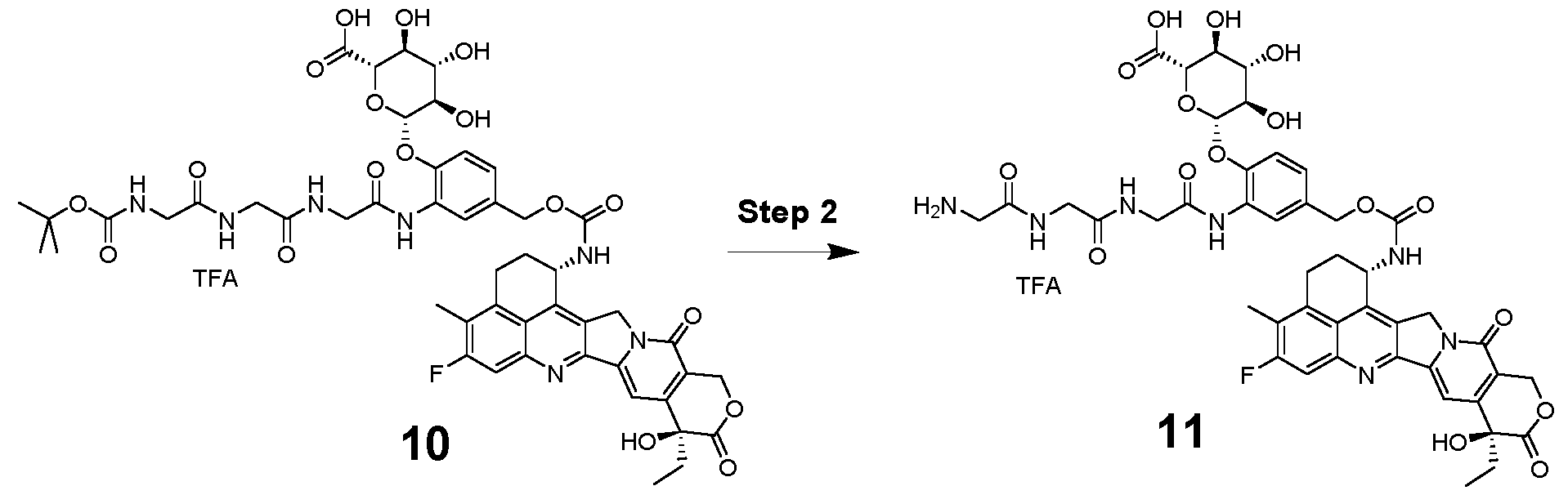 To a suspension of compound 10 (162.00 mg; 1.00 eq) in dichloromethane (5.00 ml) was added 4.0 M hydrogen chloride solution in 1,4-dioxane (682.43 µl; 20.00 eq) and it was stirred at room temperature for 3.5 hours and the reaction was monitored by LC- MS. After completion, the reaction mixture was concentrated in vacuo and the crude product was purified by prep. HPLC. The fractions containing product were combined and lyophilized to afford compound 11 trifluoroacetic acid salt as a yellow solid. Yield: 113 mg; 0.11 mmol. Precent yield: 77.0 % Analytical data: LCMS: Column: Chromolith HR RP-18e (50-4,6 mm); Mobile phase A: 0.05% HCOOH in H2O; B: 0.04% HCOOH and 1% H2O in ACN; T: 40 °C; Flow: 3,3 ml/min; MS: 100- 2000, amu positive; 1% ->100% B: 0 ->2,0min; 100% B: 2,0 ->2,5 min RT (min): 1.22; M+H: 948.0, M+2H: 474.8; Purity: 98.8% 1H NMR (700 MHz, DMSO-d6) δ 12.93 – 12.79 (m, 1H), 9.14 (s, 1H), 8.61 (t, J = 5.8 Hz, 1H), 8.41 (t, J = 5.9 Hz, 1H), 8.14 (s, 1H), 8.05 (d, J = 8.8 Hz, 1H), 8.03 – 7.93 (m, 3H), 7.79 (d, J = 10.8 Hz, 1H), 7.31 (s, 1H), 7.13 (s, 2H), 6.58 – 6.47 (m, 1H), 5.80 – 5.61 (m, 1H), 5.48 – 5.40
(m, 2H), 5.31 – 5.24 (m, 3H), 5.08 (d, J = 12.2 Hz, 1H), 5.03 (d, J = 12.3 Hz, 1H), 4.91 (d, J = 7.7 Hz, 1H), 3.96 (d, J = 5.8 Hz, 2H), 3.95 – 3.86 (m, 3H), 3.62 (q, J = 5.8 Hz, 2H), 3.43 (t, J = 9.3 Hz, 1H), 3.41 (t, J = 8.5 Hz, 1H), 3.35 (t, J = 9.0 Hz, 1H), 3.27 – 3.20 (m, 1H), 3.15 – 3.09 (m, 1H), 2.38 (s, 3H), 2.23 – 2.13 (m, 2H), 1.93 – 1.81 (m, 2H), 0.88 (t, J = 7.3 Hz, 3H). Example 3: Synthesis of a drug-linker compound with legumain-cleavable linker: Drug- linker compound 2 (DL2)
To a suspension of compound 10 (162.00 mg; 1.00 eq) in dichloromethane (5.00 ml) was added 4.0 M hydrogen chloride solution in 1,4-dioxane (682.43 µl; 20.00 eq) and it was stirred at room temperature for 3.5 hours and the reaction was monitored by LC- MS. After completion, the reaction mixture was concentrated in vacuo and the crude product was purified by prep. HPLC. The fractions containing product were combined and lyophilized to afford compound 11 trifluoroacetic acid salt as a yellow solid. Yield: 113 mg; 0.11 mmol. Precent yield: 77.0 % Analytical data: LCMS: Column: Chromolith HR RP-18e (50-4,6 mm); Mobile phase A: 0.05% HCOOH in H2O; B: 0.04% HCOOH and 1% H2O in ACN; T: 40 °C; Flow: 3,3 ml/min; MS: 100- 2000, amu positive; 1% ->100% B: 0 ->2,0min; 100% B: 2,0 ->2,5 min RT (min): 1.22; M+H: 948.0, M+2H: 474.8; Purity: 98.8% 1H NMR (700 MHz, DMSO-d6) δ 12.93 – 12.79 (m, 1H), 9.14 (s, 1H), 8.61 (t, J = 5.8 Hz, 1H), 8.41 (t, J = 5.9 Hz, 1H), 8.14 (s, 1H), 8.05 (d, J = 8.8 Hz, 1H), 8.03 – 7.93 (m, 3H), 7.79 (d, J = 10.8 Hz, 1H), 7.31 (s, 1H), 7.13 (s, 2H), 6.58 – 6.47 (m, 1H), 5.80 – 5.61 (m, 1H), 5.48 – 5.40
(m, 2H), 5.31 – 5.24 (m, 3H), 5.08 (d, J = 12.2 Hz, 1H), 5.03 (d, J = 12.3 Hz, 1H), 4.91 (d, J = 7.7 Hz, 1H), 3.96 (d, J = 5.8 Hz, 2H), 3.95 – 3.86 (m, 3H), 3.62 (q, J = 5.8 Hz, 2H), 3.43 (t, J = 9.3 Hz, 1H), 3.41 (t, J = 8.5 Hz, 1H), 3.35 (t, J = 9.0 Hz, 1H), 3.27 – 3.20 (m, 1H), 3.15 – 3.09 (m, 1H), 2.38 (s, 3H), 2.23 – 2.13 (m, 2H), 1.93 – 1.81 (m, 2H), 0.88 (t, J = 7.3 Hz, 3H). Example 3: Synthesis of a drug-linker compound with legumain-cleavable linker: Drug- linker compound 2 (DL2)
 Step 1 {4-[(2S)-3-carbamoyl-2-[(2S)-2-[(2S)-2-({[(9H-fluoren-9- yl)methoxy]carbonyl}amino)propanamido]propanamido]propanamido]phenyl}methyl 4- nitrophenyl carbonate (400 mg; 0,52 mmol; 1,00 eq.) [commercially available from Levena Biopharma US] was dissolved in N,N-Dimethylformamide (5,00 ml). Exatecan mesylate (277,30 mg; 0,52 mmol; 1,00 eq.), N-Ethyldiisopropylamine (0,27 ml; 1,57 mmol; 3,00 eq.) and 1-Hydroxybenzotriazol (HOBT) (3,52 mg; 0,03 mmol; 0,05 eq.) were added. The reaction mixture was stirred at room temperature overnight. LC/MS indicated complete conversion. The crude reaction mixture was purified via prep HPLC and lyophilized yielding 365mg (0.343 mmol) of (9H-fluoren-9-yl)methyl ((S)-1-(((S)-1-(((S)-4-amino-1-((4-(((((1S,9S)-9-ethyl-5- fluoro-9-hydroxy-4-methyl-10,13-dioxo-2,3,9,10,13,15-hexahydro-1H,12H- benzo[de]pyrano[3',4':6,7]indolizino[1,2-b]quinolin-1-yl)carbamoyl)oxy)methyl)phenyl)amino)- 1,4-dioxobutan-2-yl)amino)-1-oxopropan-2-yl)amino)-1-oxopropan-2-yl)carbamate LC/MS: [M+H] = 1064.2 Prep HPLC: Column: sunfire prep c18 obd - 75.0 g (250 bar) Solvent A : Wasser 0.1%TFA Solvent C : Solvent B : Acetonitril 0.1%TFA

Step 2
(9H-fluoren-9-yl)methyl ((S)-1-(((S)-1-(((S)-4-amino-1-((4-(((((1S,9S)-9-ethyl-5-fluoro-9- hydroxy-4-methyl-10, 13-dioxo-2,3,9, 10,13,15-hexahydro-1 H,12H- benzo[de]pyrano[3',4':6,7]indolizino[1,2-b]quinolin-1-yl)carbamoyl)oxy)methyl)phenyl)amino)- 1 ,4-dioxobutan-2-yl)amino)-1-oxopropan-2-yl)amino)-1-oxopropan-2-yl)carbamate (365 mg;
0,34 mmol; 1,00 eq.) was dissolved in N,N- (4,00 ml). Piperidine for synthesis (0,07 ml; 0,69 mmol; 2,00 eq.) was added and the reaction solution was stirred at rt for 1h.
The reaction mixture was purified via prep HPLC yielding 300mg (0.314 mmol) of 4-((S)-4- amino-2-((S)-2-((S)-2-aminopropanamido)propanamido)-4-oxobutanamido)benzyl ((1S,9S)-9- ethyl-5-fluoro-9-hydroxy-4-methyl-10, 13-dioxo-2,3,9, 10,13,15-hexahydro-1 H , 12H- benzo[de]pyrano[3',4':6,7]indolizino[1,2-b]quinolin-1-yl)carbamate.
LC/MS: [M+H]: 841.3
Prep HPLC for purification:
RediSep Saule: C18 130g
SN: E0410A0D24BE1 Lot: 262118923W
Flowrate: 75 ml/min
Condition - Volumen: 390,0 ml
Eluent: A1 WATER 0.1%TFA
Eluent: B1 ACETONITRILE 0.1%TFA
 Step 3
To a solution of 4-((S)-4-amino-2-((S)-2-((S)-2-aminopropanamido)propanamido)-4- oxobutanamido)benzyl ((1S,9S)-9-ethyl-5-fluoro-9-hydroxy-4-methyl-10,13-dioxo-
2,3,9,10,13,15-hexahydro-1 H,12H-benzo[de]pyrano[3',4':6,7]indolizino[1 ,2-b]quinolin-1- yl)carbamate (571mg; 0,60 mmol; 1 ,00 aq.) in N,N-Dimethylformamide (20ml) was added N- Ethyldiisopropylamine (203 pl; 1 ,20 mmol; 2,00 eq.) and N-Succinimidyl 3- maleimidopropionate (162 mg; 0,60 mmol; 1 ,00 eq.). The reaction mixture was then stirred for 10min and monitored via LC/MS.
The reaction mixture was purified via prep HPLC yielding 378mg (0.35 mmol) of DL2.
LC/MS: [M+H]: 992.4
Prep HPLC for purification:
RediSep column: C18 86g
SN: E0410A8B46130 Lot: 281729189W
Flowrate: 60 ml/min
Conditon - Volumen: 264,0 ml
Eluent 1 : A1 WATER 0.1%TFA
Eluent 2: B1 ACETONITRILE 0.1%TFA
Analytics for sequence:
Method Info : A: H2O + 0,05% HCOOH | B: MeCN + 0,04% HCOOH + 1 % H2O
T: 40 °C | Flow: 3,3 ml/min | MS: 100-2000 amu positive
Column: Chromolith HR RP-18e 50-4,6 mm
0% -> 100% B: 0 -> 2,0 min | 100% B: 2,0 -> 2,5 min
Example 4: Preparation of an immunoconjugate: a glucuronide-based conjugate of mAb1 (referred to as ADC1) and mAb1-M (the resulting immunoconjugate being referred to as ADC1- M), mAb4-M (the resulting immunoconjugate being referred to as ADC4-M) and mAb6-M (the resulting immunoconjugate being referred to as ADC6-M)
Although the following methods are described in the context of mAb1 (the resulting immunoconjugate being referred to as ADC1), the same method can also be used for mAb1- M (the resulting immunoconjugate being referred to as ADC1-M), mAb4-M (the resulting
immunoconjugate being referred to as ADC4-M) and for mAb6-M (the resulting immunoconjugate being referred to as ADC6-M).
4.1 Conjugation process
Antibody Preparation
The antibody mAb1 (as defined herein above) was thawed at 2 - 8°C up to 3 days prior to conjugation and stored at 2 - 8°C until use. The mAb (> 10 g) was equilibrated at room temperature on the day of conjugation prior to use. The mAb (9.6 mg/mL) was aliquoted (10.0 g, 1041.7 mL) and diluted to 5.59 mg/mL using conjugation buffer (200 mM Histidine, pH 6.5). The mAb solution was added into a 3 L Chemglass jacketed reactor and set to 25 ± 2°C while stirring at 50 rpm.
Reduction of Antibody
Added 7.0 mol eq. (9.7 mL) of 50 mM TCEP solution (50 mM TCEP in conjugation buffer) into the mAb solution vial and the reaction was allowed to proceed at 25 ± 2°C for 3 hours.
Conjugation
The drug-linker compound 1 (DL1) of formula (X) was weighed and dissolved in DMSO to prepare a 20 mM solution. 90% (148.6 mL) of the required DMSO was added to the reactor. Immediately after DMSO addition, 10.0 mol equivalents (38.2 mL) of 20 mM drug-linker solution was added to the reactor. Then, 10% (18.2 mL) of the remaining required DMSO was used to rinse the drug-linker vial to ensure total transfer. After final addition, the reaction was allowed to proceed at 25 ± 2°C for 1 hour. Total volume during conjugation was 1997.0 mL.
Note: Overall DMSO concentration of the reaction was 10% (v/v) (DMSO + Drug Linker solution).
Quench
Added 35 mol eq. (48.5 mL) of 50 mM NAC to the reactor and the reaction was allowed to proceed at 25 ± 2°C for 30 minutes.
Filtration
The filtered crude conjugate solution was transferred from the reactor and then filtered using a Millipak Gamma Gold 60 (MPGL06GH2) to give 1993.6 mL (Filter Load: 324.7 g/m2 [protein],
66.5 L/m2 [solution]) of filtered crude conjugate.
Diafiltration
The filtered crude conjugate solution was buffer exchanged (DV= 1993.6 mL) with a Pellicon 3 (30 kDa) Biomax membrane (1 x 0.11 m2, 300 LMH (550 mL/min), 16 psi TMP, actual loading
88.5 g/m2). Diafiltration buffer (10 mM Histidine, pH 5.5) was used to buffer exchange the crude conjugate for 16 diavolumes. After buffer exchanging, the solution was concentrated to > 25 mg/mL, transferred into a bottle, and the membrane flushed with diafiltration buffer. Total volume recovered from UF/DF was 361.5 mL.
Formulation
The concentrated ADC (i.e. ADC1) was diluted to 20.0 mg/mL with 112.1 mL of diafiltration buffer (10 mM Histidine, pH 5.5). The resulting solution was diluted to 15.0 mg/mL with 157.6 mL of 4X formulation buffer (10 mM Histidine, 12% (w/v) Trehalose Dihydrate, 400 mM NaCI, pH 5.5) for a final target bulk drug substance (BDS) concentration of 15.0 mg/mL.
Filtration
The final formulated ADC was filtered using a 0.2 μm Millipak Gamma Gold 40 (MPGL04GH2) filter to yield 619.6 mL (Filter Load: 464.6 g/m2 [protein], 31.0 L/m2 [solution]) ADC1 BDS. The material was packaged into HDPE bottles and stored at < -65°C.
4.2 Methods: Drug substance characterization: ADC1
Size exclusion chromatography (SEC) method
SEC Method Parameters
Wavelength 280 nm
Column Tosoh TSKgel 7.8 mm x 300 mm, 5 μm (P/N 0008541)
Mobile Phase0.14 M Potassium Phosphate Monobasic
50 mM Sodium Phosphate Monobasic
0.06 M Potassium Phosphate Dibasic
0.25 M Potassium Chloride
5% I PA
Injection Volume 20 pL
Temperature 25°C
Flow rate 0.5 mL/min
Run Time 30 min
Typical SEC chromatogram showing the purity of the stock mAb, the conjugate post UF and the final BDS: Fig. 14.
For the BDS material shown above, 1.7% HMWS and a monomeric purity of 96.9% have been reported.
Reversed-Phase HPLC (RP HPLC) method
RP HPLC Method Parameters
Wavelength 280 nm
Column PLRP-S 1000 A (50 x 2.1 mm, 8 μm) column, Agilent (P/N
PL1912-1802)
Mobile Phase A 0.1 % Formic Acid in Water with 0.01% TFA
Mobile Phase B 0.1 % Formic Acid in ACN with 0.01% TFA
Gradient
 Injection Volume 10 pL
Column Temperature 80°C
Flow rate 1.0 mL/min
Run Time 30 min
Sample Preparation Dilute sample to 2 mg/mL and add 40 pL to a micro centrifuge tube. Add 60 pL of the ~8 M Guanidine HCI, -130 mM Tris, ~1mM EDTA, pH 7.6 buffer. Add 2 pL of 500 mM DTT and vortex to mix. Incubate sample for 30 ± 2 min at 37 ± 2°C.
Typical RP-HPLC chromatogram showing the separation of light and heavy chains: Fig. 15. The chromatogram shows an overlay of the stock mAb, the crude ADC and the final BDS.
For the ADC1 BDS material above, a DAR of 7.9 was reported.
Free-drug method
Free-drug method parameters
Wavelength 254 nm
Column Phenomenex Gemini, C18, 2 x 150 mm, 3 μm (P/N 00F-4439-B0)
Mobile Phase A 0.1 % Formic acid in water
Mobile Phase B 0.1 % Formic acid in acetonitrile
Gradient

Injection Volume 10.00 pL
Column Temperature 50°C
Flow rate 0.75 mL/min
Sample Preparation Protein drop: 100 pL of Drug Substance + 250 pL of cold MeOH + 50 pL of 3 M MgCh. Spin at 20,000 rpm for 10 min
Standard Preparation Mix 20 pL of 20 mM DL1 (drug-linker compound 1 in DMSO) + 20 pL DMSO + 40 pL MeOH + 20 pL of 200 mM NAC in Diafiltration buffer. Incubate overnight to afford 4 mM DL-NAC. Dilute 4 mM DL-NAC in MeOH to afford a 4 pM DL-NAC standard.
Typical chromatogram showing the NAC standard and the free-drug levels of the final BDS: Fig. 16.
For the ADC1 BDS material shown above, residual free-drug levels below 2.4% (by molar ratio) have been reported.
Example 5: Preparation of an immunoconjugate: a peptide-based conjugate of mAb1 (referred to as ADC2)
5.1 Conjugation process
Antibody Preparation
The antibody mAb1 (as defined herein above) was thawed at 2 - 8°C up to 3 days prior to conjugation and stored at 2 - 8°C until use. The mAb (> 9.5 g) was equilibrated at room temperature on the day of conjugation prior to use. The mAb (9.6 mg/mL) was aliquoted (9.5 g, 989.6 mL) and diluted to 5.59 mg/mL using conjugation buffer (200 mM Histidine, pH 6.5). The mAb solution was added to a 3 L Chemglass jacketed reactor and set to 25 ± 2°C while stirring at 50 rpm.
Reduction of Antibody
Added 8.0 mol eq. (10.5 mL) of 50 mM TCEP solution (50 mM TCEP in conjugation buffer) into the mAb solution vial and the reaction was allowed to proceed at 25 ± 2°C for 3 hours.
Diafiltration
The reduced mAb solution was buffer exchanged against 6 DVs (DV= 1706.9 mL) using a Pellicon 3 (30 kDa) Biomax membrane (1 x 0.11 m2, 300 LMH (550 mL/min), 16 psi TMP, actual loading 86.3 g/m2. Conjugation buffer (200 mM Histidine, pH 6.5) was used to buffer exchange the reduced antibody. After buffer exchanging, the reduced mAb solution was recovered back into the reactor and the membrane flushed with conjugation buffer.
Conjugation
The drug-linker compound 2 (DL2) was weighed and dissolved in DMSO to prepare a 20 mM drug-linker solution. 90% (142.6 mL) of the required DMSO was added to the reactor. Immediately after DMSO addition, 9.5 mol equivalents (31.2 mL) of the 20 mM drug-linker solution was added to the reactor. Then, 10% (15.8 mL) of the remaining required DMSO was
used to rinse the drug-linker vial to ensure total transfer. After final addition, the reaction was allowed to proceed at 25 ± 2°C for 2 hours. Total volume during conjugation reaction was 1894.5 mL
Quench
Added 35 mol eq. (46 mL) of 50 mM NAC to the reactor and the reaction was allowed to proceed at 25 ± 2°C for 45 minutes.
Filtration
The crude conjugate solution was transferred from the reactor and filtered using a Millipak Gamma Gold 60 (MPGL06GH2) to give 1897.3 mL (Filter Load: 308.9 g/m2 [protein], 63.2 L/m2 [solution]) of filtered crude conjugate.
Diafiltration
The filtered crude conjugate solution was buffer exchanged (DV = 1897.3 mL) with a Pellicon 3 (30 kDa) Biomax membrane (1 x 0.11 m2, 300 LMH (550 mL/min), 16 psi TMP, actual loading
84.2 g/m2). The initial 12 DVs were performed using conjugation buffer (200 mM Histidine, pH 6.5) and then switched to standard diafiltration buffer (10 mM Histidine, pH 5.5) for 8 additional DVs. After buffer completing the buffer exchange, the solution was then concentrated to > 25 mg/mL, transferred into a bottle and the membrane flushed with diafiltration buffer. Total pooled volume recovered from UF/DF was 335.7 mL.
Formulation
The concentrated ADC (i.e. ADC2) was diluted to 20.0 mg/mL with 84.7 mL of Diafiltration Buffer (10mM Histidine, pH 5.5). The resulting solution was diluted with 138.6 mL of 4X Formulation Buffer (10 mM Histidine, 12% (w/v) Trehalose Dihydrate, 400 mM NaCI, pH 5.5) for a final target BDS concentration of 15.0 mg/mL.
Filtration
The final formulated ADC was aseptically filtered using a Millipak Gamma Gold 60 (MPGL06GH2) to yield 549.3 mL (Filter Load: 411.4 g/m2 [protein], 27.5 L/m2 [solution]) of ADC2 BDS. The material was packaged into HDPE bottles and stored at < -65°C.
5.2 Methods: Drug substance characterization: ADC2
Size exclusion chromatography (SEC) method
SEC Method Parameters
Wavelength 280 nm
Column Tosoh TSKgel 7.8 mm x 300 mm, 5 μm (P/N 0008541)
Mobile Phase 50 mM Sodium Phosphate Monobasic
0.4 M Sodium Perchlorate pH 6.3
Injection Volume 1 pL
Column Temperature 25°C
Flow rate 0.5 mL/min
Run Time 30 min
Typical SEC chromatogram showing the purity of the stock mAb and the final BDS: Fig. 17.
For the ADC2 BDS material shown above, 4.2% HMWS and a monomeric purity of 95.8% have been reported.
Reversed-Phase HPLC (RP HPLC) method
RP HPLC Method Parameters
Wavelength 280 nm
Column PLRP-S 1000 A (50 x 2.1 mm, 8 μm) column, Agilent (P/N
PL1912-1802)
Mobile Phase A 0.1% Formic Acid in Water with 0.01% TFA
Mobile Phase B 0.1% Formic Acid in ACN with 0.01% TFA
Gradient


Injection Volume 10 pL
Column Temperature 80°C
Flow rate 1.0 mL/min
Run Time 30 min
Sample Preparation Dilute sample to 2 mg/mL and add 40 pL to a micro centrifuge tube. Add 60 pL of the ~8 M Guanidine HCI, -130 mM Tris, ~1mM EDTA, pH 7.6 buffer. Add 2 pL of 500 mM DTT and vortex to mix. Incubate sample for 30 ± 2 min at 37 ± 2°C.
Typical RP-HPLC chromatogram showing the separation of light and heavy chains: Fig. 18. The chromatogram shows an overlay of the stock mAb and the final BDS.
For the ADC2 BDS material above, a DAR of 7.6 was reported.
Free-drug method
Free-drug method parameters
Wavelength 254 nm
Column Phenomenex Gemini, C18, 2 x 150 mm, 3 μm (P/N 00F-4439-B0)
Mobile Phase A 0.1% Formic acid in water
Mobile Phase B 0.1% Formic acid in acetonitrile
Gradient

Injection Volume 10.00 pL
Column Temperature 50°C
Flow rate 0.75 mL/min
Sample Preparation Protein drop: 100 pL of Drug Substance + 250 pL of cold MeOH + 50 pL of 3 M MgCh. Spin at 20,000 rpm for 10 min
Standard Preparation Mix 20 pL of 20 mM DL2 (drug-linker compound 2 in DMSO) + 20 pL DMSO + 40 pL MeOH + 20 pL of 200 mM NAC in Diafiltration buffer. Incubate overnight to afford 4 mM DL-NAC. Dilute 4 mM DL-NAC in MeOH to afford a 4 pM DL-NAC standard.
Typical chromatogram showing the NAC standard and the free-drug levels of the final BDS: Fig. 19.
For the ADC2 BDS material shown above, residual free-drug levels below 1 .9% (by molar ratio) have been reported.
Example 6: An analog of the ADC SAR408701
6.1 The antibody
For the purposes of further comparative experiments, an analog of Sanofi’s anti-CEACAM5 ADC SAR408701 was prepared based on a monoclonal antibody having the following sequence:
Heavy chain: SEQ ID NO: 25
Light chain: SEQ ID NO: 26
6.2 The drug-linker compound
As a drug-linker molecule to be conjugated to the above-mentioned antibody, SPDB-DM4 (obtained from Levena Biopharma) was used:
Product name: SPDB-DM4
Structure:
 Expected mass: 994.35
Observed average mass: 995.5 (Ms+IT)
Mass spectral analysis: consistent, exhibited correct MW
HPLC analysis: purity > 95%
Appearance: white powder
6.3 Conjugation
The antibody was thawed at 2 - 8°C up to 3 days prior to conjugation and stored at 2 - 8°C until use. The antibody (175 mg) was equilibrated at room temperature on the day of conjugation prior to use. The antibody (7.9 mg/mL) was diluted to 5 mg/mL using conjugation buffer (PBS pH 7.4) and a 5 mM DMSO solution (8 mol equivalents relative to the antibody) of SPDB-DM4 (Levena Biopharma). The reaction solution was mixed and incubated at 25°C for 4h.
6.4 Preparative size exclusion chromatography, desalting and filtration
The reaction mixture was purified using preparative size-exclusion chromatography. A Superdex 200 pg (50/60) column was connected to an Akta Avant 25 system (GE Healthcare) and equilibrated with PBS pH 7.4 according to the manufacturer’s instructions. Subsequently, the reaction mixture was injected and run through the column with a flowrate of 10 ml/min and PBS pH 7.4 as running buffer. ADC containing fractions were determined via UV light absorption at 280 nm, pooled and concentrated. ADC material was concentrated using 15 ml Amicon Ultra 50 kDa cutoff centrifugal devices (Merck Millipore) according to manufactures instructions. The concentrated ADC material was transferred into formulation buffer (10 mM Histidine, 130 mM Glycine, 5% Sucrose. pH 5.5) using HiPrep 26/10 desalting columns (GE Healthcare) at a flowrate of 10 ml/min on an Akta Avant 25 system (GE Healthcare) according to the manufactures instructions. The resulting ADC material was filtered using a 0.2 μm filter (Merck Millipore), aliquoted and subsequently shock frozen in liquid nitrogen. The final concentration of the ADC material was 5.82 mg/ml and the material was kept at -80°C until further use. The ADC resulting from this work is also referred to herein as “ADC SAR DM4” or, briefly, as “ADC SAR”; this ADC is an analog of SAR408701 .
Example 7: An ADC based on mAb1 and SPDB-DM4
Another ADC was prepared based on the antibody mAb1 (as described herein above) and the drug-linker compound SPDB-DM4, i.e. the same drug-linker compound as in ADC SAR DM4 described above. The ADC resulting from this work is referred to herein as “ADC mAb1 DM4” and was prepared as follows:
7.1 Materials used:
• Antibody: mAb1 , 1 mg/mL in 10 mM HEPES, pH 5.8
• Conjugation Buffer: 10 mM HEPES, pH 5.8
• Drug-linker compound: SPDB-DM4, 2 mg/mL in DMF
7.2 Method:
• Conjugation: about 30-fold molar excess of drug-linker molecule was used for conjugation (75 mL antibody + 7.1 mL SPDB-DM4 drug-linker), incubated at room temperature for 5 h with slow rocking
• Purification: ADC was buffer exchanged to 20 mM Histidine, 150 mM NaCI, pH 6.0 to remove free drug
• Formulation Buffer: 20 mM Histidine, 150 mM NaCI, pH 6.0
7.3 Purified ADC Analysis Details:
• Final Yield: 40 mg
• Concentration: 2.2 mg/mL
• DAR: 4.4
Example 8: Characterization of drug release from ADC1 and ADC2
8.1 Materials and Methods
8.1.1 Test Articles

8.1.2 Materials
All reagents and buffers were stored according to the instructions of the manufacturers and used before the batch expiration date.

8.1.3 Instruments

8.1.4 Procedures
8.1.4.1 Serum sample preparation
2 M HEPES solution: 52.1 g HEPES were dissolved in 75 mL MiliQ-water and 15 mL HCI 25%, adjusted to pH 7.55 and added up to 100 mL. This solution was mixed as 15 %v/v with serum
to obtain a stabilized serum with pH 7.3 - 7.4. Human serum from Biowest (Lot.no. S15594S4200) was thawed. 100 mL serum were mixed with 15mL 2 M HEPES buffer. Mouse serum from Biowest (Lot.no. S18169S2160) was thawed. 100mL serum were mixed with 15mL 2 M HEPES buffer. Cynomolgus serum was thawed and 8.5 mL serum were mixed with 1.5mL 2 M HEPES buffer. The pH was measured (7.37) and serum was sterile filtered. 2mL aliquots were frozen at -20°C.
The prepared serum was thawed at RT. The desired ADC protein concentration was prepared as triplicated with 180pg/mL for subsequent free payload analytics via LC-MS. After adding the ADCs to the serum, the individual batches were mixed and separated into 20 pL aliquots. Additionally, one 96h sample with 20pL for each ADC was pipetted and was used to for total work up analyses to measure recovery. Oh samples were directly frozen at - 80 °C, remaining samples were incubated at 37 °C and 5 % CO2 and reactions were stopped at 2/ 4/ 6/ 24/ 48/ 72, 96 hours incubation via storage at -80°C.
8.1.4.2 Human Liver Lysosome sample preparation pH was adjusted either to pH 5.0 or pH 4.0 using assay buffer. Lysosomal stability preparations: 80 pL Human Liver Lysosomes were prepared for triplicate measurements as exemplary shown for ADC1 (n=3): 2.76 pL ADC1 + 6 pL Human Liver Lysosomes + 71.2 pL Legumain Assay Buffer. Preparation of MeOH + PIC (1 :200): 10pL PIC III + 1990 pL MeOH. The reactions were started upon transferring the Eppendorf tubes to a Thermomix that was preheated to 37°C. Subsequently, 10 pL aliquots were drawn after 0, 1 , 2, 4, 24 and 48 hours and mixed with 40pL PIC III (1 :200).
8.2 Results
ADC stability for human, mouse and cynomolgus sera (Fig. 20). Conjugated Exatecan concentrations were calculated (initial dose -10 pM) using free Exatecan (normalized data). Similar profiles were obtained for human, cynomolgus and mouse sera. Only minor warhead release observed, most pronounced in mouse serum for ADC2 (5.9 % free of initial conj. payload at 96h) and ADC1 (1.4%).
ADC3 control stability for mouse serum and buffer (Fig. 21). Conjugated SN38 concentrations were calculated (initial dose 50 pg/mL ADC protein concentration) using free SN38 (not normalized). For both matrices, pronounced SN-38 release observed.
Payload liberation profiles for ADC1 and ADC2 in human liver lysosomes (pH 5.0) (Fig. 22). Conjugated drug concentrations were calculated using e.g. free Exatecan (initial cone. ~10 pM Exatecan), normalized data. Intermediate levels of payload release were observed for ADC1- and ADC2-cleavage mediated payload liberation (both -40% of initial total conj. Payload).
ADC catabolite profiling confirms free exatecan as lysosomal release product (Fig. 23). To confirm exatecan as major release product, ADC1 catabolite profiling study was performed in human lysosomal extracts. At this, comparison of the TIC-MS and extracted ion chromatograms at various timepoints showed that the expected exatecan catabolite was subsequently released from ADC1 during incubation (Oh, 4h, 24h). The retention time 9.33 min, the detected mass m/z 436.1671 ([M+H]+, C24H23O4N4 F) and the MS/MS spectrum of the detected catabolite are consistent with those of exatecan.
Example 9: ADC1 and ADC2 specifically kill cancer cells in vitro with high potency
Human cancer cell lines were used to assess the potential of ADC1 and ADC2 to kill cancer cells. ADC1 and ADC2 showed sub-nanomolar in vitro potency against different CEACAM5- positive and minor effect on CEACAM5-negative cell lines (Table 2 below). As shown in exemplary dose-response curves (Fig. 24A/B), ADC1 and ADC2 were very potent against CEACAM5-positive cell lines SK-CO-1 and SNll-16. In contrast, effects of ADC1 and ADC2 on antigen-negative MDA-MB-231 were limited to the highest concentrations tested (Fig. 24C).
Rituximab isotype control ADCs utilizing the respective linker and payload as for ADC1 and ADC2 showed much lower effects on SK-CO-1 cell line (Fig. 25).
In conclusion, ADC1 and ADC2 specifically kill CEACAM5 expressing human cancer cell lines in vitro with high potency.
 Table 2. Potency of ADC1 , ADC2 and free payload against multiple human cell lines. Maximal effects compared to untreated controls at the highest tested compound concentration are indicated in brackets. For each cell line, CEACAM5 expression is indicated.
Method - Viability Assay:
Cytotoxicity effects of the ADC on the cancer cell lines were measured by cell viability assays. Cells were seeded in a volume of 90 pL in 96-well plates the day before treatment. Test compounds (ADCs or free payloads) were formulated at 10-fold the starting concentration in cell culture medium. Test compounds were serial diluted (1 :4) and 10 pL of each dilution was added to the cells in triplicates. Plates were cultured at 37 °C in a CO2 incubator for six days. For cell viability measurement, Cell Titer-Gio® reagent (Promega™ Corp, Madison, Wl) was added to each well, and plates processed according to the manufacturer’s instructions. Luminescence signals were measured using a Varioskan plate reader (Thermo Fisher). Luminescence readings were converted to % viability relative to untreated cells. Data was fitted with non-linear regression analysis, using log (inhibitor) vs. response, variable slope, 4- parameter fit equation using GraphPad Prism. Data is shown as % relative cell viability vs. molar compound concentration, error bars indicating standard deviation (SD) of triplicates. Geometric mean values of IC50s derived from multiple experiments were calculated.
Using the same method as above, ADC1 and ADC2 were also compared to ADC SAR DM4 in terms of their cytotoxic effects on antigen-positive SK-CO-1 and antigen-negative MDA-MB- 231 cell line. ADC1 and ADC2 showed 2.9- and 2.7-fold higher potencies than ADC SAR DM4 against SK-CO-1 cancer cells, respectively (Fig. 26A). Non-specific effects against antigennegative MDA-MB-231 were slightly higher for ADC SAR DM4 compared to ADC1 and ADC2 (Fig. 26B). ADC SAR DM4 and ADC mAb1 DM4 showed comparable potencies against SK- CO-1 , with a slight tendency for higher potency of ADC mAb1 DM4 (Fig. 26A).
Example 10: ADC1 and ADC2 mediate potent bystander effect against antigen-negative cells in co-culture with antigen-positive cells
The potential of ADC1 and ADC2 to mediate a bystander effect against antigen-negative cells in close proximity to antigen-positive cells was evaluated in bystander assays. ADC1 and ADC2 showed a potent bystander effect against CEACAM5-negative MDA-MB-231 cells in the presence of CEACAM5-positive SK-CO-1 (Fig. 27A). The co-culture experiments were performed at an ADC concentration of 1 nM which, for ADC1 , causes maximal inhibition of CEACAM5-positive SK-CO-1 cell viability (Fig. 26A) but no effect on CEACAM5-negative MDA-MB-231 cells (Fig. 26B). In line with these findings, no non-specific effect of ADC1 or ADC2 was observed in the bystander assay setup for MDA-MB-231 only controls (Fig. 27B).
In conclusion, ADC1 and ADC2 mediate a potent bystander effect against antigen-negative cancer cells in co-culture with antigen-positive cells. These findings indicate the potential to effectively target tumors with heterogeneous target expression.
In view of utilizing the same drug-linker and Fv region, it is expected that ADC1-M, ADC2-M, ADC3-M, and ADC6-M and ADC7-M mediate similar potent bystander effects like ADC1 and ADC2.
Compared to ADC SAR, ADC1 and ADC2 mediated a much more potent bystander effect on antigen-negative cells in co-culture with antigen-positive cells (Fig. 28A, Fig. 28B). ADC mAb1 DM4 utilizing the same antibody as in ADC1 and ADC2 (i.e. mAb1) with the drug-linker molecule utilized in ADC SAR DM4 (i.e. SPDB-DM4) also showed a more pronounced bystander effect than ADC SAR DM4 (Fig. 28A, Fig. 28B). This indicated that mAb1 contributes to the higher bystander effect observed for ADC1 and ADC2 in comparison with ADC SAR utilizing a different antibody. It is therefore expected that also ADC1-M, ADC2-M, ADC3-M, ADC6-M and ADC7-M mediate more potent bystander effects than ADC SAR.
For all ADCs tested, the extent of bystander effect increased with increasing the number of antigen-positive cells added to a constant number of antigen-negative cells (compare Fig. 28A and Fig. 28B). Without wishing to be bound by theory this may be a result of more ADC being processed by the higher number of antigen-positive cells to release more free payload, which is responsible for the bystander effect on antigen-negative cells.
No non-specific effects of tested ADCs were observed on MDA-MB-231 cells alone (Fig. 28C).
Method - Bystander Assay
Cytotoxicity effects of ADCs on antigen-negative cancer cell lines in co-culture with antigenpositive cancer cell lines were measured by bystander assays. One thousand CEACAM5- negative MDA-MB-231 cells were seeded in co-culture experiments with 750 or 3000 CEACAM5-positive SK-CO-1 cells per well. As a control, 1000 MDA-MB-231 cells only were seeded in parallel. Cells were seeded in a total volume of 90 pL in 96-well plates the day before treatment. Test compounds were formulated at 10-fold the final concentration of 1 E-9 M in cell culture medium and 10 pL was added to the cells in duplicates. Plates were cultured at 37 °C in a CO2 incubator for six days.
Prior to immunofluorescence staining, medium was removed and cells were treated with 100% methanol (-20°C) for 30 minutes. After methanol removal and one PBS wash step, cells were treated with 2.5% paraformaldehyde (PFA) with 0.2% Triton X-100 in PBS for 15 minutes at room temperature. After solution removal and one PBS wash step, cells were treated with 1% BSA I 0.1 % Tween I 0.1% sodium azide in PBS for at least one hour at room temperature. Antigen-positive and antigen-negative cells were discriminated by immunofluorescence
staining with 10 pg/mL human anti-CEACAM5 (mAb1) primary antibody and 1 :2000 dilution of donkey anti-human IgG fluorescently (phycoerythrin) labeled secondary antibody (Jackson ImmunoResearch #709-116-149). Cells were identified by nuclei staining using 1 pg/mL Hoechst 33342 (Life technologies, cat# H3570) dye. Staining was carried out in 1% BSA I 0.1 % sodium azide PBS solutions for 30 minutes at room temperature. Secondary antibody staining was combined with Hoechst dye staining. Between and after staining steps, cells were washed thrice with PBS.
Plates were imaged with the confocal quantitative image cytometer CQ1 (Yokogawa® Electric Corporation, Tokyo, Japan). Analysis was adapted from the CQ1 software (Yokogawa) template “Nucleus and pseudo-Cell body” and FCS export files were analyzed using FlowJo (BD). Based on staining or absence of staining with fluorescently labeled antibody around the nucleus, antigen-positive and antigen-negative cells were distinguished and quantified. Bar graphs show the number of identified antigen-positive and antigen-negative cells per treatment condition.
Example 11 : Efficacy of ADC1 and ADC2 in a colorectal cancer (CRC) patient-derived xenograft (PDX) mouse model
Anti-tumor efficacy in vivo has been evaluated in the human patient-derived CRC xenograft model COPF217 (Shanghai LideBiotech CO., LTD). COPF217 tumor fragments were transplanted subcutaneously into the right flank of six to eight weeks old immunodeficient female mice (NU-Foxn1nu, Charles River). When tumors reached a mean volume of 165 mm3, 6 mice/group were treated once intravenously with vehicle (saline solution) or with ADC1 or ADC2 (each at a dose of 10mg/kg; day 0). Tumor length (L) and width (W) were measured with calipers and tumor volumes were calculated using the formula Lx(WA2)/2.
The single treatment with ADC1 or ADC2 at a dose of 10 mg/kg led to a significant anti-tumor effect. The two substances show a comparable effect leading to tumor stasis (Fig. 29). T reatment with ADC1 or ADC2 had no significant impact on body weight (data not shown).
Further experiments with other CRC PDX models:
In additional experiments corresponding to the one above described for COPF217, a single treatment with ADC1 also resulted in tumor stasis or tumor regression in 12 other CRC PDX models with high CEACAM5 expression.
Example 12: Efficacy of ADC1 in a non-small cell lung cancer (NSCLC) PDX mouse model
Anti-tumor efficacy in vivo has been evaluated in the human patient-derived NSCLC xenograft model LUPF160151 (Shanghai LideBiotech CO., LTD). LUPF160151 tumor fragments were transplanted subcutaneously into the right flank of six to eight weeks old immunodeficient
female mice (NU-Foxn1nu, Charles River). When tumors reached a mean volume of 180 mm3, 5 mice/group were treated once intravenously with vehicle (saline solution) or with ADC1 (6 mg/kg; day 0). T umor length (L) and width (W) were measured with calipers and tumor volumes were calculated using the formula L*(WA2)/2.
The single treatment with ADC1 at a dose of 6 mg/kg led to a significant anti-tumor effect (Fig. 30) with no impact on body weight (data not shown). The model LUPF160151 showed a heterogeneous CEACAM5 expression with CEACAM5 negative tumor cells adjacent to CEACAM5 positive tumor cells. The good efficacy of ADC1 in this model thus indicates a potent bystander effect of the ADC.
Example 13: Efficacy of ADC1 in gastric cancer PDX mouse model
Anti-tumor efficacy in vivo was evaluated in the human patient-derived gastric cancer xenograft model GAX066 (Shanghai ChemPartner Co., Ltd). Tumor fragments were transplanted subcutaneously into the right flank of immunodeficient female mice (Nu/Nu mice, Beijing Vital River Lab Animal Technology Co. Ltd, 18-22g). When tumors reached a mean volume of 220 mm3, 6 mice/group were treated once intravenously with vehicle (saline solution) or with ADC1 (3 or 10 mg/kg; Day 0). Tumor length (L) and width (W) were measured with calipers and tumor volumes were calculated using the formula L*(WA2)/2.
The single treatment with 3 or 10 mg/kg ADC1 caused a significant anti-tumor effect (Fig. 31) with no impact on body weight (data not shown). In five of six tumors, treatment with 10 mg/kg resulted in complete tumor regression.
Example 14: Efficacy of ADC1 compared to ADC3 in a pancreatic cell line derived tumor model
Efficacy of ADC1 in comparison to ADC3 has been evaluated in the human pancreatic cell line derived xenograft model HPAF-II (ATCC, CRL-1997). 5x10⁶ HPAF-II cells were injected subcutaneously into the right flank of six to eight weeks old immunodeficient female mice (Hsd:Athymic Nude-Foxn1nu, Envigo). When tumors reached a mean volume of 150 mm3, 10 mice/group were treated once intravenously with vehicle (saline solution) or ADC1 (1 mg/kg or 6mg/kg; day 0) or with ADC3 (1 mg/kg or6mg/kg; day 0). Tumor length (L) and width (W) were measured with calipers and tumor volumes were calculated using L*(WA2)/2.
The single treatment with ADC1 at a dose of 6 mg/kg led to a significant anti-tumor effect. The effect is dose-dependent, as the single treatment with 1 mg/kg only led to a mild but significant, transient anti-tumor effect. In contrast, the single treatment with same doses of ADC3 showed no significant anti-tumor effect at either dose (Fig. 32). All treatments had no significant effect on body weight (data not shown). It is expected that ADC1-M, ADC2-M, ADC3-M, ADC6-M and ADC7-M compare similarly favorably to ADC3 in this regard as does ADC1 .
Example 15: Efficacy of ADC1 compared to ADC SAR DM4 in two CRC PDX mouse models
Anti-tumor efficacy in vivo was evaluated in the human patient-derived CRC xenograft models COPF230 and REPF210 (Shanghai LideBiotech CO., LTD). Tumor fragments were transplanted subcutaneously into the right flank of six to eight weeks old immunodeficient female mice (NU-Foxn1nu, Charles River). When tumors reached a mean volume of about 170 mm3, 6 mice/group were treated once (Day 0) intravenously with vehicle (saline solution), ADC1 (6 mg/kg) or ADC SAR DM4 (6 mg/kg). Tumor length (L) and width (W) were measured with calipers and tumor volumes were calculated using the formula L*(WA2)/2.
Single treatment with ADC1 led to a significant anti-tumor effect in both PDX models COPF230 (Fig. 33) and REPF210 (Fig. 34). In contrast, a single treatment with the same dose of ADC SAR DM4 showed no anti-tumor effect in either of the CRC PDX models (Fig. 33, Fig 34). No significant effect on body weight was observed in any of the treatment groups (data not shown). It is expected that ADC1-M, ADC2-M, ADC3-M, ADC6-M and ADC7-M compare similarly favorably to ADC SAR DM4 in this regard as does ADC1.
Example 16: Efficacy of ADC1 compared to ADC SAR DM4 in a gastric PDX mouse model (GAPF313)
Anti-tumor efficacy in vivo was evaluated in the human patient-derived gastric xenograft model GAPF313 (Shanghai LideBiotech CO., LTD). Tumor fragments were transplanted subcutaneously into the right flank of six to eight weeks old immunodeficient female mice (NU- Foxnl nu, Charles River). When tumors reached a mean volume of about 180 mm3, 6 mice/group were treated 3 times every second week starting from day 0 intravenously with vehicle (saline solution), ADC1 (4 mg/kg or 7mg/kg Q2Wx3) or ADC SAR DM4 (4.7 mg/kg Q2Wx3). Tumor length (L) and width (W) were measured with calipers and tumor volumes were calculated using the formula L*(WA2)/2.
Interim data analysis of the ongoing experiment demonstrates a clear anti-tumor efficacy of ADC1 in this model and no effect of ADC SAR DM4 (Fig. 35). All treatments were well tolerated (data not shown). It is expected that ADC1-M, ADC2-M, ADC3-M, ADC6-M and ADC7-M compare similarly favorably to ADC SAR DM4 in this regard as does ADC1.
Example 17: Safety profile of ADC1 - a pilot toxicity study in cynomolgus monkeys
To investigate the safety profile, ADC1 was administered by 30-min i.v. infusion to cynomolgus monkeys, three times with a 3-week interval (on day 1 , 22 and 43), at dosages of 0, 3, 10, and 30 mg/kg, and animals were sacrificed on day 50 for gross and histopathological examination. As a result, it was found that ADC1 has a comparatively favorable safety profile in that ADC1 lacks toxicity in certain organs which are affected by toxic side effects of known ADCs.
Example 18: Expression and purification of modified antibodies and of Transglutaminase
In the following, the expression and purification of modified antibodies will be described. These modified antibodies can be used for the production of a respective antibody-drug-conjugate. The features of the modified antibodies mAb1-M, mAb2-M, mAb3-M, mAb4-M, mAb5-M, mAb6-M and mAb7-M are outlined in the following Table 4:


Table 4
DNA sequences encoding for antibodies mAb1-M, mAb2-M, mAb3-M, mAb5-M, mAb6-M and mAb7-M were synthesized and cloned onto pTT5 plasmids for recombinant expression at GeneArt (Life Technologies). Produced plasmids were used for transient transfection and recombinant protein expression in shaking flasks using the ExpiCHO expression system (GibcoTM, Thermo Fisher Scientific Inc.). Seven days post transfection, supernatants were harvested and expressed antibodies purified using a standard stepwise process including protein A affinity chromatography (HiTrap MabSelect SuRe columns, Cytiva) and sizeexclusion chromatography (HiLoad Superdex 200 pg columns, Cytiva). mAb4-M/Rituximab (F. Hoffmann-La Roche Ltd., Basel, Switzerland) was purchased in pharmacy grade quality.
Expression and purification of transglutaminase
A DNA sequence encoding for transglutaminase enzyme mTG (Seq ID 46) was synthesized and cloned onto a pET30a plasmid for recombinant expression at GeneArt (Life Technologies). An Escherichia coli BL21 (DE3) strain transformed with the generated plasmid was cultivated in shaking flasks in lysogenic broth medium supplemented with, 5 g/l glucose, 10 ml/100 ml 10x phosphate buffered saline and 30 mg/l kanamycin overnight at 28 °C and 130 rpm (50 mm throw). This culture was used to inoculate a fermenter containing 9.5 I liter growth medium (50 g/l yeast extract, 10 g/l peptone, 0.5 g/l MgSO4 x 7 H2O and 2 ml 50% Desmophen (antifoam by Rhein Chemie Rheinau) to an optical density of 0.00002. The fermenter was run at 28 °C
with 800 ll/min revolutions, 5 Nl/min aeration and pH 7.0-7.4 over night (16 h). At OD 5 the culture was induced with 0.1 mM IPTG until an OD of ~30 was reached (5-6 hours). In case of foam formation or a drop in oxygen concentration below 2 mg/ml, more Desmophen was added, or the revolutions increased to 1000 rpm, respectively. The cell mass was harvested by continuous flow-through centrifugation.
50 g of cell pellet were resuspended in 250 ml 50 mM Na-acetate,1 mM DTT, 5 mM MgCh, 25 ll/rnl Benzonase (Millipore) pH 5.5 and disrupted using a French Press. The supernatant was clarified by centrifugation and filtration (0.8/0.2 μm pore size), pH adjusted to 5.5 and loaded onto a Fractogel® SO3- (M) 85 ml column (Millipore) followed elution with a linear gradient of 20 CV from 0 - 1 M NaCI. Fractions with purified protein of expected size were identified by SDS-PAGE, pooled and dialyzed overnight at 4 °C against 50 mM Tris/HCI, 300 mM NaCI, pH 8.0. Transglutaminase was then proteolytic processed at a final concentration of 2 mg/ml using 0.5 ll/rnl Dispase®! (Sigma Aldrich) and 2 mM CaCh for 60 min at 37 °C followed by addition of 5 mM EDTA. The reaction mix was dialyzed overnight at 4°C against 50 mM sodium phosphate buffer pH 6.0, loaded onto a Fractogel® SO3- column (Millipore) and eluted with a linear gradient of 20 CV from 0 - 1 M NaCI. Fractions with efficiently cleaved and purified protein were identified by SDS-PAGE, pooled, concentrated and purified using a HiLoad Superdex 75pg size exclusion column (Cytiva) with 24 HEPES pH 7, 100 mM NaCI as a running buffer. Transglutaminase containing fractions were pooled, concentrated to >20 mg/ml, flash frozen in liquid nitrogen and stored at -80°C. Enzyme activity was determined using ZediXclusive Microbial Transglutaminase assay (Zedira).
Example 19: ADC preparation, conjugation and characterization of ADC1-M, ADC4-M and ADC6-M
19.1. Antibody Preparation and conjugation
Monoclonal antibodies (mAb) formulated in 50 mM Histidine, 100 mM NaCI, pH 6.5 were stored at -80°C. Prior conjugation, mAbs were thawed at RT and protein concentration was adjusted to 5 mg/ml via dilution with formulation buffer. Subsequently, mAbs were reduced adding ID- 12 molar equivalents excess (relative to the mAb) of TCEP and incubated for 2-3h at 37°C. 16-24 molar equivalents (relative to the mAb) of DL1 (in a 10 mM stock solution in DMSO) were added and incubated for 60 min at 25°C. The reaction mix was quenched adding 25 molar equivalents (relative to the mAb) of N-acetyl-cysteine (from a 25 mM DMSO stock solution) and incubated for 30 min at 25°C. ADCs were separated from DL1 and possible high molecular weight species (HMWS) via size exclusion chromatography (SEC). Prior to SEC purification, samples were centrifuged at 4000 x g for 2 min to remove possible precipitates. SEC was carried out using a HiLoad Superdex 200 26/60 Increase column in combination with an Akta LC system (Cytiva) at a flow rate of 2.5 ml/min and 50 mM Histidine, 100 mM NaCI,
pH 6.5 as running buffer. Fractionated samples containing the ADC material were pooled and concentrated to 6 mg/ml using Amicon Ultra 15 50K centrifugal (Millipore) followed by a final buffer exchange into 10 mM Histidine, 40 mM NaCI, 6% Trehalose, 0.05% TWEEN, pH 5.5 using HiTrap Desalting columns in combination with an Akta LC system (Cytiva). Final bulk drug substance (BDS) was filtered through a 0.22 μm sterile filter unit (Millipore) and shock frozen in liquid nitrogen till further use.
19.2. Quality attributes
ADC1-M

ADC6-M

ADC4-M

9.3 Drug substance characterization
Size exclusion chromatography (SEC) method
Monomer content and purity were assessed by size exclusion chromatography on a Waters Bio Resolve SEC mAb Column.
Wavelength 214 nm
Column Waters Bio Resolve SEC mAb Column, 200A, 2.5 μm, 4.6 x 150mm
Mobile Phase 50 mM Sodium Phosphate Monobasic
0.4 M Sodium Perchlorate, pH 6.3
Injection Volume 5 pL
Column Temperature 25°C
Flow rate 0.35 mL/min
Typical SEC chromatograms showing the purity of the input mAb and the final BDS are shown in Figure 36.
Reversed-Phase HPLC (RP HPLC) method
RP HPLC Method Parameters
Wavelength 214 nm
Column PLRP-S 1000 A (50 x 2.1 mm, 8 μm) column, Agilent
Mobile Phase A Lichrosolv H2O + 0.1 % TFA
Mobile Phase B 100% AcN + 0.1 % TFA
Gradient 30-45 % B in 7.5 min
Injection Volume 10 pL
Column Temperature 65°C
Flow rate 1.0 mL/min
Sample Preparation 40 pl sample (2.2 mg/ml) were mixed with 4 pl 0.5 M TCEP and incubated for 5 min at RT. Afterwards, 40 pl of 0.5 M lodoacetamide were added and incubated for 15 min at 37°C. 10 pl of this reaction was injected.
Typical RP-HPLC chromatograms illustrating DAR determination of the final BDS are shown in Figure 37.
Free-drug method Wavelength 254 nm Column Phenomenex Gemini, C18, 2 x 150 mm, 3 μm (P/N 00F-4439-B0) Mobile Phase A 0.1% Formic acid in water Mobile Phase B 0.1% Formic acid in acetonitrile Gradient
 Injection Volume 10.00 μL Column Temperature 50°C Flow rate 0.75 mL/min Sample Preparation Protein drop: 100 μL of Drug Substance + 250 μL of cold MeOH + 50 μL of 3 M MgCl2. Spin at 20,000 rpm for 10 min Standard PreparationMix 20 μL of 20 mM DL (MSC2702209A in DMSO) + 20 μL DMSO + 40 μL MeOH + 20 μL of 200 mM NAC in Diafiltration buffer. Incubate overnight to afford 4 mM DL-NAC. Dilute 4 mM DL-NAC in MeOH to afford a 4 μM DL-NAC standard. Endotoxin characterization Endotoxin was determined by kinetic chromogenic LAL assay using an Endosafe PTS endotoxin system (Charles River). Buffers and antibodies were diluted 10-fold in LAL reagent water. The ADCs were diluted 10-fold in LAL reagent water. All samples were analyzed on 0.01 – 1 EU/mL cartridges. The EU/mL value was converted to EU/mg by dividing by the ADC [P] mg/mL. Example 20: ADC preparation, conjugation and characterization of ADC7-M, ADC2-M, ADC5-M and ADC3-M 20.1. Antibody Preparation and conjugation Monoclonal antibodies (mAb) were stored at -80°C. Prior conjugation, mAbs were thawed at RT and buffer was exchanged to 24 mM HEPES, pH 7.0 using HiTrap Desalting columns in combination with an Äkta liquid chromatography (LC) system (Cytiva). To couple the drug linker (DL) to the antibody, a microbial transglutaminase (mTG) was used. The reaction setup was as follows: 5 mg/ml mAb, 5 molar equivalents of DL1-M per conjugation site, 20 U/ml mTG, 7 % DMSO, 24 mM HEPES, pH 7.0. The reaction was carried out at 37°C for 18 h. ADCs were separated from DL and mTG via size exclusion chromatography (SEC). Prior to SEC purification, NaCl concentration of the samples was adjusted to 100 mM using a 5 M NaCl stock solution. SEC was carried out using a HiLoad Superdex 20026/60 Increase column in combination with an Äkta LC system (Cytiva) at a flow rate of 2.5 ml/min and 10 mM Histidine, 100 mM NaCl, pH 5.5 as running buffer. Fractionated samples containing the ADC material were pooled and concentrated to 8 mg/ml using Amicon Ultra 1550K centrifugal (Millipore) followed by a final buffer exchange into 10 mM Histidine, 40 mM NaCl, 6% Trehalose, 0.05%
TWEEN, pH 5.5 using HiTrap Desalting columns in combination with an Äkta LC system (Cytiva). Final bulk drug substance (BDS) was filtered through a 0.22 µm sterile filter unit (Millipore) and shock frozen in liquid nitrogen till further use. 20.2 Quality attributes ADC7-M
Injection Volume 10.00 μL Column Temperature 50°C Flow rate 0.75 mL/min Sample Preparation Protein drop: 100 μL of Drug Substance + 250 μL of cold MeOH + 50 μL of 3 M MgCl2. Spin at 20,000 rpm for 10 min Standard PreparationMix 20 μL of 20 mM DL (MSC2702209A in DMSO) + 20 μL DMSO + 40 μL MeOH + 20 μL of 200 mM NAC in Diafiltration buffer. Incubate overnight to afford 4 mM DL-NAC. Dilute 4 mM DL-NAC in MeOH to afford a 4 μM DL-NAC standard. Endotoxin characterization Endotoxin was determined by kinetic chromogenic LAL assay using an Endosafe PTS endotoxin system (Charles River). Buffers and antibodies were diluted 10-fold in LAL reagent water. The ADCs were diluted 10-fold in LAL reagent water. All samples were analyzed on 0.01 – 1 EU/mL cartridges. The EU/mL value was converted to EU/mg by dividing by the ADC [P] mg/mL. Example 20: ADC preparation, conjugation and characterization of ADC7-M, ADC2-M, ADC5-M and ADC3-M 20.1. Antibody Preparation and conjugation Monoclonal antibodies (mAb) were stored at -80°C. Prior conjugation, mAbs were thawed at RT and buffer was exchanged to 24 mM HEPES, pH 7.0 using HiTrap Desalting columns in combination with an Äkta liquid chromatography (LC) system (Cytiva). To couple the drug linker (DL) to the antibody, a microbial transglutaminase (mTG) was used. The reaction setup was as follows: 5 mg/ml mAb, 5 molar equivalents of DL1-M per conjugation site, 20 U/ml mTG, 7 % DMSO, 24 mM HEPES, pH 7.0. The reaction was carried out at 37°C for 18 h. ADCs were separated from DL and mTG via size exclusion chromatography (SEC). Prior to SEC purification, NaCl concentration of the samples was adjusted to 100 mM using a 5 M NaCl stock solution. SEC was carried out using a HiLoad Superdex 20026/60 Increase column in combination with an Äkta LC system (Cytiva) at a flow rate of 2.5 ml/min and 10 mM Histidine, 100 mM NaCl, pH 5.5 as running buffer. Fractionated samples containing the ADC material were pooled and concentrated to 8 mg/ml using Amicon Ultra 1550K centrifugal (Millipore) followed by a final buffer exchange into 10 mM Histidine, 40 mM NaCl, 6% Trehalose, 0.05%
TWEEN, pH 5.5 using HiTrap Desalting columns in combination with an Äkta LC system (Cytiva). Final bulk drug substance (BDS) was filtered through a 0.22 µm sterile filter unit (Millipore) and shock frozen in liquid nitrogen till further use. 20.2 Quality attributes ADC7-M
 ADC2-M
 ADC3-M
 ADC5-M
 20.3. Methods: Drug substance characterization Size exclusion chromatography (SEC) method Monomer content and purity were assessed by size exclusion chromatography on a TOSOH TSKgel column. Injection volumes ranged from 1 – 5 μl (up to 10 μg protein). Wavelength 214 nm Column Tosoh TSKgel 7.8 mm x 300 mm, 5 μm Mobile Phase 50 mM Sodium Phosphate Monobasic, 0.4 M Sodium Perchlorate, pH 6.3 Injection Volume 1-5 μL Column Temperature 25°C Flow rate 0.5 mL/min Typical SEC chromatograms showing the purity of the input mAb and the final BDS are shown in Figure 38. Reversed-Phase HPLC (RP HPLC) method RP HPLC Method Parameters Wavelength 214 nm Column PLRP-S 1000 Å (50 x 2.1 mm, 8 μm) column, Agilent Mobile Phase A Lichrosolv H2O + 0.1 % TFA Mobile Phase B 100% AcN + 0.1 % TFA
Gradient 30-45 % B in 7.5 min Injection Volume 10 μL Column Temperature 65°C Flow rate 1.0 mL/min Sample Preparation 40 µl sample (2.2 mg/ml) were mixed with 4 µl 0.5 M TCEP and incubated for 5 min at RT. Afterwards, 40 µl of 0.5 M Iodoacetamide were added and incubated for 15 min at 37°C.10 µl of this reaction was injected. Typical RP-HPLC chromatograms illustrating DAR determination of the final BDS are shown in Figure 39. Free-drug method Wavelength 254 nm Column Phenomenex Gemini, C18, 2 x 150 mm, 3 μm (P/N 00F-4439-B0) Mobile Phase A 0.1% Formic acid in water Mobile Phase B 0.1% Formic acid in acetonitrile Gradient
20.3. Methods: Drug substance characterization Size exclusion chromatography (SEC) method Monomer content and purity were assessed by size exclusion chromatography on a TOSOH TSKgel column. Injection volumes ranged from 1 – 5 μl (up to 10 μg protein). Wavelength 214 nm Column Tosoh TSKgel 7.8 mm x 300 mm, 5 μm Mobile Phase 50 mM Sodium Phosphate Monobasic, 0.4 M Sodium Perchlorate, pH 6.3 Injection Volume 1-5 μL Column Temperature 25°C Flow rate 0.5 mL/min Typical SEC chromatograms showing the purity of the input mAb and the final BDS are shown in Figure 38. Reversed-Phase HPLC (RP HPLC) method RP HPLC Method Parameters Wavelength 214 nm Column PLRP-S 1000 Å (50 x 2.1 mm, 8 μm) column, Agilent Mobile Phase A Lichrosolv H2O + 0.1 % TFA Mobile Phase B 100% AcN + 0.1 % TFA
Gradient 30-45 % B in 7.5 min Injection Volume 10 μL Column Temperature 65°C Flow rate 1.0 mL/min Sample Preparation 40 µl sample (2.2 mg/ml) were mixed with 4 µl 0.5 M TCEP and incubated for 5 min at RT. Afterwards, 40 µl of 0.5 M Iodoacetamide were added and incubated for 15 min at 37°C.10 µl of this reaction was injected. Typical RP-HPLC chromatograms illustrating DAR determination of the final BDS are shown in Figure 39. Free-drug method Wavelength 254 nm Column Phenomenex Gemini, C18, 2 x 150 mm, 3 μm (P/N 00F-4439-B0) Mobile Phase A 0.1% Formic acid in water Mobile Phase B 0.1% Formic acid in acetonitrile Gradient
 Injection Volume 10.00 μL Column Temperature 50°C Flow rate 0.75 mL/min Sample Preparation Protein drop: 100 μL of Drug Substance + 250 μL of cold MeOH + 50 μL of 3 M MgCl2. Spin at 20,000 rpm for 10 min Standard PreparationMix 20 μL of 20 mM DL (MSC2702209A in DMSO) + 20 μL DMSO + 40 μL MeOH + 20 μL of 200 mM NAC in Diafiltration buffer. Incubate overnight to afford 4 mM DL-NAC. Dilute 4 mM DL-NAC in MeOH to afford a 4 μM DL-NAC standard. Endotoxin characterization Endotoxin was determined by kinetic chromogenic LAL assay using an Endosafe PTS endotoxin system (Charles River). Buffers and antibodies were diluted 10-fold in LAL reagent water. The ADCs were diluted 10-fold in LAL reagent water. All samples were analyzed on
0.01 - 1 Ell/mL cartridges. The Ell/mL value was converted to Ell/mg by dividing by the ADC [P] mg/mL.
Injection Volume 10.00 μL Column Temperature 50°C Flow rate 0.75 mL/min Sample Preparation Protein drop: 100 μL of Drug Substance + 250 μL of cold MeOH + 50 μL of 3 M MgCl2. Spin at 20,000 rpm for 10 min Standard PreparationMix 20 μL of 20 mM DL (MSC2702209A in DMSO) + 20 μL DMSO + 40 μL MeOH + 20 μL of 200 mM NAC in Diafiltration buffer. Incubate overnight to afford 4 mM DL-NAC. Dilute 4 mM DL-NAC in MeOH to afford a 4 μM DL-NAC standard. Endotoxin characterization Endotoxin was determined by kinetic chromogenic LAL assay using an Endosafe PTS endotoxin system (Charles River). Buffers and antibodies were diluted 10-fold in LAL reagent water. The ADCs were diluted 10-fold in LAL reagent water. All samples were analyzed on
0.01 - 1 Ell/mL cartridges. The Ell/mL value was converted to Ell/mg by dividing by the ADC [P] mg/mL.
Example 21 : ADC1-M and ADC2-M specifically kill cancer cells in vitro with high potency
Human cancer cell lines were used to assess the potential of ADC1-M and ADC2-M to kill cancer cells. ADC1-M and ADC2-M showed sub-nanomolar and sub-nanomolar to single digit nanomolar in vitro potency against different CEACAM5-positive cell lines, respectively (Table 3). In contrast, effects of ADC1-M and ADC2-M were minor on the CEACAM5-negative cell line MDA-MB-231 (Table 3). As shown in exemplary dose-response curves, ADC1-M and ADC2-M were very potent against CEACAM5-positive cell lines SK-CO-1, SNll-16, MKN-45 and LS174T (Fig. 40a-d & Fig. 41a-d). In contrast, ADC1-M and ADC2-M had only minor effects on antigen-negative MDA-MB-231 cell viability (Fig. 40e & Fig. 41e).
Isotype control ADCs utilizing the same linker payloads as ADC1-M and ADC2-M showed much lower effects on the tested CEACAM5-positive cell lines (Fig. 40 & Fig. 41).
In conclusion, ADC1-M and ADC2-M specifically kill CEACAM5 expressing human cancer cell lines in vitro with high potency.

Table 3. Potency of ADC1-M, ADC2-M and free payload against multiple human cell lines. Maximal effects compared to untreated controls at the highest tested compound concentration are indicated in brackets. Cut-off for potency reporting -50% effect, otherwise “n/a” stated. For each cell line, CEACAM5 expression is indicated.
Method - Viability Assay:
Cytotoxicity effects of the ADC on the cancer cell lines were measured by cell viability assays. Cells were seeded in a volume of 90 pL in 96-well plates the day before treatment. Test compounds (ADCs or free payloads) were formulated at 10-fold the starting concentration in cell culture medium. Test compounds were serial diluted (1 :4) and 10 pL of each dilution was added to the cells in triplicates. Plates were cultured at 37 °C in a CO2 incubator for six days. For cell viability measurement, Cell Titer-Gio® reagent (Promega™ Corp, Madison, Wl) was added to each well, and plates processed according to the manufacturer’s instructions. Luminescence signals were measured using a Varioskan plate reader (Thermo Fisher). Luminescence readings were converted to % viability relative to untreated cells. Data was fitted with non-linear regression analysis, using log (inhibitor) vs. response, variable slope, 4- parameter fit equation using Genedata Screener or GraphPad Prism. Data is shown as % relative cell viability vs. molar compound concentration, error bars indicating standard deviation (SD) of duplicates or triplicates. Geometric mean values of IC50s derived from multiple experiments were calculated.
ADC1-M and ADC2-M were also compared to ADC SAR DM4 in terms of their cytotoxic effects on antigen-positive SK-CO-1 and antigen-negative MDA-MB-231 cell lines. ADC1-M and ADC2-M showed similar potency as ADC SAR DM4 against SK-CO-1 cancer cells (Fig. 40a and Fig. 41a compared to Fig. 26a). Non-specific effects against antigen-negative MDA-MB- 231 were higher for ADC SAR DM4 compared to ADC1-M and ADC2-M (Fig. 40e and Fig. 41 e compared to Fig. 26b) and the difference was more pronounced for ADC2-M than ADC1-M.
ADC1-M and ADC2-M were also generated utilizing an antibody backbone lacking YTE mutation. Both these ADCs (ADC6-M and ADC7-M) showed comparable results like respective ADCs with YTE mutation (ADC1-M and ADC2-M).
Example 23: Pharmacokinetic studies of ADCs
Pharmacokinetic studies in human FcRn transgenic (276 hemizygous model) mice were performed, following single intravenous administration of 3 mg/kg of ADC1 , ADC1-M, ADC6- M, ADC7-M, ADC2-M. Samples were taken at 1 , 24, 48, 72, 144, 168, 240, 336 and 504 h after dose, from 6 animals per treatment group for ADC1-M, ADC6-M, ADC7-M and ADC2-M, and at 0.08 (about 5 minutes), 4, 24, 48, 72, 168, 240, 336 and 504 h after dose for ADC1 from 9 animals. Data were pooled for PK analysis, see Fig. 42 for plasma concentrations per treatment. Back extrapolation of plasma concentration at time 0 (CO) resulted in slightly lower than theoretical maximal concentration for ADC1-M, ADC7-M, and ADC2-M and close to the theoretical maximum for ADC6-M and ADC1 (assuming dilution in 40 mL/kg plasma volume). The plasma concentration decreased with a biphasic profile: steep plasma concentration decrease was observed up to 24 h followed by a slower decrease up to the last sampling time
(504 h) in all molecule groups except for animals treated with ADC6-M that was quantifiable up to 336 h.
The longest terminal half-life (t1/2) value was calculated for ADC2-M (190 h) a Lala-YTE- mutant with DAR=4, followed by ADC7-M (122 h) a Lala-mutant with DAR=4, ADC1-M (64.5 h) a Lala-YTE-mutant and DAR=8 and ADC6-M (33.8 h) a Lala-mutant with DAR=8 and finally ADC1 (29.6 h) an lgG1.4 with DAR=8. The % of extrapolated ALICinf was lower than 20% allowing reliable calculation of AUCO-inf and derived parameters (Cl, Vz and Vss). The highest AUCO-inf and the lowest Cl values were calculated for ADC2-M (Lala-YTE-mutant, DAR=4) followed by ADC7-M, ADC6-M, ADC1-M, and ADC1 in transgenic mice. TheAUCO-inf and Cl values ranged from 1360000 to 10200000 h*ng/mL and from 0.293 to 1.16 mL/h/kg respectively. No relevant differences in the volume of distribution (Vss) were observed with Vss ranging from 49.8 to 113 mL/kg.
In conclusion, after single iv administration of 3 mg/kg in transgenic mice the best PK profile was shown by ADC2-M with the highest plasma exposure, lowest Cl and longest t1/2. PK profiles improved by moving from lgG1.4 to a Lala-mutant, further by including a YTE-mutant the strongest effect was observed by reducing DAR from 8 to 4.
Table 5: PK parameters for Ceacam5 back-up molecules after 3 mg/kg i.v. administration. The antibody-drug-conjugate (ADC) and drug-to-antibody ratio (DAR) is indicated as well.

Example 24: Efficacy of ADC1-M and ADC2-M in a pancreatic cell line derived tumor model
Efficacy of ADC1-M and ADC2-M has been evaluated in the human pancreatic, cell line derived xenograft model BxPC3 (ATCC, CRL-1687). 5x10⁶ BxPC3 cells were injected subcutaneously into the right flank of six to eight weeks old immunodeficient female mice (Hsd:Athymic Nude- Foxnl nu, Envigo). When tumors reached a mean volume of 85 mm3, 10 mice/group were treated once intravenously with vehicle (saline solution) or ADC1-M (5mg/kg; day 0) or with ADC2-M (5 mg/kg or 10mg/kg; day 0). Tumor length (L) and width (W) were measured with calipers and tumor volumes were calculated using L*(WA2)/2.
The single treatment with ADC1-M at a dose of 5 mg/kg and with ADC2-M at a dose of 5 mg/kg or 10mg/kg led to a significant anti-tumor effect. Both ADCs have comparable efficacy in this model (Fig. 43). The treatments had no significant effect on body weight (data not shown).
Example 25: Preparation of ADC8, an analog of the ADC labetuzumab govitecan
25.1 The antibody
For the purposes of further comparative experiments, a further analog of the ADC labetuzumab govitecan was prepared based on a monoclonal antibody having the following sequence:
Heavy chain: DIQLTQSPSS LSASVGDRVT ITCKASQDVG TSVAWYQQKP GKAPKLLIYW TSTRHTGVPS RFSGSGSGTD FTFTISSLQP EDIATYYCQQ YSLYRSFGQG TKVEIKRTVA APSVFIFPPS DEQLKSGTAS VVCLLNNFYP REAKVQWKVD NALQSGNSQE SVTEQDSKDS TYSLSSTLTL SKADYEKHKV YACEVTHQGL SSPVTKSFNR GECEVQLVES GGGVVQPGRS LRLSCSASGF DFTTYWMSWV RQAPGKGLEW IGEIHPDSST INYAPSLKDR FTISRDNAKN TLFLQMDSLR PEDTGVYFCA SLYFGFPWFA YWGQGTPVTV SSASTKGPSV FPLAPSSKST SGGTAALGCL VKDYFPEPVT VSWNSGALTS
GVHTFPAVLQ SSGLYSLSSV VTVPSSSLGT QTYICNVNHK PSNTKVDKRV EPKSCDKTHT CPPCPAPELL GGPSVFLFPP KPKDTLMISR TPEVTCVVVD VSHEDPEVKF
NWYVDGVEVH NAKTKPREEQ YNSTYRVVSV LTVLHQDWLN GKEYKCKVSN
KALPAPIEKT ISKAKGQPRE PQVYTLPPSR EEMTKNQVSL TCLVKGFYPS DIAVEWESNG
QPENNYKTTP PVLDSDGSFF LYSKLTVDKS RWQQGNVFSC SVMHEALHNH YTQKSLSLSP GK (SEQ ID NO: 62)
Light chain: DIQLTQSPSS LSASVGDRVT ITCKASQDVG TSVAWYQQKP GKAPKLLIYW TSTRHTGVPS RFSGSGSGTD FTFTISSLQP EDIATYYCQQ YSLYRSFGQG TKVEIKRTVA APSVFIFPPS DEQLKSGTAS VVCLLNNFYP REAKVQWKVD NALQSGNSQE SVTEQDSKDS TYSLSSTLTL SKADYEKHKV YACEVTHQGL SSPVTKSFNR GEC (SEQ ID NO: 63)
25.2 The drug-linker compound
As a drug-linker molecule to be conjugated to the above-mentioned antibody, a molecule of the following structure was used:

This drug-linker molecule was purchased from SyntaBio LLC, 10239 Flanders Ct, San Diego, CA 92121. Lot No. S041070422.
25.3 Conjugation
The monoclonal antibody (mAb) was thawed at 2 - 8°C up to 3 days prior to conjugation and stored at 2 - 8°C in PBS pH 6.8 until further use. On the day of conjugation, the pH of the mAb solution was adjusted by addition of 0.5 M Tris, 0.025 M EDTA, pH 8.5 to a final concentration of 5% (v/v). After pH adjustment, the mAb was reduced using 10 molar equivalents of TCEP and an incubation at 20°C for 120 min. Subsequently, the mAb solution was diluted 1 :1 with 20 mM Histidine, 80 mM NaCI, pH 5.5, the DMSO concentration was adjusted to 10% (v/v) and 16 molar equivalents of the above-mentioned drug-linker were added to start the reaction. The reaction was incubated at 20°C for 60 min and was finally quenched by addition of 100 mM NAC (n-acetyl-cysteine). Afterwards, the conjugated mAb (i.e. the ADC) was processed via preparative size exclusion chromatography.
25.4 Preparative size exclusion chromatography, desalting and filtration
The reaction mixture was purified using preparative size-exclusion chromatography. A GE HiLoad 26/60 Superdex S200 column was connected to an Akta Avant 25 system (GE Healthcare) and equilibrated with 20 mM Histidine, 80 mM NaCI, pH 5.5 according to the manufacturers’ instructions. Subsequently, the reaction mixture was injected and run through the column with a flowrate of 5 ml/min using 20 mM Histidine, 80 mM NaCI, pH 5.5 as running buffer. ADC-containing fractions were determined via UV light absorption at 280 nm, pooled and concentrated. ADC material was concentrated using Vivaspin VS2022 devices (Sartorius UK Ltd.) according to manufacturer’s instructions. The concentrated ADC material was transferred into formulation buffer (10mM Histidine 100 mM NaCI, 3% trehalose, 0.05% (w/v) PS20, pH 5.5) using HiPrep 26/10 desalting columns (GE Healthcare) at a flowrate of 10 ml/min on an Akta Avant 25 system (GE Healthcare) according to the manufacturer’s
instructions. The final ADC material was filtered using a 0.2 μm filter (0.2 μm PES filters, Merck Millipore), aliquoted and subsequently shock frozen in liquid nitrogen. Final concentration of the ADC material (drug substance) was 7.7 mg/ml. The material was kept at -80°C until further use. The ADC resulting from this work is referred to herein as “ADC8”; this ADC is an analog of labetuzumab govitecan.
25.5 Further characterization of ADC8 drug substance
The ADC8 drug substance obtained above was further analyzed by (a) size exclusion chromatography (SEC), showing a monomeric purity of 99.3%, (b) reversed-phase HPLC (RP HPLC), showing a DAR of 7.7, and (c) an RP HPLC-based free-drug method, showing residual free-drug levels below 0.02% (by molar ratio).
Example 26: ADC1-M, ADC2-M, ADC6-M and ADC7-M kill cancer cells with higher specificity than ADC SAR DM4 and ADC8
Using the same method as described in Example 21 , human cancer cell lines were used to compare effects of ADC1-M, ADC2-M, ADC6-M and ADC7-M, as well as ADC SAR DM4 and ADC8, on cancer cells with CEACAM5 expression relative to effects on cancer cells lacking CEACAM5 expression. A fold reduction in IC50, defined as a SPECIFICITY FACTOR, was calculated by dividing the IC50 against CEACAM5-negative MDA-MB-231 cells by the IC50 against each CEACAM5-positive cell line (see Table 6). The larger the value of the SPECIFICITY FACTOR is, the more specific is the tested ADC. ADC1-M, ADC2-M, ADC6-M and ADC7-M showed much lower IC50s in the CEACAM5-positive SK-CO-1 , SNU-16, and MKN-45 cells than in the CEACAM5-negative MDA-MB-231 cells, which resulted in SPECIFICITY FACTORS in the range of 116 to 874. SPECIFICITY FACTORS for ADC2-M and ADC7-M are likely underestimated due to the lack of effect on MDA-MB-231 cells in the tested concentration range (as shown for ADC2-M in Table 3 and Figure 41e). For this reason, for ADC2-M and ADC7-M only, the IC50 was set to the highest tested concentration of 100 nM to calculate SPECIFICITY FACTORS. In contrast, ADC SAR DM4 and ADC8 showed lower SPECIFICITY FACTORS in the range of 26 to 73 and 1.8 to 4.8, respectively. In conclusion, a more selective killing of CEACAM5-positive cancer cells by ADC1-M, ADC2-M, ADC6-M and ADC7-M compared with ADC SAR DM4 and ADC8 in vitro was shown.

Table 6. Relative potency increases of ADCs in CEACAM5-positive cell lines compared to CEACAM5-negative MDA-MB-231 cell line (SPECIFICITY FACTOR). IC50 in MDA-MB-231 was divided by IC50 in each of the three CEACAM5-positive cell lines to calculate the respective SPECIFICITY FACTOR for each ADC (the theoretical factor of 1 for MDA-MB-231 being reported only for illustration). Calculations were based on IC50 geometric means of 2 to 4 individual experiments.
Example 27: ADC1-M, ADC2-M, ADC6-M, ADC7-M mediate a more potent bystander effect than ADC SAR DM4 against antigen-negative cells in co-culture with antigenpositive cells
Using the same method as in Example 10, the potential of ADC1-M, ADC2-M, ADC6-M, ADC7- M and ADC SAR DM4 to mediate a bystander effect against antigen-negative cells in close proximity to antigen-positive cells was evaluated in bystander assays. ADC1-M, ADC2-M, ADC6-M and ADC7-M showed a potent bystander effect against CEACAM5-negative MDA- MB-231 cells in the presence of CEACAM5-positive SK-CO-1 (Fig. 44). The co-culture experiments were performed at an ADC concentration of 1 nM which, for ADC1 , causes maximal inhibition of CEACAM5-positive SK-CO-1 cell viability (Fig. 26A) but no effect on CEACAM5-negative MDA-MB-231 cells alone (Fig. 26B). In line with these findings, no nonspecific effect of ADC1-M, ADC2-M, ADC6-M, ADC7-M and ADC SAR DM4 was observed in the bystander assay setup for MDA-MB-231 only controls.
In conclusion, ADC1-M, ADC2-M, ADC6-M, ADC7-M mediate a potent bystander effect against antigen-negative cancer cells in co-culture with antigen-positive cells. These findings indicate the potential to effectively target tumors with heterogeneous target expression.
Compared to ADC SAR DM4, ADC1-M, ADC2-M, ADC6-M and ADC7-M mediated a much more potent bystander effect on antigen-negative cells in co-culture with antigen-positive cells (Fig. 44).
For all ADCs tested, the extent of bystander effect increased with increasing the number of antigen-positive cells added to a constant number of antigen-negative cells. Without wishing to be bound by theory, this may be a result of more ADC being processed by the higher number of antigen-positive cells to release more free payload, which is responsible for the bystander effect on antigen-negative cells.
No non-specific effects of tested ADCs were observed on MDA-MB-231 cells alone for all tested ADCs at 1 E-9M testing concentration in bystander assays.
Example 28: Efficacy of ADC1-M and ADC3-M compared to ADC8
Efficacy of ADC1-M and ADC3-M in comparison to ADC8 was evaluated in the human pancreatic adenocarcinoma, cell line-derived xenograft model HPAF-II (ATCC, CRL-1997). Six to eight weeks old immunodeficient female mice (Hsd:Athymic Nude-Foxn1nu, Envigo) were injected subcutaneously in the right flank with 5x10⁶ HPAF-II cells. When tumors reached a mean volume of 150 mm3, 10 mice/group were treated once intravenously with ADC1-M (1 mg/kg or 6mg/kg) or with ADC3-M (1 mg/kg or 6mg/kg) or with ADC8 (1 mg/kg or 6mg/kg). Tumor length (L) and width (W) were measured with calipers and tumor volumes were calculated using LxWA2/2.
The single treatment with ADC1-M or ADC3-M at a dose of 1 or 6 mg/kg led to a significant anti-tumor effect in comparison to the vehicle control. The effect is dose-dependent, as the single treatment with 1 mg/kg only led to a minor and temporary anti-tumor effect, while 6 mg/kg showed a much stronger anti-tumor effect. In contrast, the single treatment with ADC8 showed no significant anti-tumor effect at either dose (Fig. 45). All treatments had no significant effect on body weight (data not shown).
Example 29: Efficacy of ADC1-M and ADC3-M compared to ADC SAR DM4 in a CRC PDX mouse model
Anti-tumor efficacy of ADC1-M and ADC3-M in comparison to ADC SAR DM4 was in the human CRC xenograft model COPF230. The study was performed at Lide Biotech (Shanghai). Six to eight weeks old immunodeficient female mice (Crl: NU-Foxn1 nu) were transplanted subcutaneously in the right flank with COPF230 tumor fragments. When tumors reached a mean volume of 165 mm3 6 mice/group were treated once intravenously with vehicle (saline
solution) or with ADC1-M, ADC3-M or ADC SAR DM4 (each 6 mg/kg). Tumor length (L) and width (W) were measured with calipers and tumor volumes were calculated using LxWA2/2.
The single treatment with 6 mg/kg ADC1-M or ADC3-M led to a comparable and significant anti-tumor effect in comparison to vehicle control group. In contrast, single treatment with 6 mg/kg ADC SAR DM4 demonstrated only minor anti-tumor effect (Fig 46). All treatments had no significant impact on body weight (data not shown).
Example 30: Efficacy of ADC1-M andADC3-M compared to ADC SAR DM4 in a GC PDX mouse model
Anti-tumor efficacy of ADC1-M and ADC3-M in comparison to ADC SAR DM4 has been evaluated in the human GC xenograft model GAPF313. The study was performed at Lide Biotech (Shanghai). Six to eight weeks old immunodeficient female mice (Crl: NU-Foxn1nu) were transplanted subcutaneously in the right flank with GAPF313 tumor fragments. When tumors reached a mean volume of 176 mm3 6 mice/group were treated intravenously every other week (3 cycles) with vehicle (saline solution) or with 4 mg/kg ADC1-M or ADC3-M or 4.7 mg/kg ADC SAR DM4. Tumor length (L) and width (W) were measured with calipers and tumor volumes were calculated using LxWA2/2.
The multiple treatment with ADC1-M or ADC3-M led to a comparable and significant anti-tumor effect in comparison to the vehicle control. ADC SAR DM4 is significantly less effective in comparison to ADC1-M and ADC3-M (Fig 47). All treatments had no significant impact on body weight (data not shown).
Claims
1. An isolated antibody which binds to human CEACAM5 protein; and wherein the isolated antibody comprises
(i) at least one light chain constant region (CL) that comprises a sequence selected from the group consisting of GGTLQSPP, LLQGA, GGLLQGPP, TLQSG, TLQSPP and TLQSA and preferably comprising this sequence at the C-terminus of said light chain constant region; and/or
(ii) at least one heavy chain constant region (CH) comprising one or more of the following amino acid substitutions:
(a) L234A and L235A (LALA mutation);
(b) L234A and L235A and P329G (LALA-PG mutation);
(c) L235A and G237A (LAGA mutation);
(d) M252Y and S254T and T256E (YTE mutation);
(e) K222R; and wherein Eu numbering is used for said amino acid substitutions; and wherein said isolated antibody comprises a CDR1-H consisting of the amino acid sequence of SEQ ID NO: 3, a CDR2-H consisting of the amino acid sequence of SEQ ID NO: 4, a CDR3-H consisting of the amino acid sequence of SEQ ID NO: 5, a CDR1-L consisting of the amino acid sequence of SEQ ID NO: 6, a CDR2-L consisting of the amino acid sequence of SEQ ID NO: 7, and a CDR3-L consisting of the amino acid sequence of SEQ ID NO: 8.
2. The isolated antibody of claim 1, wherein said isolated antibody comprises framework regions FR1 , FR2, FR3, FR4, FR5, FR6, FR7 and FR8 having the structure FR1 - CDR1-H - FR2 - CDR2-H - FR3 - CDR3-H - FR4 and FR5 - CDR1-L - FR6 - CDR2-L - FR7 - CDR3- L - FR8; wherein FR1 consists of SEQ ID NO: 54, FR2 consists of SEQ ID NO: 55, FR3 consists of SEQ ID NO: 56, FR4 consists of SEQ ID NO: 57, FR5 consists of SEQ ID NO: 58, FR6 consists of SEQ ID NO: 59, FR7 consists of SEQ ID NO: 60 and FR8 consists of SEQ ID NO: 61.
3. An isolated antibody which binds to human CEACAM5 protein; and wherein the isolated antibody comprises
(i) at least one light chain constant region (CL) that comprises a sequence selected from the group consisting of GGTLQSPP, TLQSPP, TLQSA and TLQSG, and preferably comprising this sequence at the C-terminus of said light chain constant region; and/or
(ii) at least one heavy chain constant region (CH) comprising one or more of the following amino acid substitutions:
(a) L234A and L235A (LALA mutation);
(b) L234A and L235A and P329G (LALA-PG mutation);
(c) L235A and G237A (LAGA mutation);
(d) M252Y and S254T and T256E (YTE mutation);
(e) K222R; and wherein Eu numbering is used for said amino acid substitutions; and wherein preferably said isolated antibody comprises a CDR1-H consisting of the amino acid sequence of SEQ ID NO: 3, a CDR2-H consisting of the amino acid sequence of SEQ ID NO: 4, a CDR3-H consisting of the amino acid sequence of SEQ ID NO: 5, a CDR1-L consisting of the amino acid sequence of SEQ ID NO: 6, a CDR2-L consisting of the amino acid sequence of SEQ ID NO: 7, and a CDR3-L consisting of the amino acid sequence of SEQ ID NO: 8; and wherein said isolated antibody comprises framework regions FR1 , FR2, FR3, FR4, FR5, FR6, FR7 and FR8 having the structure FR1 - CDR1-H - FR2 - CDR2-H - FR3 - CDR3-H - FR4 and FR5 - CDR1-L - FR6 - CDR2-L - FR7 - CDR3-L - FR8; wherein FR1 consists of SEQ ID NO: 54, FR2 consists of SEQ ID NO: 55, FR3 consists of SEQ ID NO: 56, FR4 consists of SEQ ID NO: 57, FR5 consists of SEQ ID NO: 58, FR6 consists of SEQ ID NO: 59, FR7 consists of SEQ ID NO: 60 and FR8 consists of SEQ ID NO: 61.
4. The isolated antibody of any one of claims 1-3, wherein both heavy chain constant regions (CH) comprise one or more of said amino acid substitutions (a) through (e) and/or wherein both light chain constant regions comprise said sequence GGTLQSPP.
5. The isolated antibody of any one of claims 1-3, wherein at least one heavy chain constant region (CH) comprises the amino acid sequence of SEQ ID NO: 31 or SEQ ID NO: 32 and/or wherein at least one of said light chain constant regions (CL) comprises the amino acid sequence of SEQ ID NO: 33.
6. The isolated antibody of any one of claims 1-3, wherein said heavy chain constant regions (CH) and light chain constant regions (CL) have any of the following sequence combinations:
(1) both CH comprise a sequence of SEQ ID NO: 31 and both CL comprise a sequence of SEQ ID NO: 12; or
(2) both CH comprise a sequence of SEQ ID NO: 31 and both CL comprise a sequence of SEQ ID NO: 33; or
(3) both CH comprise a sequence of SEQ ID NO: 32 and both CL comprise a sequence of SEQ ID NO: 33; or
(4) both CH comprise a sequence of SEQ ID NO: 50 and both CL comprise a sequence of SEQ ID NO: 12; or
(5) both CH comprise a sequence of SEQ ID NO: 50 and both CL comprise a sequence of SEQ ID NO: 33; or
(6) one CH comprise a sequence of SEQ ID NO: 31 and one CL comprise a sequence of SEQ ID NO: 12; or
(7) one CH comprise a sequence of SEQ ID NO: 31 and one CL comprise a sequence of SEQ ID NO: 33; or
(8) one CH comprise a sequence of SEQ ID NO: 32 and one CL comprise a sequence of SEQ ID NO: 33; or
(9) one CH comprise a sequence of SEQ ID NO: 50 and one CL comprise a sequence of SEQ ID NO: 12; or
10) one CH comprise a sequence of SEQ ID NO: 50 and one CL comprise a sequence of SEQ ID NO: 33.
7. Antibody according to any one of claims 1 to 6, which comprises a heavy chain variable region (VH) comprising an amino acid sequence that is at least 85 % identical to the amino acid sequence of SEQ ID NO: 9 and a light chain variable region (VL) comprising an amino acid sequence that is at least 85 % identical to the amino acid sequence of SEQ ID NO: 10.
8. Antibody according to claim 7, which comprises a heavy chain variable region (VH) comprising the amino acid sequence of SEQ ID NO: 9 and a light chain variable region (VL) comprising the amino acid sequence of SEQ ID NO: 10.
9. Antibody according to any one of claims 1 to 8, which comprises
(i) a heavy chain (HC) comprising the amino acid sequence of SEQ ID NO: 34 and a light chain
(LC) comprising the amino acid sequence of SEQ ID NO: 14; or
(ii) a heavy chain (HC) comprising the amino acid sequence of SEQ ID NO: 34 and a light chain (LC) comprising the amino acid sequence of SEQ ID NO: 36; or
(iii) a heavy chain (HC) comprising the amino acid sequence of SEQ ID NO: 35 and a light chain (LC) comprising the amino acid sequence of SEQ ID NO: 36; or
(iv) a heavy chain (HC) comprising the amino acid sequence of SEQ ID NO: 51 and a light chain (LC) comprising the amino acid sequence of SEQ ID NO: 14; or
(v) a heavy chain (HC) comprising the amino acid sequence of SEQ ID NO: 51 and a light chain (LC) comprising the amino acid sequence of SEQ ID NO: 36.
10. An isolated antibody which competes for binding to A2-B2 domain of human CEACAM5 protein with an antibody comprising a heavy chain variable region (VH) of the amino acid sequence of SEQ ID NO: 9 and a light chain variable region (VL) of the amino acid sequence of SEQ ID NO: 10 and wherein said heavy chain constant regions (CH) and light chain constant regions (CL) are as defined in any one of claims 1-9.
11 . Antibody according to claim 10, wherein the antibody also competes for binding to A2- B2 domain of Macaca fascicularis CEACAM5 protein with an antibody comprising a heavy chain variable region (VH) of the amino acid sequence of SEQ ID NO: 9 and a light chain variable region (VL) of the amino acid sequence of SEQ ID NO: 10 and wherein said heavy chain constant regions (CH) and light chain constant regions (CL) are as defined in any one of claims 1-9.
12. Antibody according to any one of claims 1 to 11 , wherein the antibody does not significantly cross-react with human CEACAM1 , human CEACAM6, human CEACAM7, human CEACAM8 and Macaca fascicularis CEACAM6.
13. Antibody according to any one of claims 1 to 12, wherein the antibody is an antibody fragment.
14. Antibody according to claim 13, wherein the antibody is an antibody fragment selected from the group consisting of Fv, Fab, F(ab')2, Fab', dsFv, (dsFv)2, scFv, sc(Fv)2, and diabodies.
15. Antibody according to any one of claims 1 to 14, which is a bispecific or a multispecific antibody.
16. An isolated antibody which binds to human CEACAM5 protein and which
consists of two identical heavy chains (HC) comprising the amino acid sequence of SEQ ID NO: 34 and two identical light chains (LC) comprising the amino acid sequence of SEQ ID NO: 14; or which consists of two identical heavy chains (HC) comprising the amino acid sequence of SEQ ID NO: 34 and two identical light chains (LC) comprising the amino acid sequence of SEQ ID NO: 36; or which consists of two identical heavy chains (HC) comprising the amino acid sequence of SEQ ID NO: 35 and two identical light chains (LC) comprising the amino acid sequence of SEQ ID NO: 36; or which consists of two identical heavy chains (HC) comprising the amino acid sequence of SEQ ID NO: 51 and two identical light chains (LC) comprising the amino acid sequence of SEQ ID NO: 14; or which consists of two identical heavy chains (HC) comprising the amino acid sequence of SEQ ID NO: 51 and two identical light chains (LC) comprising the amino acid sequence of SEQ ID NO: 36.
17. An isolated nucleic acid comprising a nucleic acid sequence encoding an antibody according to any one of claims 1 to 16.
18. A host cell which has been transformed with a nucleic acid according to claim 17.
19. An immunoconjugate comprising an antibody according to any one of claims 1 to 16 covalently linked via a linker to at least one growth inhibitory agent.
20. Immunoconjugate according to claim 19, wherein the growth inhibitory agent is a cytotoxic drug or a radioactive moiety.
21. Immunoconjugate according to claim 19 or 20, wherein the growth inhibitory agent is selected from a group consisting of chemotherapeutic agents, enzymes, antibiotics, toxins such as small molecule toxins or enzymatically active toxins, toxoids, vincas, taxanes, maytansinoids or maytansinoid analogs, tomaymycin or pyrrolobenzodiazepine derivatives, cryptophycin derivatives, leptomycin derivatives, auristatin or dolastatin analogs, prodrugs, topoisomerase I inhibitors, topoisomerase II inhibitors, DNA alkylating agents, anti-tubulin agents, CC-1065 and CC-1065 analogs.
22. Immunoconjugate according to any one of claims 19 to 21 , wherein the growth inhibitory agent is exatecan.
23. Immunoconjugate according to any one of claims 19 to 22, wherein linker is a cleavable linker.
24. Immunoconjugate according to any one of claims 19 to 23, wherein the linker is a linker cleavable in an endosome of a mammalian cell.
25. Immunoconjugate according to any one of claims 19 to 24, wherein the linker is a linker cleavable by the human enzyme glucuronidase.
26. Immunoconjugate according to any one of claims 19 to 25, wherein the immunoconjugate has the following formula (II) or formula (HA):


 n
n
(HA), wherein S is a sulfur atom of the antibody, and wherein n is a number of [(linker)-(growth inhibitory agent)] moieties covalently linked to the antibody.
27. Immunoconjugate according to claim 26, wherein n is between 1 and 10 and preferably
28. Immunoconjugate according to any one of claims 19 to 26, wherein the immunoconjugate has the following formula (IV) or formula (IVA):
(IVA), wherein S is a sulfur atom of the antibody, and wherein n is a number of [(linker)— (exatecan)] moieties covalently linked to the antibody.
29. Immunoconjugate according to claim 28, wherein the immunoconjugate has formula (IVA) and n is between 1 and 10 and preferably 4.
30. Immunoconjugate according to any one of claims 19 to 29, wherein the S is a sulfur atom of a cysteine of the antibody.
31. Immunoconjugate according to claim 30, wherein the cysteine of the antibody is one of the cysteines capable of forming an interchain disulfide bond.
32. Immunoconjugate according to any one of claims 26 to 31 , wherein n is between 7 and 8 for formulas (ll)and (IV); and wherein n is between 3 and 5 for formulas (HA) and (IVA).
33. Immunoconjugate according to any one of claims 26 to 32, wherein n is between 7.5 and 8.0 for formulas (ll)and (IV); and wherein n is between 3.5 and 4.5 for formulas (HA) and (IVA) and preferably n is 4 for formulas (I I A) and (IVA).
34. A pharmaceutical composition comprising an antibody according to any one of claims 1 to 16 or an immunoconjugate according to any one of claims 19 to 33, further comprising a pharmaceutically acceptable carrier, diluent and/or excipient.
35. An antibody according to any one of claims 1 to 16 or an immunoconjugate according to any one of claims 19 to 33 or a pharmaceutical composition according to claim 34 for use as a medicament.
36. An antibody according to any one of claims 1 to 16 or an immunoconjugate according to any one of claims 19 to 33 or a pharmaceutical composition according to claim 34 for use in the treatment of cancer.
37. Antibody, immunoconjugate or pharmaceutical composition for the use according to claim 36, wherein the cancer is a CEACAM5 expressing cancer.
38. Antibody, immunoconjugate or pharmaceutical composition for the use according to claim 36 or 37, wherein the cancer is a colorectal cancer, gastric cancer, lung cancer, pancreatic cancer, esophageal cancer or prostate cancer.
39. Method of treating cancer, comprising administering to a subject an antibody according to any one of claims 1 to 16 or an immunoconjugate according to any one of claims 19 to 33 or a pharmaceutical composition according to claim 34.
40. Method according to claim 39, wherein the cancer is a CEACAM5 expressing cancer.
41 . Method according to claim 39 or 40, wherein the cancer is a colorectal cancer, gastric cancer, lung cancer, pancreatic cancer, esophageal cancer or prostate cancer.
42. Method for detecting CEACAM5 expression ex vivo in a biological sample from a subject using an antibody according to any one of claims 1 to 16.
43. Method according to claim 42, wherein the antibody is labelled with a detectable molecule.
44. Use of an antibody according to any one of claims 1 to 16 for detecting CEACAM5 expression ex vivo in a biological sample from a subject.
45. Use according to claim 44, wherein the antibody is labelled with a detectable molecule.
Applications Claiming Priority (2)
| Application Number | Priority Date | Filing Date | Title |
|---|---|---|---|
| EP22161191.6 | 2022-03-09 | ||
| EP22161191 | 2022-03-09 |
Publications (1)
| Publication Number | Publication Date |
|---|---|
| WO2023170240A1 true WO2023170240A1 (en) | 2023-09-14 |
Family
ID=80685229
Family Applications (2)
| Application Number | Title | Priority Date | Filing Date |
|---|---|---|---|
| PCT/EP2023/056081 WO2023170240A1 (en) | 2022-03-09 | 2023-03-09 | Anti-ceacam5 antibodies and conjugates and uses thereof |
| PCT/EP2023/056080 WO2023170239A1 (en) | 2022-03-09 | 2023-03-09 | Methods and tools for conjugation to antibodies |
Family Applications After (1)
| Application Number | Title | Priority Date | Filing Date |
|---|---|---|---|
| PCT/EP2023/056080 WO2023170239A1 (en) | 2022-03-09 | 2023-03-09 | Methods and tools for conjugation to antibodies |
Country Status (3)
| Country | Link |
|---|---|
| AR (1) | AR128745A1 (en) |
| TW (1) | TW202346354A (en) |
| WO (2) | WO2023170240A1 (en) |
Citations (45)
| Publication number | Priority date | Publication date | Assignee | Title |
|---|---|---|---|---|
| US244A (en) | 1837-06-30 | Edward flint | ||
| US5204A (en) | 1847-07-24 | james cantelo | ||
| US4179337A (en) | 1973-07-20 | 1979-12-18 | Davis Frank F | Non-immunogenic polypeptides |
| WO1981001145A1 (en) | 1979-10-18 | 1981-04-30 | Univ Illinois | Hydrolytic enzyme-activatible pro-drugs |
| US4301144A (en) | 1979-07-11 | 1981-11-17 | Ajinomoto Company, Incorporated | Blood substitute containing modified hemoglobin |
| EP0125023A1 (en) | 1983-04-08 | 1984-11-14 | Genentech, Inc. | Recombinant immunoglobulin preparations, methods for their preparation, DNA sequences, expression vectors and recombinant host cells therefor |
| US4496689A (en) | 1983-12-27 | 1985-01-29 | Miles Laboratories, Inc. | Covalently attached complex of alpha-1-proteinase inhibitor with a water soluble polymer |
| EP0173494A2 (en) | 1984-08-27 | 1986-03-05 | The Board Of Trustees Of The Leland Stanford Junior University | Chimeric receptors by DNA splicing and expression |
| US4640835A (en) | 1981-10-30 | 1987-02-03 | Nippon Chemiphar Company, Ltd. | Plasminogen activator derivatives |
| WO1987002671A1 (en) | 1985-11-01 | 1987-05-07 | International Genetic Engineering, Inc. | Modular assembly of antibody genes, antibodies prepared thereby and use |
| US4670417A (en) | 1985-06-19 | 1987-06-02 | Ajinomoto Co., Inc. | Hemoglobin combined with a poly(alkylene oxide) |
| WO1987005330A1 (en) | 1986-03-07 | 1987-09-11 | Michel Louis Eugene Bergh | Method for enhancing glycoprotein stability |
| EP0239400A2 (en) | 1986-03-27 | 1987-09-30 | Medical Research Council | Recombinant antibodies and methods for their production |
| WO1988007378A1 (en) | 1987-03-09 | 1988-10-06 | Cancer Research Campaign Technology Ltd. | Improvements relating to drug delivery systems |
| US4791192A (en) | 1986-06-26 | 1988-12-13 | Takeda Chemical Industries, Ltd. | Chemically modified protein with polyethyleneglycol |
| US4861719A (en) | 1986-04-25 | 1989-08-29 | Fred Hutchinson Cancer Research Center | DNA constructs for retrovirus packaging cell lines |
| US4975278A (en) | 1988-02-26 | 1990-12-04 | Bristol-Myers Company | Antibody-enzyme conjugates in combination with prodrugs for the delivery of cytotoxic agents to tumor cells |
| WO1991009967A1 (en) | 1989-12-21 | 1991-07-11 | Celltech Limited | Humanised antibodies |
| EP0519596A1 (en) | 1991-05-17 | 1992-12-23 | Merck & Co. Inc. | A method for reducing the immunogenicity of antibody variable domains |
| US5202238A (en) | 1987-10-27 | 1993-04-13 | Oncogen | Production of chimeric antibodies by homologous recombination |
| US5208020A (en) | 1989-10-25 | 1993-05-04 | Immunogen Inc. | Cytotoxic agents comprising maytansinoids and their therapeutic use |
| US5225539A (en) | 1986-03-27 | 1993-07-06 | Medical Research Council | Recombinant altered antibodies and methods of making altered antibodies |
| US5278056A (en) | 1988-02-05 | 1994-01-11 | The Trustees Of Columbia University In The City Of New York | Retroviral packaging cell lines and process of using same |
| EP0592106A1 (en) | 1992-09-09 | 1994-04-13 | Immunogen Inc | Resurfacing of rodent antibodies |
| WO1994011026A2 (en) | 1992-11-13 | 1994-05-26 | Idec Pharmaceuticals Corporation | Therapeutic application of chimeric and radiolabeled antibodies to human b lymphocyte restricted differentiation antigen for treatment of b cell lymphoma |
| WO1994019478A1 (en) | 1993-02-22 | 1994-09-01 | The Rockefeller University | Production of high titer helper-free retroviruses by transient transfection |
| WO1995014785A1 (en) | 1993-11-23 | 1995-06-01 | Rhone-Poulenc Rorer S.A. | Composition for the in vivo production of therapeutic products |
| WO1996002576A1 (en) | 1994-07-13 | 1996-02-01 | Chugai Seiyaku Kabushiki Kaisha | Reconstituted human antibody against human interleukin-8 |
| US5500362A (en) | 1987-01-08 | 1996-03-19 | Xoma Corporation | Chimeric antibody with specificity to human B cell surface antigen |
| US5530101A (en) | 1988-12-28 | 1996-06-25 | Protein Design Labs, Inc. | Humanized immunoglobulins |
| WO1996022378A1 (en) | 1995-01-20 | 1996-07-25 | Rhone-Poulenc Rorer S.A. | Cells for the production of recombinant adenoviruses |
| US5565332A (en) | 1991-09-23 | 1996-10-15 | Medical Research Council | Production of chimeric antibodies - a combinatorial approach |
| WO1997010354A1 (en) | 1995-09-11 | 1997-03-20 | Kyowa Hakko Kogyo Co., Ltd. | ANTIBODY AGAINTS α-CHAIN OF HUMAN INTERLEUKIN 5 RECEPTOR |
| US5821337A (en) | 1991-06-14 | 1998-10-13 | Genentech, Inc. | Immunoglobulin variants |
| WO1998045322A2 (en) | 1997-04-10 | 1998-10-15 | Royal Netherlands Academy Of Arts And Sciences | Diagnosis method and reagents |
| US5859205A (en) | 1989-12-21 | 1999-01-12 | Celltech Limited | Humanised antibodies |
| US5882877A (en) | 1992-12-03 | 1999-03-16 | Genzyme Corporation | Adenoviral vectors for gene therapy containing deletions in the adenoviral genome |
| US6013516A (en) | 1995-10-06 | 2000-01-11 | The Salk Institute For Biological Studies | Vector and method of use for nucleic acid delivery to non-dividing cells |
| WO2004091668A1 (en) | 2003-04-15 | 2004-10-28 | Algeta As | Thorium-227 for use in radiotherapy of soft tissue disease |
| US20050003403A1 (en) | 2003-04-22 | 2005-01-06 | Rossi Edmund A. | Polyvalent protein complex |
| WO2008010101A2 (en) | 2006-07-18 | 2008-01-24 | Sanofi-Aventis | Antagonist antibody against epha2 for the treatment of cancer |
| WO2009032661A1 (en) | 2007-08-29 | 2009-03-12 | Sanofi-Aventis | Humanized anti-cxcr5 antibodies, derivatives thereof and their uses |
| EP2050764A1 (en) | 2007-10-15 | 2009-04-22 | sanofi-aventis | Novel polyvalent bispecific antibody format and uses thereof |
| WO2014079886A1 (en) * | 2012-11-20 | 2014-05-30 | Sanofi | Anti-ceacam5 antibodies and uses thereof |
| WO2021121204A1 (en) * | 2019-12-16 | 2021-06-24 | 江苏恒瑞医药股份有限公司 | Anti-cea antibody-exatecan analog conjugate and pharmaceutical use thereof |
Family Cites Families (4)
| Publication number | Priority date | Publication date | Assignee | Title |
|---|---|---|---|---|
| US5204244A (en) | 1987-10-27 | 1993-04-20 | Oncogen | Production of chimeric antibodies by homologous recombination |
| US6660510B2 (en) | 2001-12-17 | 2003-12-09 | Food Industry Research And Development | Transglutaminase gene of Streptoverticillium ladakanum and the transglutaminase encoded therefrom |
| AU2018337671B2 (en) | 2017-09-19 | 2021-03-04 | Paul Scherrer Institut | Transglutaminase conjugation method and linker |
| AU2020243056A1 (en) | 2019-03-19 | 2021-09-09 | Paul Scherrer Institut | Transglutaminase conjugation method with a glycine based linker |
-
2023
- 2023-03-09 AR ARP230100586A patent/AR128745A1/en unknown
- 2023-03-09 WO PCT/EP2023/056081 patent/WO2023170240A1/en unknown
- 2023-03-09 WO PCT/EP2023/056080 patent/WO2023170239A1/en unknown
- 2023-03-09 TW TW112108802A patent/TW202346354A/en unknown
Patent Citations (47)
| Publication number | Priority date | Publication date | Assignee | Title |
|---|---|---|---|---|
| US5204A (en) | 1847-07-24 | james cantelo | ||
| US244A (en) | 1837-06-30 | Edward flint | ||
| US4179337A (en) | 1973-07-20 | 1979-12-18 | Davis Frank F | Non-immunogenic polypeptides |
| US4301144A (en) | 1979-07-11 | 1981-11-17 | Ajinomoto Company, Incorporated | Blood substitute containing modified hemoglobin |
| WO1981001145A1 (en) | 1979-10-18 | 1981-04-30 | Univ Illinois | Hydrolytic enzyme-activatible pro-drugs |
| US4640835A (en) | 1981-10-30 | 1987-02-03 | Nippon Chemiphar Company, Ltd. | Plasminogen activator derivatives |
| EP0125023A1 (en) | 1983-04-08 | 1984-11-14 | Genentech, Inc. | Recombinant immunoglobulin preparations, methods for their preparation, DNA sequences, expression vectors and recombinant host cells therefor |
| US4816567A (en) | 1983-04-08 | 1989-03-28 | Genentech, Inc. | Recombinant immunoglobin preparations |
| US4496689A (en) | 1983-12-27 | 1985-01-29 | Miles Laboratories, Inc. | Covalently attached complex of alpha-1-proteinase inhibitor with a water soluble polymer |
| EP0173494A2 (en) | 1984-08-27 | 1986-03-05 | The Board Of Trustees Of The Leland Stanford Junior University | Chimeric receptors by DNA splicing and expression |
| US4670417A (en) | 1985-06-19 | 1987-06-02 | Ajinomoto Co., Inc. | Hemoglobin combined with a poly(alkylene oxide) |
| WO1987002671A1 (en) | 1985-11-01 | 1987-05-07 | International Genetic Engineering, Inc. | Modular assembly of antibody genes, antibodies prepared thereby and use |
| WO1987005330A1 (en) | 1986-03-07 | 1987-09-11 | Michel Louis Eugene Bergh | Method for enhancing glycoprotein stability |
| US5225539A (en) | 1986-03-27 | 1993-07-06 | Medical Research Council | Recombinant altered antibodies and methods of making altered antibodies |
| EP0239400A2 (en) | 1986-03-27 | 1987-09-30 | Medical Research Council | Recombinant antibodies and methods for their production |
| US4861719A (en) | 1986-04-25 | 1989-08-29 | Fred Hutchinson Cancer Research Center | DNA constructs for retrovirus packaging cell lines |
| US4791192A (en) | 1986-06-26 | 1988-12-13 | Takeda Chemical Industries, Ltd. | Chemically modified protein with polyethyleneglycol |
| US5500362A (en) | 1987-01-08 | 1996-03-19 | Xoma Corporation | Chimeric antibody with specificity to human B cell surface antigen |
| WO1988007378A1 (en) | 1987-03-09 | 1988-10-06 | Cancer Research Campaign Technology Ltd. | Improvements relating to drug delivery systems |
| US5202238A (en) | 1987-10-27 | 1993-04-13 | Oncogen | Production of chimeric antibodies by homologous recombination |
| US5278056A (en) | 1988-02-05 | 1994-01-11 | The Trustees Of Columbia University In The City Of New York | Retroviral packaging cell lines and process of using same |
| US4975278A (en) | 1988-02-26 | 1990-12-04 | Bristol-Myers Company | Antibody-enzyme conjugates in combination with prodrugs for the delivery of cytotoxic agents to tumor cells |
| US5530101A (en) | 1988-12-28 | 1996-06-25 | Protein Design Labs, Inc. | Humanized immunoglobulins |
| US5585089A (en) | 1988-12-28 | 1996-12-17 | Protein Design Labs, Inc. | Humanized immunoglobulins |
| US5208020A (en) | 1989-10-25 | 1993-05-04 | Immunogen Inc. | Cytotoxic agents comprising maytansinoids and their therapeutic use |
| US5859205A (en) | 1989-12-21 | 1999-01-12 | Celltech Limited | Humanised antibodies |
| WO1991009967A1 (en) | 1989-12-21 | 1991-07-11 | Celltech Limited | Humanised antibodies |
| EP0519596A1 (en) | 1991-05-17 | 1992-12-23 | Merck & Co. Inc. | A method for reducing the immunogenicity of antibody variable domains |
| US5821337A (en) | 1991-06-14 | 1998-10-13 | Genentech, Inc. | Immunoglobulin variants |
| US5565332A (en) | 1991-09-23 | 1996-10-15 | Medical Research Council | Production of chimeric antibodies - a combinatorial approach |
| EP0592106A1 (en) | 1992-09-09 | 1994-04-13 | Immunogen Inc | Resurfacing of rodent antibodies |
| WO1994011026A2 (en) | 1992-11-13 | 1994-05-26 | Idec Pharmaceuticals Corporation | Therapeutic application of chimeric and radiolabeled antibodies to human b lymphocyte restricted differentiation antigen for treatment of b cell lymphoma |
| US5882877A (en) | 1992-12-03 | 1999-03-16 | Genzyme Corporation | Adenoviral vectors for gene therapy containing deletions in the adenoviral genome |
| WO1994019478A1 (en) | 1993-02-22 | 1994-09-01 | The Rockefeller University | Production of high titer helper-free retroviruses by transient transfection |
| WO1995014785A1 (en) | 1993-11-23 | 1995-06-01 | Rhone-Poulenc Rorer S.A. | Composition for the in vivo production of therapeutic products |
| WO1996002576A1 (en) | 1994-07-13 | 1996-02-01 | Chugai Seiyaku Kabushiki Kaisha | Reconstituted human antibody against human interleukin-8 |
| WO1996022378A1 (en) | 1995-01-20 | 1996-07-25 | Rhone-Poulenc Rorer S.A. | Cells for the production of recombinant adenoviruses |
| WO1997010354A1 (en) | 1995-09-11 | 1997-03-20 | Kyowa Hakko Kogyo Co., Ltd. | ANTIBODY AGAINTS α-CHAIN OF HUMAN INTERLEUKIN 5 RECEPTOR |
| US6013516A (en) | 1995-10-06 | 2000-01-11 | The Salk Institute For Biological Studies | Vector and method of use for nucleic acid delivery to non-dividing cells |
| WO1998045322A2 (en) | 1997-04-10 | 1998-10-15 | Royal Netherlands Academy Of Arts And Sciences | Diagnosis method and reagents |
| WO2004091668A1 (en) | 2003-04-15 | 2004-10-28 | Algeta As | Thorium-227 for use in radiotherapy of soft tissue disease |
| US20050003403A1 (en) | 2003-04-22 | 2005-01-06 | Rossi Edmund A. | Polyvalent protein complex |
| WO2008010101A2 (en) | 2006-07-18 | 2008-01-24 | Sanofi-Aventis | Antagonist antibody against epha2 for the treatment of cancer |
| WO2009032661A1 (en) | 2007-08-29 | 2009-03-12 | Sanofi-Aventis | Humanized anti-cxcr5 antibodies, derivatives thereof and their uses |
| EP2050764A1 (en) | 2007-10-15 | 2009-04-22 | sanofi-aventis | Novel polyvalent bispecific antibody format and uses thereof |
| WO2014079886A1 (en) * | 2012-11-20 | 2014-05-30 | Sanofi | Anti-ceacam5 antibodies and uses thereof |
| WO2021121204A1 (en) * | 2019-12-16 | 2021-06-24 | 江苏恒瑞医药股份有限公司 | Anti-cea antibody-exatecan analog conjugate and pharmaceutical use thereof |
Non-Patent Citations (21)
| Title |
|---|
| "GenBank", Database accession no. AAA51967.1 |
| "Therapeutic Antibodies and Protocols", vol. 525, 2009, SPRINGER SCIENCE, pages: 445 |
| "UniProt", Database accession no. P06731 |
| ALMAGROFRANSSON, FRONT BIOSCI., vol. 13, 2008, pages 1619 - 1633 |
| EBRAHIMMNEJAD ET AL., EXP CELL RES, vol. 260, 2000, pages 365 |
| EDELMAN ET AL., PROC. NATL. ACAD. SCI. USA, vol. 63, 1969, pages 78 - 85 |
| GAZZANO-SANTORO ET AL., JOURNAL OF IMMUNOLOGICAL METHODS., vol. 202, no. 2, March 1997 (1997-03-01), pages 1035 - 1038,1570-1580 |
| GOLDFREEDMAN, J EXP MED, vol. 121, 1965, pages 439 |
| HATAKEYAMA ET AL.: "Novel protein isoforms of carcinoembryonic antigen are secreted from pancreatic, gastric and colorectal cancer cells", BMC RESEARCH NOTES, vol. 6, 2013, pages 381, XP021165261, DOI: 10.1186/1756-0500-6-381 |
| JESPERS ET AL., BIOTECHNOLOGY, vol. 12, 1994, pages 899 |
| KABAT, E.A. ET AL.: "National Institutes of Health (U.S.) Office of the Director. Sequences of Proteins of Immunological Interest", 1991, DIANE PUBLISHING: COLLINGDALE |
| LEFRANC ET AL., DEV. COMP. IMMUNOL., vol. 27, no. 1, 2003, pages 55 - 77 |
| NEEDLEMANWUNSCH, J. MOL. BIOL., vol. 48, 1970, pages 443 |
| SAMBROOK, J.RUSSELL, D.W: "Molecular Cloning: A Laboratory Manual", vol. 1, 2001, CSHL PRESS |
| SCHLOTHAUER ET AL.: "Novel human IgG1 and IgG4 Fc-engineered antibodies with completely abolished immune effector functions", PROTEIN ENGINEERING, DESIGN AND SELECTION, vol. 29, no. 10, October 2016 (2016-10-01), pages 457 - 466, XP055414310, DOI: 10.1093/protein/gzw040 |
| SCHMIDT ET AL., CANCER IMMUNOL. IMMUNOTHER, vol. 57, 2008, pages 1879 |
| SHARKEY ET AL., CANCER RESEARCH, vol. 50, 1990, pages 2823 |
| SHARKEY ET AL., CANCER RESEARCH, vol. 55, 1995, pages 5935 |
| SHITARA K ET AL., J IMMUNOL METHODS., vol. 167, no. 1-2, 3 January 1994 (1994-01-03), pages 271 - 8 |
| STRICKLAND ET AL., J PATHOL, vol. 218, 2009, pages 380 |
| ZHAO ET AL., J IMMUNOL METHODS, vol. 293, 2004, pages 207 |
Also Published As
| Publication number | Publication date |
|---|---|
| TW202346354A (en) | 2023-12-01 |
| WO2023170239A1 (en) | 2023-09-14 |
| AR128745A1 (en) | 2024-06-12 |
Similar Documents
| Publication | Publication Date | Title |
|---|---|---|
| US20220340682A1 (en) | Anti-ceacam5 antibodies and uses thereof | |
| US20240010749A1 (en) | Anti-ceacam5 antibodies and conjugates and uses thereof | |
| JP2020141682A (en) | Antibodies that bind to AXL | |
| KR102649942B1 (en) | Antibody-drug conjugates targeting claudin 18.2 | |
| JP2018535674A (en) | ASCT2-specific binding molecules and uses thereof | |
| EP2938358A2 (en) | Anti-lamp1 antibodies and antibody drug conjugates, and uses thereof | |
| WO2023170240A1 (en) | Anti-ceacam5 antibodies and conjugates and uses thereof | |
| AU2023230951A1 (en) | Anti-gd2 antibodies, immunoconjugates and therapeutic uses thereof | |
| CN117999101A (en) | EGFR-targeting Fc antigen binding fragment-drug conjugates |
Legal Events
| Date | Code | Title | Description |
|---|---|---|---|
| 121 | Ep: the epo has been informed by wipo that ep was designated in this application |
Ref document number: 23710342 Country of ref document: EP Kind code of ref document: A1 |