Category:Differential interference contrast microscopic images
Jump to navigation
Jump to search
Subcategories
This category has only the following subcategory.
Media in category "Differential interference contrast microscopic images"
The following 100 files are in this category, out of 100 total.
-
1-1-1 Pits from Aluminum Alloying.jpg 2,161 × 1,627; 492 KB
-
10.1371 journal.pbio.1000005.g002-L.gif 569 × 432; 160 KB
-
12862 2018 1224 Fig1 HTML.webp 1,418 × 1,346; 263 KB
-
12862 2018 1224 Fig1h.jpg 420 × 545; 18 KB
-
12862 2018 1224 Fig1i.jpg 518 × 297; 14 KB
-
41467 2016 Article BFncomms10543 Fig1 HTML.webp 946 × 1,104; 81 KB
-
41467 2016 Article BFncomms10543 Fig1a HTML.png 444 × 322; 108 KB
-
41467 2023 40657 Fig1c.jpg 428 × 536; 81 KB
-
41598 2015 Article BFsrep14735 Fig1-Glaucocystis.webp 1,575 × 662; 72 KB
-
41598 2015 Article BFsrep14735 Fig1a-Glaucocystis geitleri.jpg 776 × 653; 95 KB
-
41598 2015 Article BFsrep14735 Fig1b-Glaucocystis nostochinearum.jpg 779 × 655; 56 KB
-
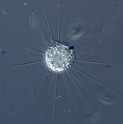
Actinophrys sol.jpg 960 × 960; 194 KB
-
Al photoresist pattern developed via Nomarski DIC.jpg 2,124 × 1,620; 536 KB
-
-
Arcella sp.jpg 512 × 512; 89 KB
-
Ascus.gif 136 × 140; 14 KB
-
Asymmetrical and Spherical.JPG 1,800 × 853; 474 KB
-
Asymmetry-of-Early-Endosome-Distribution-in-C.-elegans-Embryos-pone.0000493.s001.ogv 7.0 s, 548 × 198; 3.11 MB
-
Aurantiochytrium limacinum SR21.jpg 1,056 × 783; 167 KB
-
Botryococcus braunii.jpg 1,024 × 768; 277 KB
-
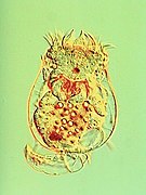
Brachionuscalyciflorus.jpg 2,157 × 2,880; 537 KB
-
Cefftriaxon antibiotic.jpg 2,074 × 2,776; 1.74 MB
-
Cercomonas sp.jpg 512 × 512; 73 KB
-
Chlorarachnion reptans.jpg 640 × 480; 104 KB
-
Closterium sp Div.jpg 640 × 480; 87 KB
-
Closterium sp.jpg 640 × 480; 76 KB
-
Coccoliths PL DIC.jpg 1,024 × 512; 83 KB
-
-
Cross-cut bone.jpg 2,776 × 2,074; 2.3 MB
-
Cryptosporidium muris.jpg 600 × 326; 12 KB
-
Crystals of IgG antibodies 02.jpg 2,776 × 2,074; 1.76 MB
-
Crystals of IgG antibodies.jpg 2,074 × 2,776; 1.71 MB
-
-
Diatomite DIC.jpg 640 × 640; 181 KB
-
Diatoms PhC DIC.jpg 1,024 × 512; 157 KB
-
Dinobryon sp.jpg 640 × 480; 115 KB
-
Dopamin.jpg 3,543 × 4,742; 4.13 MB
-
Elastic-Coupling-of-Nascent-apCAM-Adhesions-to-Flowing-Actin-Networks-pone.0073389.s007.ogv 18 s, 640 × 480; 3.26 MB
-
Elastic-Coupling-of-Nascent-apCAM-Adhesions-to-Flowing-Actin-Networks-pone.0073389.s008.ogv 1 min 52 s, 224 × 224; 1.47 MB
-
Elastic-Coupling-of-Nascent-apCAM-Adhesions-to-Flowing-Actin-Networks-pone.0073389.s009.ogv 26 s, 420 × 420; 1.55 MB
-
Elastic-Coupling-of-Nascent-apCAM-Adhesions-to-Flowing-Actin-Networks-pone.0073389.s012.ogv 7.1 s, 632 × 544; 6.41 MB
-
Etoposide 01.jpg 2,074 × 2,776; 2.27 MB
-
Euglena sp (250 19) Euglena sp. (protozoan).jpg 3,752 × 2,401; 1.56 MB
-
Exophiala phaeomuriformis.jpg 562 × 562; 224 KB
-
Fert6.JPG 577 × 823; 52 KB
-
Fert7.JPG 602 × 864; 55 KB
-
Glaucocystis nostochinearum.jpg 438 × 413; 34 KB
-
Glaucocystis sp.jpg 640 × 480; 123 KB
-
Glucose crystal.jpg 3,600 × 2,690; 2.25 MB
-
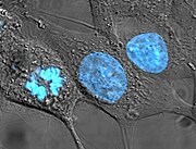
HeLa cells stained with Hoechst 33258.jpg 459 × 350; 30 KB
-
Hydrodictyon reticulatum.jpg 640 × 480; 122 KB
-
Hypermastigida.jpg 640 × 480; 141 KB
-
Hypomyces chrysospermus.jpg 236 × 334; 17 KB
-
Image DIC.tif 1,357 × 959; 2.71 MB
-
-
-
Mabthera crystals.jpg 2,776 × 2,074; 1.99 MB
-
Macrophage.jpg 1,280 × 1,024; 279 KB
-
Micrasterias radiata.jpg 640 × 480; 111 KB
-
Mikrofoto.de-Pleurosigma angulatum-3.jpg 1,000 × 667; 276 KB
-
Mikrofoto.de-Pleurosigma angulatum-4.jpg 1,000 × 667; 321 KB
-
Mikrofoto.de-Pleurosigma angulatum-5.jpg 1,000 × 667; 278 KB
-
Mikrofoto.de-Pleurosigma angulatum-6.jpg 1,000 × 667; 302 KB
-
Mikrofoto.de-Pleurosigma angulatum-7.jpg 1,000 × 667; 302 KB
-
Nuclearia sp Nikko (cropped).jpg 550 × 469; 100 KB
-
Nuclearia sp Nikko.jpg 640 × 640; 121 KB
-
Orciraptor attacking Actinotaenium.webp 613 × 452; 72 KB
-
Orciraptor-gr1a.jpg 1,688 × 740; 235 KB
-
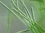
Oscillatoria sp.jpg 640 × 480; 120 KB
-
Parasite140120-fig3 Acanthamoeba keratitis Figure 3A.png 2,080 × 2,102; 2.38 MB
-
Parasite140120-fig3 Acanthamoeba keratitis Figure 3B.png 2,032 × 2,102; 2.71 MB
-
Parasite140120-fig3 Acanthamoeba keratitis.png 4,200 × 2,102; 5.19 MB
-
Parasite140120-fig4 Acanthamoeba keratitis Figure 4A.png 1,951 × 1,985; 765 KB
-
Parasite140120-fig4 Acanthamoeba keratitis Figure 4B.png 1,960 × 1,985; 741 KB
-
Parasite140120-fig4 Acanthamoeba keratitis Figure 4C.png 1,911 × 1,985; 709 KB
-
Parasite140120-fig4 Acanthamoeba keratitis.png 5,950 × 1,985; 2.2 MB
-
Parasite140120-fig5 Acanthamoeba keratitis.png 827 × 827; 979 KB
-
Partially etched silicon dioxide via Nomarski DIC.jpg 2,113 × 1,550; 755 KB
-
Pediastrum duplex.jpg 512 × 512; 118 KB
-
Perforated Actinotaenium cell with lid.webp 791 × 351; 30 KB
-
Phytoplankton Lake Chuzenji.jpg 640 × 512; 174 KB
-
Polykrikos and Strombidium.jpg 640 × 480; 129 KB
-
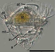
Pomatoceros lamarckii trochophore.jpg 243 × 229; 37 KB
-
Raphidiophrys contractilis.jpg 523 × 525; 100 KB
-
-
-
S cerevisiae under DIC microscopy.jpg 1,560 × 1,560; 610 KB
-
SAS-1-Is-a-C2-Domain-Protein-Critical-for-Centriole-Integrity-in-C.-elegans-pgen.1004777.s017.ogv 28 s, 480 × 320; 3.27 MB
-
Scenedesmus dimorphus.jpg 512 × 512; 96 KB
-
Shmoo yeast S cerevisiae.jpg 847 × 847; 184 KB
-
Synechococcus PCC 7002 DIC.jpg 1,687 × 1,491; 523 KB
-
Synura spp.jpg 1,200 × 960; 160 KB
-
Syringe.png 640 × 480; 318 KB
-
-
Trachelomonas sp.jpg 640 × 480; 66 KB
-
-
-
Водоросли пресноводного водоема 2.jpg 1,650 × 1,650; 677 KB


