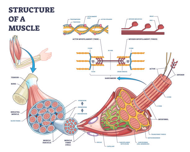
Structure of muscle with isolated myosin and actin closeup outline diagram. Labeled educational arm bone muscular inner parts detailed description with sarcomere magnification vector illustration.
Structure of muscle with isolated myosin and actin closeup outline diagram. Labeled educational arm bone muscular inner parts detailed description with sarcomere magnification vector illustration.
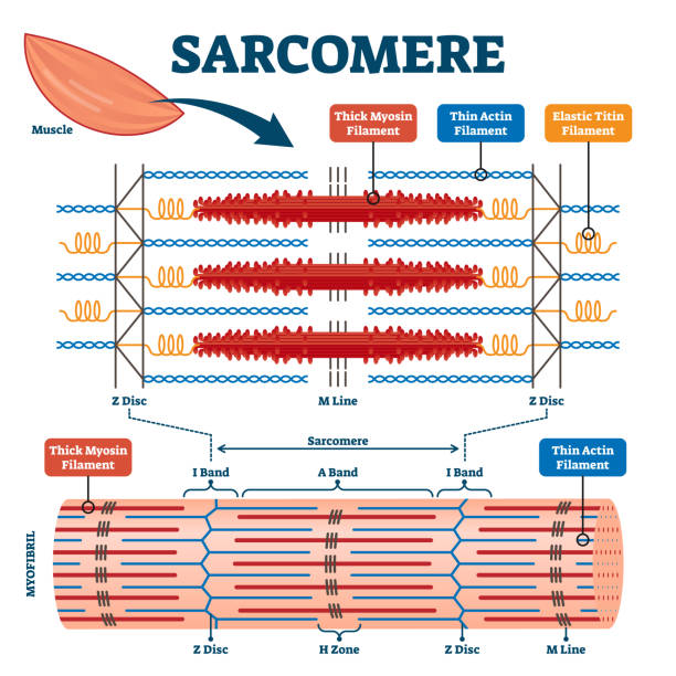
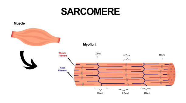
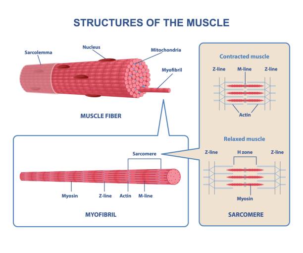
Sarcomere muscular biology scheme vector illustration. Myosin filaments, discs, lines and bands. Myofibril detailed labeled diagram. Sports educational health information. Muscular system anatomy.
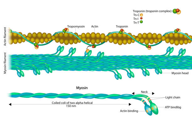
Actin filament and Myosin filament. Structure Myosin. Muscle Actin myosin interaction. Troponin or troponin complex.
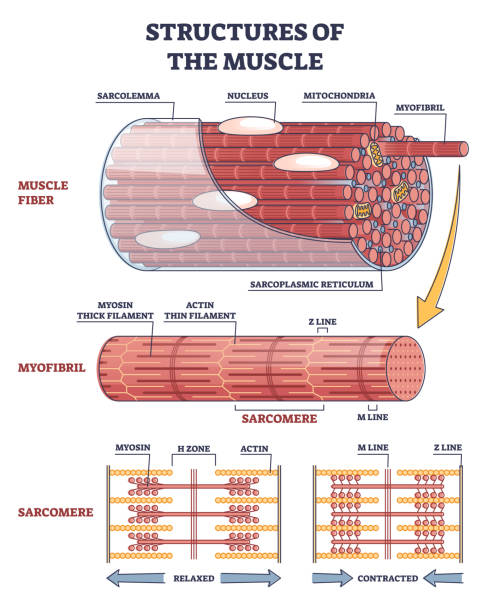
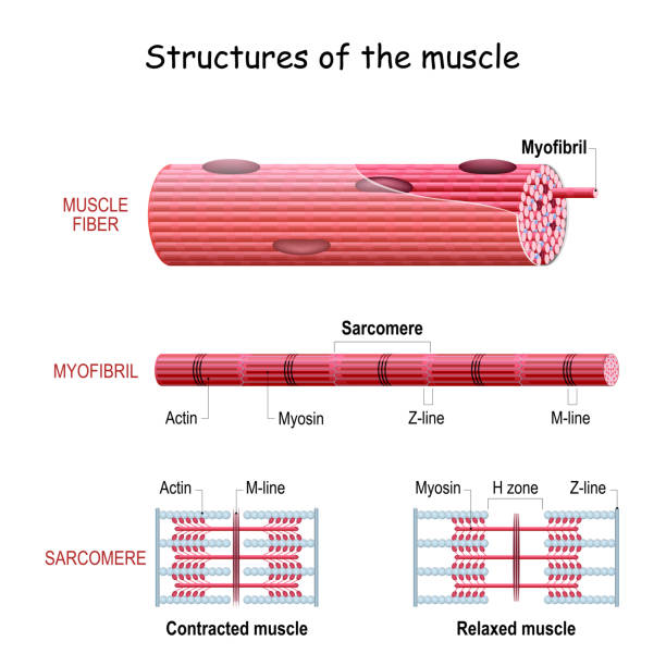
Structures of muscle with fiber, myofibril and sarcomere contraction outline diagram. Labeled educational isolated parts closeup description from anatomical and physiology sides vector illustration.
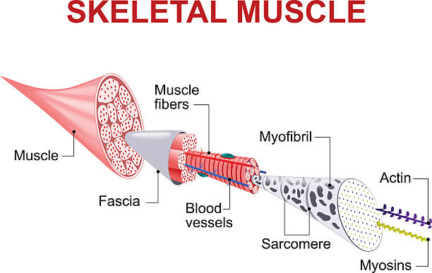
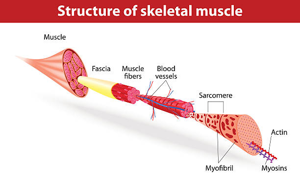
Skeletal muscles are organs of the vertebrate muscular system that are mostly attached by tendons to bones of the skeleton. The muscle cells of skeletal muscles are much longer than in the other types of muscle tissue, and are often known as muscle fibers

Muscle tissue is a soft tissue that composes muscles in animal bodies, and gives rise to muscles' ability to contract.
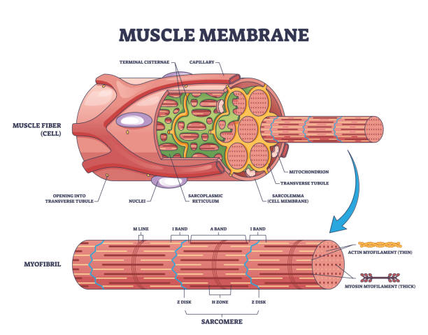
Muscle membrane or sarcolemma anatomical structure outline diagram. Labeled educational microscopic closeup with myofibril and fiber detailed description vector illustration. Band and zones scheme.
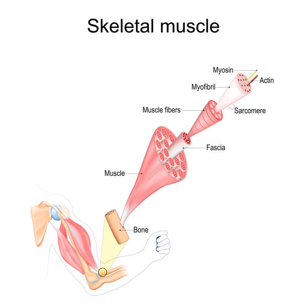
Skeletal Muscle anatomy. structure of Muscle fibers from Fascia and Tendon to Actin and Myosin. Vector poster
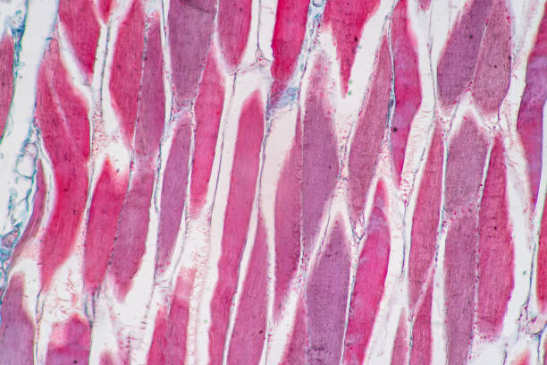
Characteristics of anatomy and Histological sample Striated (Skeletal) muscle of mammal Tissue under the microscope.

3D Isometric Flat Vector Conceptual Illustration of Structures Of Muscle , Medical Educational Diagram

High magnification light micrograph of striated skeletal muscle fibers stained with a silver method. The sarcomeres display alternating A (dark) and I (light) bands. A clear H zone stands out in the center of A bands.

Smooth muscle tissue. Anatomy of a relaxed and contracted smooth muscle cell

Anatomy of a Skeletal muscle fiber. Myofibril structure include Myosin, Z-line, M-line, Actin filaments, and Sarcomere. Isometric flat vector Illustration
Colorful structure skeletal muscle scheme on white background. Muscles contract by sliding myosin and filaments along each other. Myofibril with thin and thick filament. Flat vector illustration
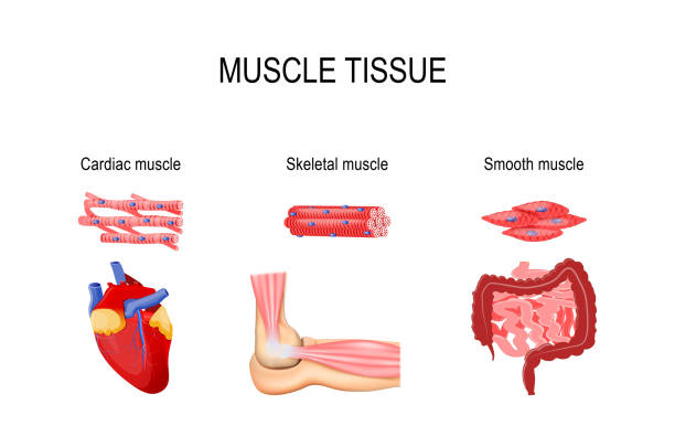
Types of muscle tissue. Skeletal muscle (elbow joint), smooth (gastrointestinal tract) and cardiac muscle (heart). Human internal organs and Muscle cells. vector illustration for medical, educational and science use
Each skeletal muscle fiber has many bundles of myofilaments. Each bundle is called a myofibril. This is what gives the muscle its striated appearance. The contractile units of the cells are called sarcomeres.
Structure Skeletal Muscle. myofibril with thin and thick filament. close up of a sarcomere. Muscles contract by sliding the myosin and actin filaments along each other. Biomedical Science. Mechanism of mechanical contraction
Vector illustration. Muscle Tissues. Each skeletal muscle fiber has many bundles of myofilaments. Each bundle is called a myofibril. This is what gives the muscle its striated appearance. The contractile units of the cells are called sarcomeres.

Transmission electron microscope micrographs showing the aspect of muscle fiber sarcomere components in longitudinal (below) and transverse (above) sections at the Z-line, I and A bands and H zone
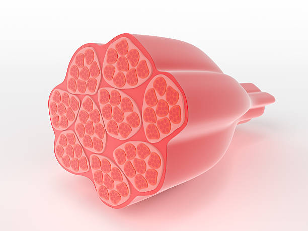
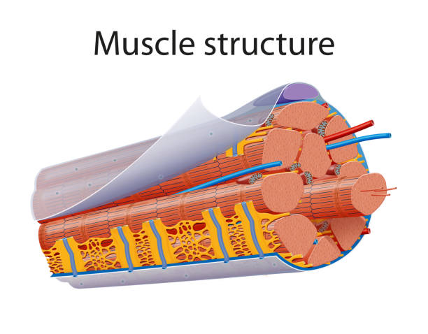
Structure of skeletal or voluntary muscle showing a cross-section through the cylindrical multi-nucleate fibres narrowing down to the attachment point which would attach to a bone in the body

Types of muscle tissue. Skeletal muscle, smooth muscle, cardiac muscle. Vector scheme.
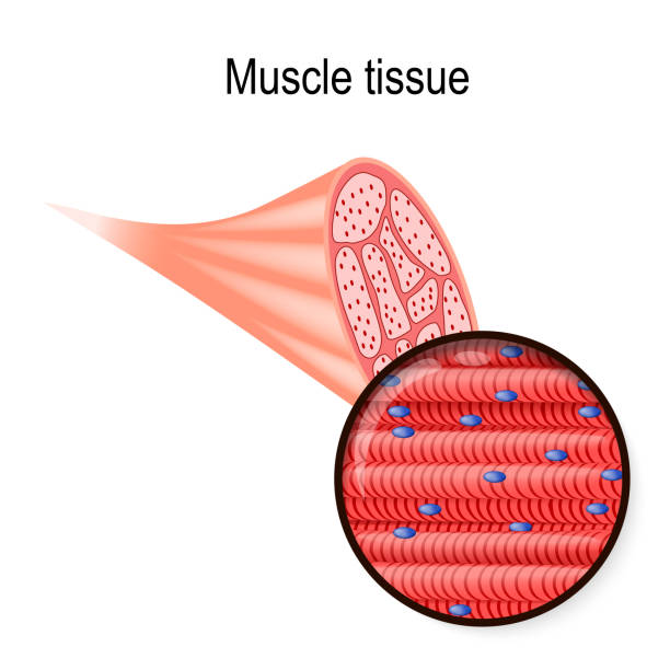
skeletal muscle. Tissue and fiber. Part of the biceps and close-up of muscle fibers. Vector illustration for biological, medical, science and educational use.

Muscular Health Concept. Tiny Doctors Characters at Huge Board with Infographics Presenting Skeletal, Cardiac and Smooth Musculature. Medicine, Muscles Anatomy. Cartoon People Vector Illustration

Skeletal muscle cells are tubular. They have multiple nuclei. Skeletal muscle is striated, it has an alternating pattern of light and darks bands.

Common cells of the human body illustration

Illustration of the muscles of the human eye
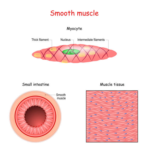
Structure of smooth muscle fibers. anatomy of Myocyte. Background of smooth muscle tissue. Set of vectors illustrations for education, sports and medical use.
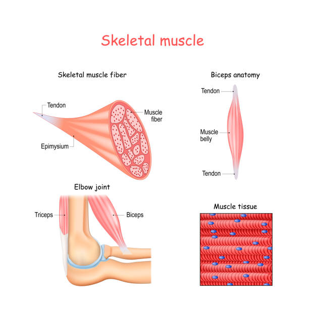
Structure of skeletal muscle fibers. Biceps and Triceps. Biceps anatomy. Background of muscle tissue. Set of vectors illustrations for education, sports and medical use.
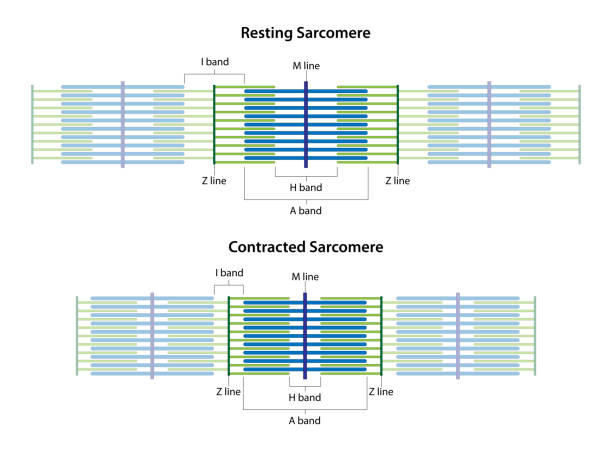
Sarcomeres in different functional stages: resting and contracted. Sarcomere showing the location of the I band, A band, H band, M line, and Z lines.

Structure of Cardiac muscle fibers. anatomy of cardiomyocyte. Background of heart muscle tissue. Set of vectors illustrations for education, sports and medical use.

Types of muscle tissue. Skeletal muscle, smooth muscle, cardiac muscle.

Human cardiac muscle cells on a white background

Tiny Doctor Character Presenting Intestines Smooth Musculature on Huge Screen with Infographics, Man Eating Fast Food Having Problem with Belly or Stomach Muscles. Cartoon People Vector Illustration

3D Isometric Flat Vector Conceptual Illustration of Muscle Fiber Types, Skeletal Muscle Structure

The structure of the muscle is fusiform. Infographics. Vector illustration on isolated background.

3D Isometric Flat Vector Conceptual Illustration of Skeletal Muscles, Anatomical Structure

Muscle relaxation, stretching, and contraction. Close-up of a Skeletal muscle fiber. Isometric flat vector Illustration

Characteristics of anatomy and Histological sample Striated (Skeletal) muscle of mammal Tissue under the microscope.

Characteristics of anatomy and Histological sample Striated (Skeletal) muscle of mammal Tissue under the microscope.

Characteristics of anatomy and Histological sample Striated (Skeletal) muscle of mammal Tissue under the microscope.

False colour transmission electron microscope (TEM) micrograph of a striated muscle fiber showing myofibrils (Z line, pink; I band, yellow; A band, green; H band, blue) and mitochondria (red).

False colour transmission electron microscope (TEM) micrograph of a striated muscle fiber showing myofibrils (Z line, red; I band, light blue; A band, brown; H band, blue) and mitochondria (green).

Characteristics of anatomy and Histological sample Striated (Skeletal) muscle of mammal Tissue under the microscope.

Cardiac muscle fibers' structure. Heart muscle tissue, anatomy of cardiomyocyte. Didactic scheme of anatomy of human muscular system. Involuntary control cells. Flat vector illustration

Muscle tissue. Close-up of skeletal muscle in biceps, cardiac muscle in heart, and smooth muscle in small intestine and colon. Myocytes. Vector illustration

Sarcolemma, structure of muscle fiber. Educational closeup anatomical diagram. Cell membrane surrounding a skeletal muscle fiber or a cardiomyocyte. Flat vector illustration

False colour transmission electron micrograph (TEM) showing cross-sectioned muscle myofibrils at the A-band (green) and at the I-band (blue) level, and mitochondria (red). Basal lamina (pink)
© 2025 iStockphoto LP. The iStock design is a trademark of iStockphoto LP. Browse millions of high-quality stock photos, illustrations, and videos.
Do Not Sell or Share