Spinal Mechanisms for Sensorimotor Integration
Vestibular System
Watch: The Vestibular System | Narrated animation | Quiz
Making Sense of Our Senses
Animation of semicircular canals and otolith receptors
[Vestibular system & Spaceflight. PBS, 4/11/97, 2:00 a.m.]
Key Takeaways
Two types of vestibular receptors signal information about rotation and tilt of the head
1. Semicircular canals signal angular acceleration of the head
2. Otolith receptors signal linear acceleration and static tilt of the head
• Utricle: horizontal plane
• Saccule: vertical plane
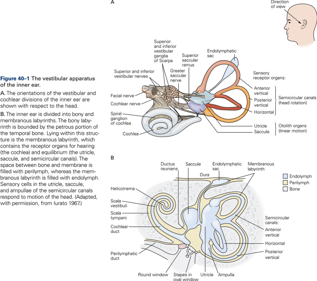
Location of the semicircular canals and otolith receptors with respect to the head
The semicircular canals sense head rotation
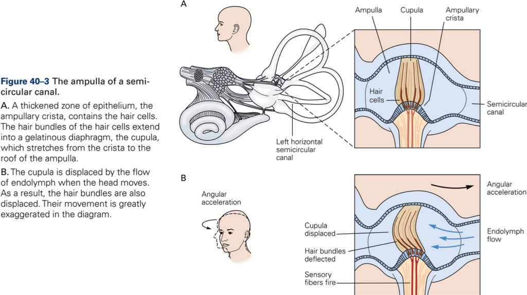
Three semicircular canals are arranged in planes perpendicular to one another. They are filled with fluid called endolymph. Each canal has a thickened portion called ampulla that contains specialized receptor cells, the vestibular hair cells. Within the ampulla, the hair cells lie on the ampullary crest. The ampullary crest is covered by a gelatinous mass called the cupula. When the head is rotated, the rotatory force exerted on the cupula by the fluid produces a displacement of the sensory hairs on the receptor cells.
Vestibular hair cells transduce mechanical stimuli into receptor potentials
The hairs are differentiated into 40-70 stereocilia and a single kinocillium. The stereocilia are of varying length and are arranged according to length, with the longest (kinocillium) on one end of the hair bundle. Hair cells release transmitter continuously and therefore fire action potentials continuously. Bending hair cells in the direction of the kinocilium depolarizes the cell, and thus increases firing rate, whereas bending away from the kinocilium hyperpolarizes the cell, and thus reduces the firing rate.
Example: head turns to the left. Hairs in left horizontal canal bend towards kinocilium, which increases firing rate in afferent VIIIth nerve fibers. Hairs in right horizontal canal bend away from kinocilium, which decreases firing rate in afferent VIIIth nerve fibers.
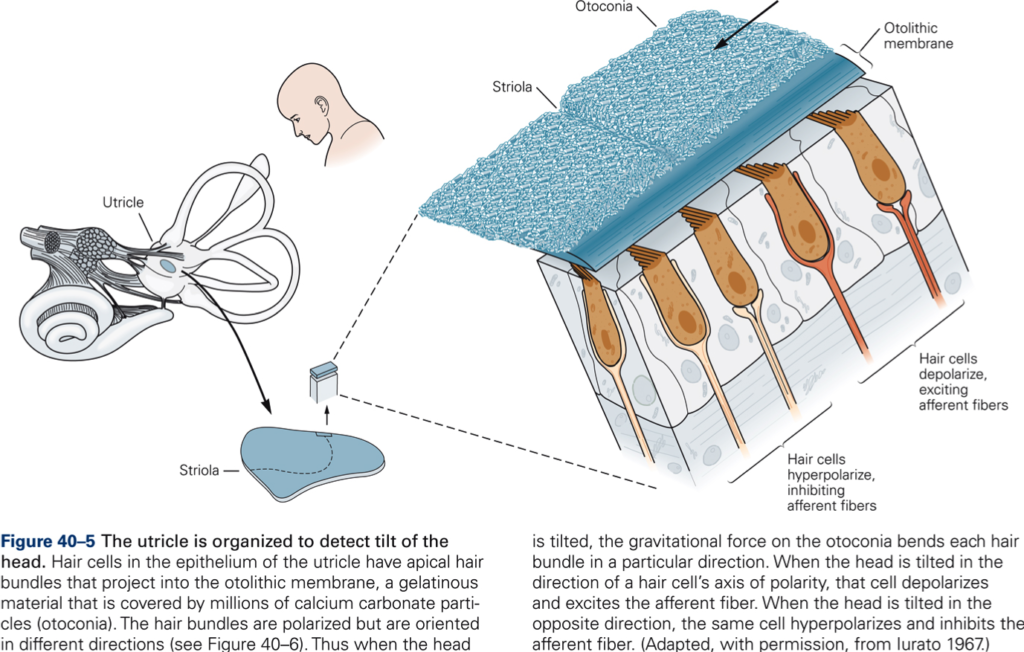
The utricle is organized to detect tilt of the head
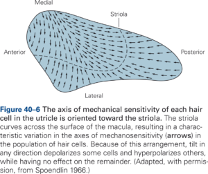
Within each otolith receptor, hair cells are located in an area called the macula. The macula is covered with gelatinous mass in which are embedded crystals of calcium carbonate, called otoliths. The macula of the utricle is oriented in the horizontal plane, that of saccule in the vertical plane. Both utricle and saccule detect static head tilt by bending of hair cells by otoliths. They also respond to linear acceleration of the head, utricle to linear acceleration in the horizontal plane, saccule to linear acceleration in the vertical plane. Different hair cells in the macula of the utricle are arranged in different directions so that the utricle can detect tilt of the head in any direction. All utricular hair cells together provide accurate information about head position.
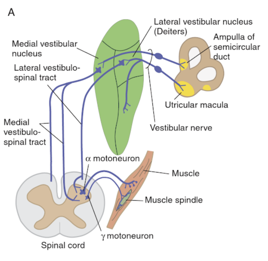
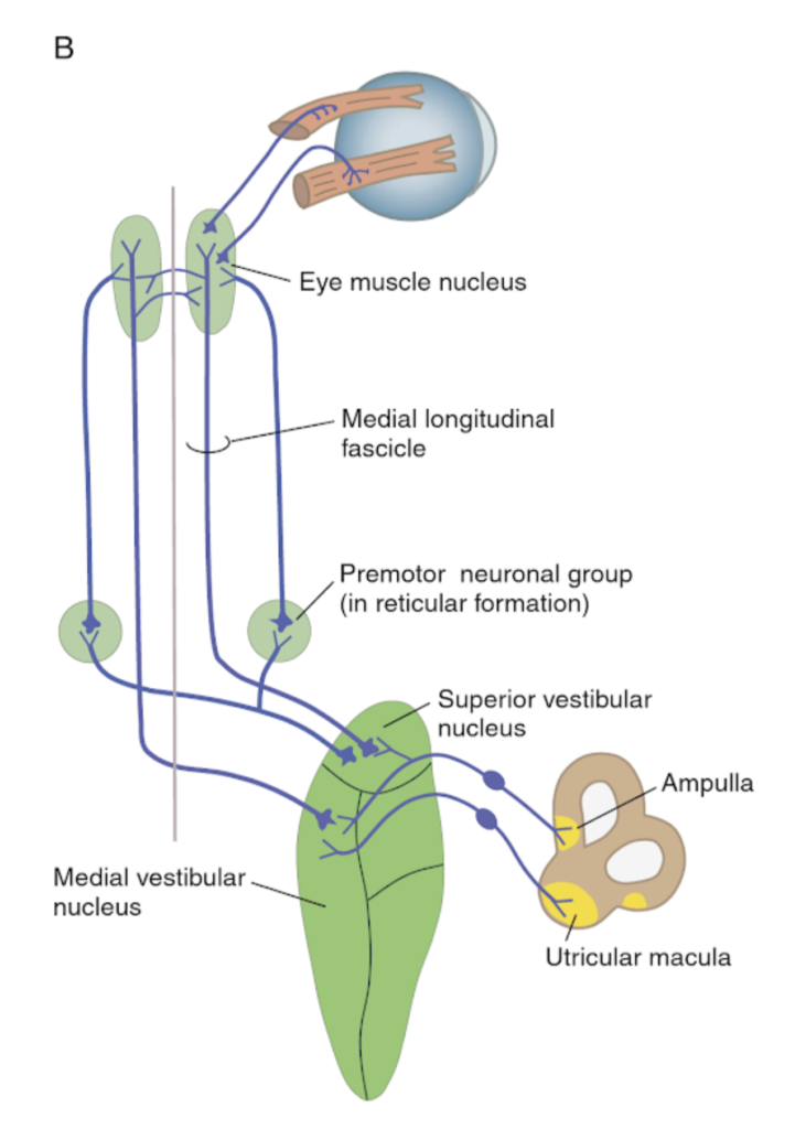
Central connections of receptors to vestibular nuclei
Lateral vestibular nucleus (Deiters)
- Vestibuloocular reflex (VOR)
- Posture (limb muscles via lateral vestibular tract)
Medial and superior vestibular nuclei
- Vestibuloocular reflex (VOR)
- Posture (neck muscles via medial longitudinal fasciculus, MLF)
Inferior vestibular nucleus
- contributes to vestibulospinal and vestibuloreticular pathways
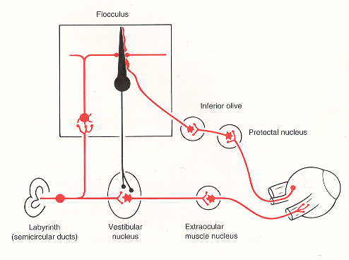
The vestibuloocular reflex (VOR) aligns eyes and head to stabilize images on the retina

