Thoracic cage
The thoracic cage is part of the axial skeleton (also known as the rib cage), and consists of 24 ribs, the sternum, costal cartilage, and the 12 thoracic vertebrae. The thoracic cage makes up the skeleton for the thoracic wall, and provides the attachments needed for the muscles of the neck, thorax, upper abdomen, and back. It protects the heart, lungs, and thoracic viscera and it helps with breathing respiration.
Functions
The primary functions of the thoracic cage are protect to the heart and lungs, support the clavicle and scapula, plus perform hemopoiesis. The thoracic cage helps the lungs with the inhalation that is accomplished when the muscular diaphragm which is at the floor of the thoracic cavity,contracts and flattens, while contraction of intercostal muscles lift the thoracic cage in and out.[1]
Parts
Thoracic Cage is made up of the 12 thoracic vertebrae, 24 ribs that are in 12 pairs, costal cartilage, and the sternum. Together with the skin, associated fasica, and the muscles they all make up the thoracic wall.[2]
Ribs

Ribs are curved and flattened bones that contribute to the wall of the thorax. The ribs articulate posteriorly with T1-T12 thoracic vertebrae, most attach anteriorly by means of their coastal cartlidges. A typical rib is twisted along its axis and has a sharp bend in the shaft called the coastal angle. Atypical rib, Ribs 1-2 are short,flat, and tilted forward more than other ribs. There are three types of ribs that make up the thoracic cage: true ribs, false ribs, and floating ribs.
True ribs are the first seven pairs of rib bones (beginning at the top of the sternum) are called. They connect to the spine at the back, and connect to the sternum by costal cartilage in the front. Costal cartilage is elastic and allows the ribcage to expand during respiration.
False ribs are the next three pairs of rib bones (8, 9 and 10). Like the true ribs, false ribs are connected to the spine at the back. The primary difference in true ribs and false ribs comes in where the false ribs connect in the front. Instead of connecting to the sternum, false ribs actually connect to the lowest true ribs.
Floating ribs are the last two pairs of ribs (11 and 12) are the smallest of all of the rib bones. They get the name "floating rib" because they are connected to the spin at the back, but are not connected to anything at the front, which is why they are called floating ribs. [3]
Sternum
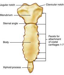
The sternum is a flat and elongated bone that forms the middle of the anterior part of the thoracic cage. The sternum consists of 3 parts those parts are the manubrium, the body of the sternum, and the xiphoid process.
The manubrium is the thickest of the three parts of the sternum. It articulates with the coastal cartilage of the very first rib, and also the superior half of the articular surface of the costal cartilage of the second rib.
The body of the sternum is much longer, narrower, and thinner than the manubrium that extends down to the front of the chest wall. It is located between both the manubrium and the xiphoid process. The body artiuculates with the coastal cartlidges from the second to the seventh ribs.
The xiphoid process is the smallest component in the sternum. It is a small cartilaginous structure that can vary in shape from pointed to blunt.[4]
Thoracic Vertebrae
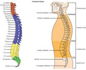
The thoracic vertebrae is a group of 12 small bones that make form the sapine of the thorax. The vertebrae is independent, have vetebral arches, and have several processes for muscular and articular connections. They have coastal facets on their traverse processes for articulation with the turbacles of the ribs.[1]
Coastal Cartlidge
The costal cartilage are segments of cartilage that connect the sternum to the ribs and help to extend the ribs into a forward motion. This cartilage also contributes to elasticity within the walls of the thorax, allowing the chest to expand during respiration. There are twelve costal cartilage sections. Each has two cartilages, extremities, and borders. Seven pairs of the costal cartilage are connected to the sternum. Two of the costal cartilage sections are pointed, ending in the walls of the abdomen. Three pairs of costal cartilage are articulated with the preceding ribs. The anterior surfaces are convex, while the posterior surfaces are concave. The borders are superior and inferior in nature. The superior section is concave, while the inferior is convex. The eleventh and twelfth costal cartilage segments are pointed and are free of attachments.[5]
References
- ↑ 1.0 1.1 [/www.kenhub.com/en/library/anatomy/thoracic-cage] "Kenhub". Web. Accessed on OCtober 2, 2017. Unknown Author. Cite error: Invalid
<ref>tag; name "Kenhub" defined multiple times with different content - ↑ [1] Visible Body. Web. last accessed October 6, 2017. Unknown author
- ↑ [2] "Unit IV". Web. Accessed on October 3, 2017. Unknown Author.
- ↑ [3] "Kenhub". Web. Accessed on October 2, 2017. Unknown Author.
- ↑ [4] Health Guide Info. Web. last accessed October 3, 2017. Unknown author
| ||||||||||||||||||||
