Heart
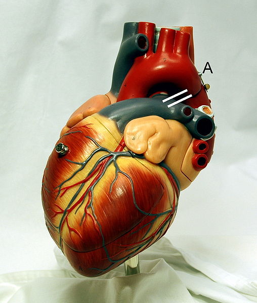
The heart is an organ of the circulatory system responsible for pumping blood. It receives incoming blood from the other organs of the body in vessels called veins and pumps it back out in arteries. It is one of the most important organs in the entire human body because it supplies all the cells, tissues, and organs with a constant supply of life sustaining nutrients and carries away waste. The heart is a powerful muscle that works continuously without fatiguing. It weighs about 10.5 ounces and is cone-shaped. The three layers which are epicardium, myocardium, and endocardium make up the outer wall of the human heart. The cardiac muscle which is the average heart’s muscle contracts and slows down about 50 to 80 times per minute. Moreover, there are various heart diseases which affect people's health problems severally like coronary artery heart disease, cardiomyopathies, and Hypertensive heart diseases. The heart diseases can be expressed by various symptoms such as chest pain, difficulty breathing, heartbeat problems, dizziness, nausea, or sweating. People who have a heart disease should change their lifestyle or should see the doctor. [1]
Structure

The three layers which are epicardium, myocardium, and endocardium, make up the outer wall of the human heart. The heart is composed of four chambers, two atria and two ventricles. Among the four cavities, two of these cavities are atria and the other two are ventricles. In the left side of the heart, there is one ventricle and one atrium. The other ventricle and atrium located at the right side of the heart. The ventricles have thick walls and the atria have thin walls. On the other hand, both the right and the left atria have auricles which refer to the entire atrium. [2]. The Septum divides into the right and left side of the heart. A valve attaches to the ventricles. The largest blood vessel is called the aorta which carries blood in the heart. The other largest blood vessel which is called the pulmonary artery attaches to the lungs. The superior vena cava and the inferior vena cava are the largest veins that carry the blood. [3]
Function
The atrium's (singular of atria) meaning is the entrance, and the ventricle's meaning is the belly. Thus, the two atria are the entrances to the heart and two ventricles are bellies of the heart. After the two atria receive the chambers, two ventricles discharge the chambers. Some large veins like pulmonary veins, the superior vena cava, and inferior vena cava bring blood into the heart. The right and left atrium are separated by the muscular wall they both share which is called the interventricular septum. The right atrium leads the blood to the right ventricle by the right atrioventricular canal which helps to regulate how blood flows through the canal. Blood from the left atrium is separated by the left atrioventricular valve which helps to open valve in various response. The papillary muscles usually connect to the valve via chordae tendineae.[2]
The Flow of Blood Through the Heart
Blood flows down to the left atrioventricular canal and the left ventricle. After that, all of the blood in the left ventricle flows out of the aorta, which only flows in one direction. At first, it enters the left atrium. It then travels into the left atrioventricular canal and the left atrioventricular valve. After that, it enters into left ventricle, passing through the aortic semilunar valve and exits the aorta. After the blood comes back from the inferior vena cava and the coronary sinus, it travels into the right atrium, the right atrioventricular canal, the right atrioventricular valve, and right ventricle. And then it passes through the pulmonary semilunar valve and the pulmonary trunk. After that, the blood just goes to lungs again.[4]
Disease
Heart disease is term to describe various diseases that can affect your heart. The heart disease can express various symptoms like chest pain, breath difficulty, heartbeat problem, dizziness, nausea, or sweating. [5]
Coronary artery heart disease
Coronary heart disease is that plaque which consists of cholesterol, fat, and other things. It builds up in the coronary arteries. When it is in that situation, it is hard to get blood back to their heart muscle. It can be caused by a heart attack or other serious problem. Coronary heart disease is a very affective disease, that can affect both men and women. Many people can prevent this disease by maintaining and regulating their life style.[6]
Cardiomyopathy
Cardiomyopathy is a heart muscle disease. There are two different types of cardiomyopathy: extrinsic cardiomyophathies and intrinsic cardiomyophathies. Most cardiomyophthies are usually extrinsic cardiomyophathies.
Extrinsic cardiomyopathies is disease that outside of the myocardium.
- Alcoholic cardiomyopathy.
- Nutritional diseases.
- Ischemic cardiomyopathy.
- Hypertensive cardiomyopathy.
- Myocardiodystrophy.[7]
Intrinsic cardiomyopathies is a disease that causes weakness of the muscle of the heart.
- Dilated cardiomyopathy(DCM)- There is a problem with the heart's pumping funtion.
- Hypertrophic cardiomyopathy (HCM)- There is a problem with the heart's muscle.
- Noncompaction Cardiomyopathy- There is a problem with the heart's left ventricle wall.
- Restrictive cardiomyopathy (RCM)- There is a problem with the heart's ventricle.[7]
Hypertensive heart disease
Hypertension is the high blood pressure in the heart.
There are various treatments for heart disease. People who have heart disease should change their lifestyle. Nowadays, many people don't exercise due to their busy life. Although many people usually eat vitamins or minerals for their health, it is better to exercise regularly at least 30 minutes every day to prevent heart disease. Moreover, people who have heart disease should reduce their intake of fat and sodium. If it is not enough, they should go to see a doctor. The doctors may make your heart better. There are few more options like surgery or other medical procedures.[8]
Origin
Even the evolutionists can see design in the heart
| “ | We conclude that there is a design in the evolution of the venous connections of the heart, pectinate muscles, atrioventricular valves,' left ventricular tendons, outflow tracts, and great arteries. One neglected aspect in the study of evolution is that of anticipation. Fish atria and ventricles appear to have a built-in provision for becoming updated to the human 4-chambered structure. This transformation is achieved in stages: the truncus yields the great arteries, appropriate shifting takes place in the great arteries, the left ventricle decreases in sponginess and increases in the size of its lumen, the chordopapillary apparatusbecomes more sophisticated, the coronary circulation undergoes changes, and the ventricular septal defect closes. This evolutionary progression points to a master design and plan for countless millennia. (Solomon Victor, Vijaya M. Nayak, Raveen Rajasingh, "Evolution of the Ventricles," Texas Heart Institute Journal, Vol.26(3):168-75 (1999), internal citations removed.) | ” |
Video
See also
References
- ↑ Human heart Wikipedia, Wikimedia Foundation, 9 May, 2011.
- ↑ 2.0 2.1 Wile, Jay L., and Shannon, Marilyn M. The Human Body: Fearfully and Wonderfully Made!. Cincinnati: Apologia Educational Ministries, Inc., 2001. Print.(Pg 339)
- ↑ Structure of the Human Heart Unknown, The Franklin Institute, 2011.
- ↑ Heart Blood Jennifer Heisler, About.com, August 06,2009.
- ↑ Heart disease definition Mayo Clinic Staff, Mayo Foundation, 12 January, 2011.
- ↑ What Is Coronary Artery Disease?National Heart Lung and Blood Institute, February 2009.
- ↑ 7.0 7.1 7.2 Heart disease Wikipedia, Wikimedia Foundation, 26 April, 2011.
- ↑ Heart Disease TreatmentsMayo Clinic Staff, Mayo Foundation, 12 January, 2011.
Additional Information
| ||||||||||||||||||||
