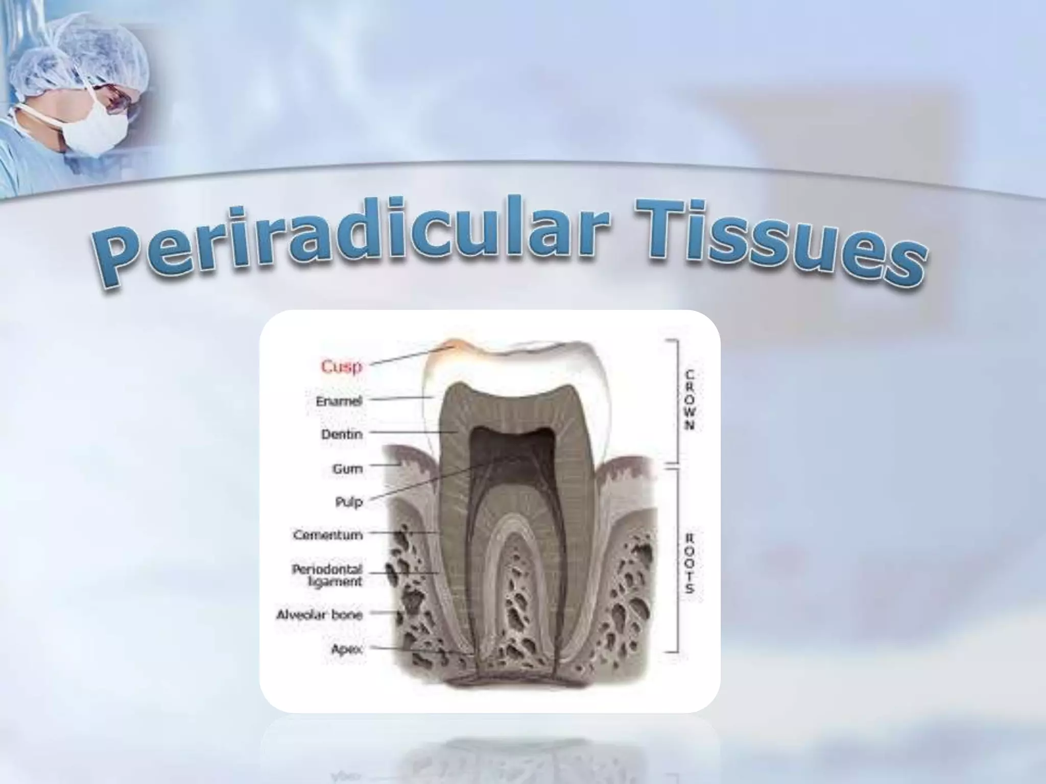
This document discusses the anatomy and histopathology of the periodontium, which consists of cementum, periodontal ligament, and alveolar bone. It describes the different types of cementum and cells found in the periodontal ligament. Chronic periapical lesions are discussed, including their etiology, clinical features, classifications, and examples such as chronic apical periodontitis and periapical granuloma. Treatment options are mentioned for various pathological conditions like symptomatic apical periodontitis.
This document provides an overview of pulp and periapical pathologies. It begins by defining the dental pulp and pulpitis. It then covers causes of pulp disease including physical, chemical, and bacterial factors. It classifies pulp disease and discusses factors affecting the pulp response. It details the pathways of bacterial invasion of the pulp and describes different types of reversible and irreversible pulpitis. It also discusses chronic hyperplastic pulpitis, gangrenous necrosis of the pulp, and diseases of the periapical tissues like acute and chronic apical periodontitis and periapical abscess.
Teeth don’t possess regenerative ability found in most other tissues. Therefore, once enamel & dentin are lost as a result of caries, trauma or wear, restorative material must be used, to reestablish form & function.
Teeth require preparation to receive restoration & these preparations must be based on fundamental principles, which are discussed in this presentation, from which basic criteria can be developed to help predict the success of prosthodontic treatment.
The document defines and describes periodontal pockets. It notes that periodontal pockets can be classified based on their location relative to the alveolar bone as either suprabony or infrabony. Suprabony pockets have bone loss horizontally while infrabony pockets have bone loss vertically. The document also discusses the pathogenesis of pocket formation, clinical features, histopathology, and diagnosis and probing of periodontal pockets.
Splinting is one of the oldest forms of aids to periodontal therapy. By redistribution of forces on the affected teeth the splint minimizes the effects caused by loss of support. Splinting teeth to each other allows weakened teeth to be supported by neighbouring teeth. This presentation reviews the rationale, techniques, advantages and ill effects of stabilization of teeth by splinting as an aid to periodontal therapy. With the acceptance and clinical predictability of adhesive procedures, the use of conservative bonding techniques to splint teeth offers a useful alternative to more invasive restorative procedures. Loss of tooth-supporting structures results in tooth mobility. Increased tooth mobility adversely affects function, aesthetics, and the patient’s comfort. Splints are used to overcome all these problems. When faced with the dilemma of how to manage periodontally compromised teeth, splinting of mobile teeth to stronger adjacent teeth is a viable option. This prolongs the life expectancy of loose teeth, gives stability for the periodontium to reattach, and improves comfort, function and aesthetics.
This document discusses various classifications and types of periapical diseases including symptomatic and asymptomatic apical periodontitis, acute alveolar abscess, phoenix abscess, persistent apical periodontitis, chronic alveolar abscess, radicular cyst, condensing osteitis, and different types of external and internal root resorption. It provides definitions, causes, symptoms, diagnostic features and treatment options for each condition.
An inlay may cap none, or may cap all but one cusp.
Sturdevant’s 4th ed. page579
Inlays may be used as single-tooth restorations for proximo-occlusal or gingival lesions with minimal to moderate extensions
Shillingburg page 1
An inlay may be defined as a restoration which has been constructed out of mouth from gold, porcelain, or other material & then cemented into the prepared cavity of a tooth.
William McGehee pg410
Local Anesthesia in Oral and Maxillofacial SurgerySapna Vadera
Local anaesthesia is a loss of sensation in a circumscribed area without loss of consciousness. The document discusses the history of local anaesthesia from ancient times to modern developments. It also covers the desirable properties, mechanisms of action, classifications, pharmacology and clinical aspects of local anaesthetics. The summary provides a high-level overview of the key topics covered in the document relating to the definition, history, properties and mechanisms of local anaesthetics.
This document discusses Necrotizing Ulcerative Gingivitis (NUG), also known as trench mouth. It defines NUG as a microbial disease of the gingiva caused by an impaired host response. Key clinical features include necrosis of gingival tissue and pain. Diagnosis is based on these clinical findings and microscopic examination. Management involves reducing the microbial load, removing necrotic tissue, treating any systemic conditions, and supportive periodontal therapy. Prognosis is generally good with treatment but recurrence is possible without ongoing maintenance of oral hygiene.
4.furcation involvement and its treatmentpunitnaidu07
This document discusses furcation involvement in multi-rooted teeth. It begins with introductions and definitions, then describes the anatomy of furcated teeth. Several classifications of furcation involvement are presented based on horizontal and vertical bone loss. Potential etiologies include dental plaque, local anatomic factors like furcation dimensions and root concavities, developmental anomalies, trauma, caries, and pulpal pathology. Diagnosis and various treatment options are also covered, along with prognostic factors and conclusions.
Direct pulp capping involves placing a protective material directly over an exposed dental pulp to encourage healing and formation of reparative dentin. It is a conservative treatment alternative to root canal therapy to save a tooth's vitality when the exposure is small. Success rates range widely from 13-98% depending on factors like the type of exposure, quality of the restoration, and operator skill. Calcium hydroxide and mineral trioxide aggregate are commonly used capping materials that induce healing, but newer options like Biodentine show promise as well. Future trends may involve techniques like lasers, gene therapy, or stem cells to further improve pulp capping outcomes.
This document provides an overview of endodontics. It defines endodontics as the branch of dentistry concerned with the morphology, physiology, and pathology of the dental pulp and periradicular tissues. The scope of endodontics includes treating diseases of the pulp, such as pulpal inflammation and infection, through both nonsurgical and surgical root canal treatment and periradicular surgery. Pathogenesis is discussed, noting that while physical and chemical factors can induce inflammation, microbial infection is essential for progression of pulpal and periradicular disease. Routes of canal infection and the focal infection theory are also summarized.
- Endodontic instruments have evolved over time to have standardized sizes and tapers. Ingle and LeVine suggested standardizing diameters that increase by 0.05mm while maintaining a constant taper.
- Instruments are now numbered 6-140 based on tip diameter in hundredths of a millimeter. The diameter increases 0.32mm over the first 16mm of the instrument.
- Instruments can be hand operated, low-speed, engine-driven, or ultrasonic/sonic and are used for cleaning and shaping root canals.
The document discusses the dentinogenic concept introduced by John P. Frush and Roland D. Fisher. This concept states that the form of one's teeth is determined by factors like sex, age, and personality. Masculine features include prominent, square teeth while feminine features include smaller, rounded teeth. Personality is also a factor, with vigorous people having squarer teeth and delicate people having smaller, symmetrically arranged teeth. Age affects features like shade, wear, and the shape of the smiling line and canines. Dentinogenic restorations aim to incorporate these factors to create natural-looking teeth according to one's sex, personality, and age.
The document discusses cysts of the jaws, including their classification and pathogenesis. It focuses on odontogenic cysts and developmental cysts. Specifically, it describes a dentigerous cyst as an odontogenic cyst that surrounds the crown of an impacted tooth, caused by fluid accumulation between the reduced enamel epithelium and enamel surface, resulting in a cyst enclosing the tooth crown. Dentigerous cysts usually involve permanent teeth, often third molars or cuspids. They present as well-defined radiolucencies associated with unerupted teeth on imaging.
This document discusses various flap techniques used in periodontal surgery. It defines flaps as sections of gingiva and mucosa surgically separated from underlying tissues to provide access to bone and roots. Full and partial thickness flaps are classified based on the depth of tissue reflection. Techniques include the modified Widman flap, undisplaced flap, apically displaced flap, papilla preservation flap, and techniques for distal molar surgery. Healing after flap surgery is described in stages from initial clot formation to establishment of new connective tissue attachment after 4 weeks.
1. Gingival recession is the exposure of root surface caused by an apical shift in gingival position. It can be classified as visible, hidden, localized, or generalized.
2. Miller and Atkin & Sullivan classified gingival recession defects based on their location and amount of bone loss. Common causes of recession include age, faulty brushing technique, tooth malposition, gingival inflammation, abnormal frenal attachment, and masochistic habits.
3. Recession can be treated non-surgically through modifying risks or surgically through pedicle or free soft tissue grafts to cover exposed root surfaces and reduce sensitivity.
This document discusses the potential for a dental caries vaccine. It begins by defining dental caries and explaining why it is a major public health problem. It then covers how the immune system works and classifications of immunity. Key aspects of the microbiology of dental caries are explained, focusing on Streptococcus mutans and its antigenic determinants. The document discusses the need for a caries vaccine, potential routes of administration including mucosal and systemic routes, and advantages and disadvantages of passive immunization approaches. It concludes by considering the public health perspective on a potential caries vaccine and analyzing whether it could help reduce the global burden of dental caries.
This document discusses gingival inflammation and gingivitis. It begins by defining inflammation and describing the cardinal signs. It then outlines the stages of gingivitis from initial to established to advanced/periodontitis. Microorganisms attached to teeth secrete enzymes that damage tissues and widen junctional epithelium, allowing bacterial products to access connective tissue and activate immune cells. Studies showed that not practicing oral hygiene led to plaque buildup and gingivitis within 10-21 days. Gingivitis is characterized by redness, swelling, bleeding and is prevalent worldwide. The document discusses features, course, distribution and systemic influences of gingival inflammation.
This document discusses red and white lesions of the oral cavity, focusing on oral candidiasis. It describes the various types of oral candidiasis including pseudomembranous, erythematous, chronic plaque-type, and median rhomboid glossitis. Predisposing factors, clinical findings, diagnosis, treatment with antifungal medications or surgery, and prognosis are summarized for each type. Chronic hyperplastic candidiasis may require long-term antifungal therapy or surgery due to risk of recurrence. Overall prognosis is generally good if predisposing factors can be addressed.
This document summarizes different techniques for pulp capping including direct and indirect pulp capping. It discusses the history of various materials used for pulp capping such as calcium hydroxide and mineral trioxide aggregate. Key points covered include the indications and contraindications for pulp capping, the ideal properties of capping materials, and the histological response after pulp capping including zone formation. The document provides an overview of pulp capping procedures and factors influencing their success.
- Trauma from occlusion occurs when occlusal forces exceed the adaptive capacity of the periodontium, causing injury. It can be acute or chronic.
- The magnitude, direction, duration, and frequency of forces impact the periodontium's ability to adapt. Excessive pressure or tension can damage tissues.
- Primary trauma from occlusion is caused by changes in occlusal forces, while secondary trauma occurs when reduced bone support impairs the tissues' resistance to normal forces.
- The periodontium responds to trauma in three stages - injury, repair through new tissue formation, and adaptive remodeling to better withstand forces. Trauma can cause reversible damage if forces are reduced, or lead to irreversible injury if
1. The document discusses electronic apex locators (EALs), which are devices used to determine the working length of a root canal without radiography.
2. It describes the six generations of EALs, from first-generation resistance-based devices to sixth-generation adaptive apex locators. Key EAL models are provided for each generation.
3. The mechanisms of EALs are explained, noting they measure changes in resistance or impedance as a file tip approaches the apex. Accuracy and limitations of each generation are summarized.
Necrotizing gingivostomatitis is an inflammatory condition affecting the gingiva and mouth that is caused by fusiform bacteria and spirochetes. It is characterized by painful, red gingiva with crater-like sores between teeth, bleeding gums, bad breath, and fever. Examination of affected tissue shows many bacteria and white blood cells. Histologically, there is gingival ulceration and necrosis with an inflammatory cell infiltrate. Treatment involves cleaning the mouth with antiseptics followed by scaling and antibiotics to resolve the infection.
In this lecture I explain in step-by-step fashion the basics of Indirect Pulp Capping Procedure. a photo guide is attached to the guide to aid in better understanding of the topic
The document discusses focal infection and inflammation in the dental pulp. It describes how microorganisms from infected dental sites can spread through the bloodstream and lymphatic system, potentially causing infection elsewhere. When the pulp becomes inflamed in response to injury or infection, it undergoes degenerative or proliferative changes. The "FISH" study identified four zones of tissue reaction around an infected area: the zone of infection, contamination, irritation, and stimulation. Kronfeld's mountain pass theory applied this concept to explain the inflammatory response in the periapex as a defensive reaction analogous to a military mobilization.
This document discusses the management of deep dental caries. It defines affected dentin as softened but not infected, while infected dentin is both softened and contaminated with bacteria. Various treatment modalities are described depending on whether the pulp is exposed and vital or non-vital. Factors like remaining dentin thickness and choice of restorative material influence pulpal response and reactionary dentin deposition. Materials used for pulp capping include calcium hydroxide, MTA, Biodentine and glass ionomer cements. Indirect and direct pulp capping techniques are also outlined.
This document provides an overview of abscesses of the periodontium, specifically focusing on periodontal abscesses. It defines a periodontal abscess and classifies them based on location, course, number, affected tissue, and cause. Periodontal abscesses are most prevalent in molar sites and those with pre-existing periodontal pockets. They can be caused by factors like untreated periodontitis, foreign bodies, or changes after periodontal procedures or antibiotics. The pathogenesis involves bacterial entry triggering an inflammatory response that leads to tissue destruction and pus formation.
this seminar is talking about one of the most important topics for any dentist in the world (pulp and periapical diseases)
i hope it will be helpful for you
The document provides an overview of periapical diseases and their classification. It begins with an introduction to periapical diseases and their causes. The document then classifies periapical diseases into symptomatic and asymptomatic categories. Within each category, it describes specific conditions such as symptomatic apical periodontitis, acute alveolar abscess, chronic alveolar abscess, radicular cyst, and condensing osteitis. For each condition, it discusses causes, symptoms, diagnosis, differential diagnosis, and treatment. The document provides a comprehensive review of different periapical diseases and conditions that can affect the tissues around the root apex.
The document discusses pulp and periapical diseases. It begins by describing the pathobiology of the periapex and classifying pulpoperipaical pathoses. It then discusses the classifications of Franklin Weine and the WHO for pulpoperipaical lesions. Key lesions discussed include acute and chronic apical periodontitis, periapical granuloma, periapical cyst, condensing osteitis, and Ludwig's angina. Differential diagnoses and clinical signs and symptoms are also covered.
This document discusses osteomyelitis, an inflammatory process that affects bones. It begins by defining osteomyelitis and listing predisposing factors. It then discusses various classifications of osteomyelitis including acute suppurative, chronic, diffuse sclerosing, focal sclerosing, proliferative periostitis, and alveolar osteitis. For each classification, it provides details on clinical features, pathogenesis, radiographic findings, and treatment approaches.
This document provides an overview of periradicular diseases, including the normal periradicular tissues of cementum, periodontal ligament, and alveolar process. It describes various periradicular pathologies such as acute apical periodontitis, periapical abscess, chronic apical periodontitis, and periapical cyst. Acute apical periodontitis presents as pain and swelling, while chronic apical periodontitis often causes no symptoms. A periapical abscess forms when an acute infection leads to suppuration, and a periapical cyst is a localized pathological sac formation in the periapical region. The document discusses the histopathology, clinical features, diagnosis, and treatment of these
Diseases of periradicular tissues by dr ramesh bhartiRamesh Bharti
This document discusses periradicular lesions of pulpal origin. It defines acute apical periodontitis as a painful inflammation of the periodontium resulting from trauma, irritation or infection through the root canal, regardless of whether the pulp is vital or non-vital. Causes include occlusal trauma, foreign objects between teeth, blows to teeth, and bacterial diffusion from non-vital pulps. Symptoms are pain and tenderness, and diagnosis is based on history and percussion sensitivity. Treatment aims to determine the cause and relieve symptoms. Chronic apical periodontitis may develop without symptoms and be preceded by acute conditions.
- Diseases of the pulp and periapical tissues can result from caries, trauma, or other injuries that lead to inflammation and necrosis. This summary will discuss pulpitis, periapical diseases, and osteomyelitis.
- Pulpitis can be focal/reversible, acute, or chronic and results from inflammation of the pulp in response to injuries or irritants. Acute pulpitis causes severe pain while chronic pulpitis may be asymptomatic.
- Periapical diseases like apical periodontitis, periapical granulomas, cysts, and abscesses occur when inflammation spreads from the pulp through the root canals into surrounding tissues. Left untreated, periapical abscesses
Periapical radiolucencies can have many causes, both benign and malignant. They are often classified as either anatomical pseudoperiapical radiolucencies, which do not contact the tooth apex, or true periapical radiolucent lesions, which do. Common true lesions include periapical granulomas, radicular cysts, and periapical abscesses. Periapical granulomas appear as well-defined radiolucencies, while radicular cysts can cause tooth displacement if left untreated. Management depends on the diagnosis and may involve root canal treatment, extraction, or surgery. Differential diagnosis considers conditions like osteomyelitis, dentigerous cysts,
This document provides information on various types of pulpal and periapical diseases. It discusses the etiology, signs and symptoms, pathogenesis, diagnosis, and treatment of different conditions including acute and chronic apical periodontitis, apical abscesses, granulomas, cysts, condensing osteitis, and root resorption. Microorganisms commonly associated with these diseases include streptococcus, peptostreptococcus, and provotella. Diagnosis involves clinical examination, vitality testing, and radiographic examination to identify features such as bone loss, lesions, or sinus tracts.
This document outlines diseases of the oral cavity and upper airways. It begins with an outline of topics including diseases of the teeth/supporting structures, inflammatory/reactive lesions of the oral cavity, infections, oral manifestations of systemic diseases, precancerous and cancerous lesions, and odontogenic cysts and tumors. It then provides more detailed descriptions of several common conditions including dental caries, gingivitis, periodontitis, aphthous ulcers, irritation fibroma, pyogenic granuloma, peripheral ossifying fibroma, herpes simplex virus, oral candidiasis, hairy leukoplakia, leukoplakia/erythroplakia, squamous cell carcinoma
This document discusses osteomyelitis of the jaws, including predisposing factors, pathogenesis, classification, clinical presentation, radiographic features, and management. It notes that osteomyelitis typically occurs due to spread of an odontogenic infection or trauma. Predisposing factors include age, immunosuppression, drugs, local factors like osteoporosis, and malnutrition. Management involves both medical approaches like antibiotics and surgical approaches like incision and drainage, debridement, and sequestrectomy. The document also discusses a recent study finding that pentoxifylline and tocopherol used as an adjunct for more than 3 months can help increase bone density and decrease inflammation in osteomyelitis.
This document discusses chronic infections of the jaws, including:
- Types of infections like periostitis, osteitis, and osteomyelitis and their characteristics.
- Causative factors like bacteria, trauma, and dental infections.
- Classification systems based on features like pathogenesis, clinical presentation, and anatomy.
- Presentation and management of specific types like acute suppurative osteomyelitis, chronic sclerosing osteomyelitis, and Garre's osteomyelitis.
- Role of predisposing conditions like diabetes and factors like poor vascularity that increase risk.
This document discusses the sequelae of pulpitis, including necrotic pulp, acute and chronic apical periodontitis, periapical abscess, periapical granuloma, periapical cyst, osteomyelitis, cellulitis, and periostitis. It describes the clinical features, radiographic appearance, and treatment for each condition. Non-surgical management of apical lesions includes conservative root canal treatment, decompression techniques, aspiration and irrigation, use of calcium hydroxide, lesion sterilization and repair therapy, and the apexum procedure.
This document provides information on describing gingival characteristics. It discusses the normal color, size, consistency, contour, surface texture, and position of gingiva. It describes changes seen in these characteristics due to various inflammatory and non-inflammatory conditions. Treatment approaches for conditions that alter gingival characteristics are also summarized, such as procedures for depigmentation and techniques for treating gingival recession.
This document discusses different types of periapical abscesses, including acute periapical abscesses, phoenix abscesses, and chronic alveolar abscesses. It describes the etiology, symptoms, diagnosis, and treatment of each. Bacteria entering the pulp through breaks in dentin are the most common cause of these periradicular tissue lesions. Acute periapical abscesses present with rapid onset pain and swelling, while chronic alveolar abscesses are generally asymptomatic but can be detected by sinus tracts or radiographs. Treatment involves drainage, antibiotics if needed, and resolving the pulpal infection through root canal treatment or extraction.
The document discusses diseases of the dental pulp, including pulpitis, pulp degeneration, and necrosis. It defines reversible and irreversible pulpitis, and describes their causes, signs, and treatments. Reversible pulpitis can be treated by removing irritants, while irreversible pulpitis may require root canal treatment or extraction. Pulp degeneration includes calcific, atrophic, and fibrous changes. Necrosis is the death of pulp tissue from issues like trauma, infection, or treatment. Necrotic pulp is typically treated with root canal therapy.
This document discusses clinical and microscopic changes that occur in gingivitis. It notes that gingivitis is characterized by inflammation of the gingiva caused by plaque bacteria. Key signs include redness, bleeding, changes in consistency from firm to soggy. Microscopically, there is thinning of sulcular epithelium and dilation of blood vessels. The document also outlines factors that can affect gingival features like color, contour, size, surface texture and position in health and disease.
Title: Regulation of Tubular Reabsorption – A Comprehensive Overview
Description:
This lecture provides a detailed and structured explanation of the mechanisms regulating tubular reabsorption in the kidneys. It explores how different physiological and hormonal factors influence glomerular filtration and reabsorption rates, ensuring fluid and electrolyte balance in the body.
🔍 Who Should Read This?
This presentation is designed for:
✔️ Medical Students (MBBS, BDS, Nursing, Allied Health Sciences) preparing for physiology exams.
✔️ Medical Educators & Professors looking for structured teaching material.
✔️ Healthcare Professionals (doctors, nephrologists, and physiologists) seeking a refresher on renal physiology.
✔️ Postgraduate Students & Researchers in the field of medical sciences and physiology.
📌 What You’ll Learn:
✅ Local Regulation of Tubular Reabsorption
✔️ Glomerulo-Tubular Balance – its mechanism and clinical significance
✔️ Net reabsorptive forces affecting peritubular capillaries
✔️ Role of peritubular hydrostatic and colloid osmotic pressures
✅ Hormonal Regulation of Tubular Reabsorption
✔️ Effects of Aldosterone, Angiotensin II, ADH, and Natriuretic Peptides
✔️ Clinical conditions like Addison’s disease & Conn Syndrome
✔️ Mechanisms of pressure natriuresis and diuresis
✅ Nervous System Regulation
✔️ Sympathetic Nervous System activation and its effects on sodium reabsorption
🩺 Clinical Correlations & Case Discussions
✔️ How renal regulation is altered in hypertension, hypotension, and proteinuria
✔️ Comparison of Glomerulo-Tubular Balance vs. Tubulo-Glomerular Feedback
This presentation provides detailed diagrams, flowcharts, and calculations to enhance understanding and retention. Whether you are studying, teaching, or practicing medicine, this lecture will serve as a valuable resource for mastering renal physiology.
📢 Keywords for Easy Search:
#Physiology #RenalPhysiology #TubularReabsorption #GlomeruloTubularBalance #HormonalRegulation #MedicalEducation #Nephrology
Presentació que va acompanyar la demostració pràctica de metge d'Innovació José Ferrer sobre el projecte Benestar de BSA, nom d'IDIAP Pere Gol, el 5 de març de 2025 a l'estand de XarSMART al Mobible Word Congress.
Local Anesthetic Use in the Vulnerable PatientsReza Aminnejad
Local anesthetics are a cornerstone of pain management, but their use requires special consideration in vulnerable groups such as pediatric, elderly, diabetic, or obese patients. In this presentation, we’ll explore how factors like age and physiology influence local anesthetics' selection, dosing, and safety. By understanding these differences, we can optimize patient care and minimize risks.
PERSONALITY DEVELOPMENT & DEFENSE MECHANISMS.pptxPersonality and environment:...ABHAY INSTITUTION
Personality theory is a collection of ideas that explain how a person's personality develops and how it affects their behavior. It also seeks to understand how people react to situations, and how their personality impacts their relationships.
Key aspects of personality theory
Personality traits: The characteristics that make up a person's personality.
Personality development: How a person's personality develops over time.
Personality disorders: How personality theories can be used to study personality disorders.
Personality and environment: How a person's personality is influenced by their environment.
legal Rights of individual, children and women.pptxRishika Rawat
A legal right is a claim or entitlement that is recognized and protected by the law. It can also refer to the power or privilege that the law grants to a person. Human rights include the right to life and liberty, freedom from slavery and torture, freedom of opinion and expression, the right to work and education
Rabies Bali 2008-2020_WRD Webinar_WSAVA 2020_Final.pptxWahid Husein
A decade of rabies control programmes in Bali with support from FAO ECTAD Indonesia with Mass Dog Vaccination, Integrated Bite Case Management, Dog Population Management, and Risk Communication as the backbone of the programmes
Acute & Chronic Inflammation, Chemical mediators in Inflammation and Wound he...Ganapathi Vankudoth
A complete information of Inflammation, it includes types of Inflammation, purpose of Inflammation, pathogenesis of acute inflammation, chemical mediators in inflammation, types of chronic inflammation, wound healing and Inflammation in skin repair, phases of wound healing, factors influencing wound healing and types of wound healing.
Non-Invasive ICP Monitoring for NeurosurgeonsDhaval Shukla
This presentation delves into the latest advancements in non-invasive intracranial pressure (ICP) monitoring techniques, specifically tailored for neurosurgeons. It covers the importance of ICP monitoring in clinical practice, explores various non-invasive methods, and discusses their accuracy, reliability, and clinical applications. Attendees will gain insights into the benefits of non-invasive approaches over traditional invasive methods, including reduced risk of complications and improved patient outcomes. This comprehensive overview is designed to enhance the knowledge and skills of neurosurgeons in managing patients with neurological conditions.
Invasive systems are commonly used for monitoring intracranial pressure (ICP) in traumatic brain injury (TBI) and are considered the gold standard. The availability of invasive ICP monitoring is heterogeneous, and in low- and middle-income settings, these systems are not routinely employed due to high cost or limited accessibility. The aim of this presentation is to develop recommendations to guide monitoring and ICP-driven therapies in TBI using non-invasive ICP (nICP) systems.
Chair and Presenters Sara A. Hurvitz, MD, FACP, Carey K. Anders, MD, FASCO, and Vyshak Venur, MD, discuss metastatic HER2-positive breast cancer in this CME/NCPD/CPE/AAPA/IPCE activity titled “Fine-Tuning the Selection and Sequencing of HER2-Targeting Therapies in HER2-Positive MBC With and Without CNS Metastases: Expert Guidance on How to Individualize Therapy Based on Latest Evidence, Disease Features, Treatment Characteristics, and Patient Needs and Preferences.” For the full presentation, downloadable Practice Aids, and complete CME/NCPD/CPE/AAPA/IPCE information, and to apply for credit, please visit us at https://bit.ly/4f8sUs7. CME/NCPD/CPE/AAPA/IPCE credit will be available until March 2, 2026.
IMMUNO-ONCOLOGY DESCOVERING THE IMPORTANCE OF CLINICAL IMUNOLOGY IN MEDICINERelianceNwosu
Periapical pathology
1. PRESENTED BY Dr EKTA GARG
MDS 1st YEAR
DEPARTMENT OF CONSERVATIVE DENTISTRY &
ENDODONTICS
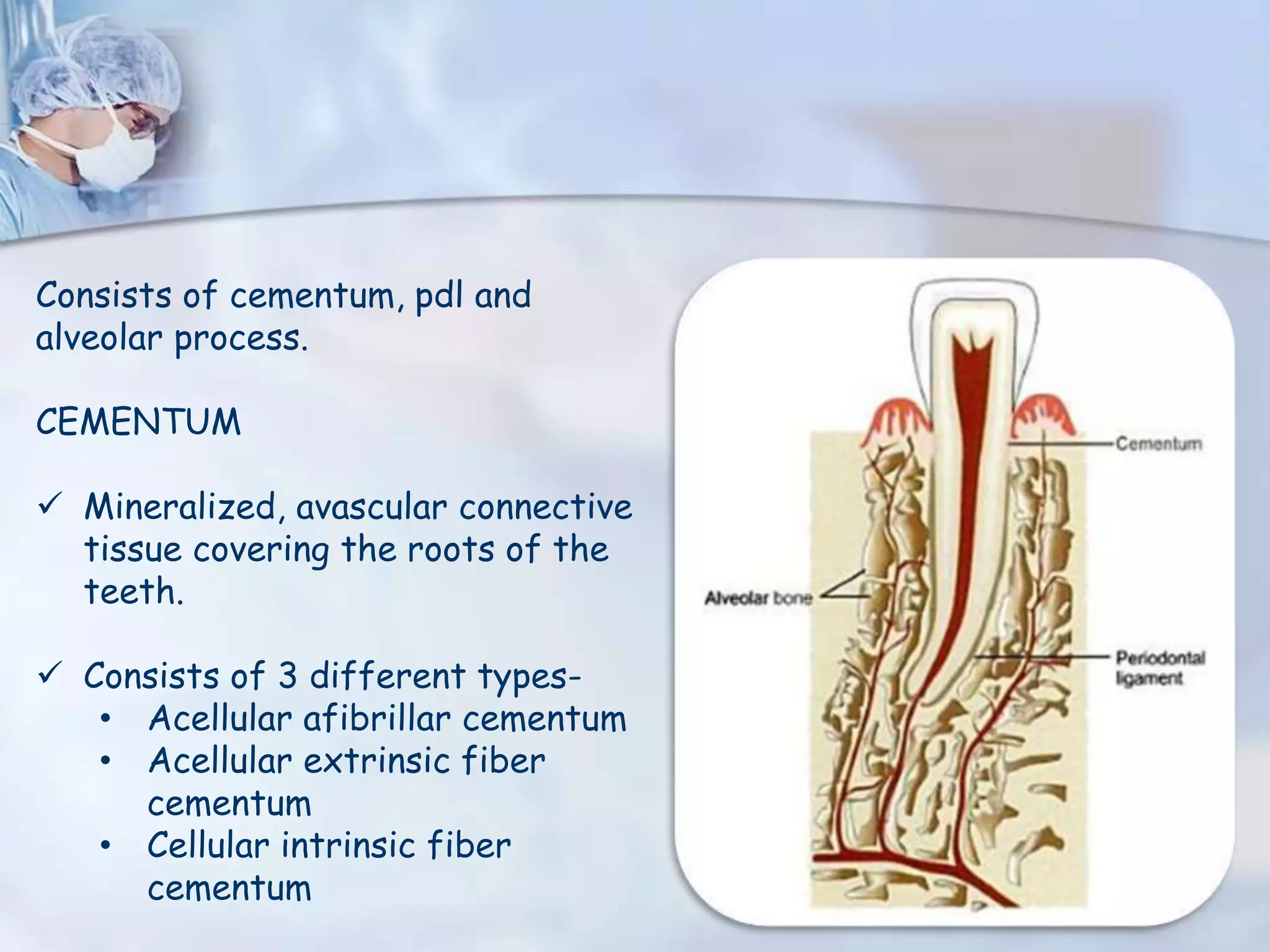
3. Consists of cementum, pdl and
alveolar process.
CEMENTUM
Mineralized, avascular connective
tissue covering the roots of the
teeth.
Consists of 3 different types-
• Acellular afibrillar cementum
• Acellular extrinsic fiber
cementum
• Cellular intrinsic fiber
cementum
4. Cemental matrix consists of growth factors such as IGF-1, FGFs, EGF,
BMPs, TGF-β and PDGF.
These factors r shown 2 b associated with cementoblast proliferation,
migration, and differentiation during cementum wound healing.
PERIODONTAL LIGAMENT
Soft, specialized connective tissue that connects the cementum to the
alveolar bone.
Contains heterogenous cell populations and extracellular matrix(ECM)
Cells include- osteoblasts, osteoclasts, fibroblasts, epithelial cell rests
of malassez, macrophages, cementoblasts, & undifferentiated
mesenchymal cells (stem cells)
5. ECM consists of collagen fibers, fibronectin, elastin, other non-
collagenous proteins, & proteoglycans.
ALVEOLAR PROCESS
Forms the bony troughs containing the roots of the teeth.
Divided into-
• Alveolar bone proper : lines the alveolus or the bony sockets
that house the roots of the teeth.
• Supporting Alveolar bone : Cancellous(spongy) bone adjacent to
the alveolar bone proper covered by 2 outer tables of compact
bone.
7. As a consequence of pathologic changes in the dental pulp, the root
canal system can harbor numerous irritants. Egress of these irritants
from infected root canals into the periradicular tissues can initiate
formation and perpetuation of PERIRADICULAR LESIONS.
Depending on the nature and quantity of these irritants, as well as the
duration of exposure of the periradicular tissues, a variety of tissue
changes can occur.
When the irritants are transient in nature, the inflammatory process is
short-lived and self-limiting.
However, with an excessive amount of irritants or persistent exposure,
the nonspecific and specific immunologic reactions can cause
destruction of periradicular tissues.
8. Radiographically, these lesions appear as radiolucent areas around
the portal(s) of exit of the main canal or lateral and/or accessory
canals.
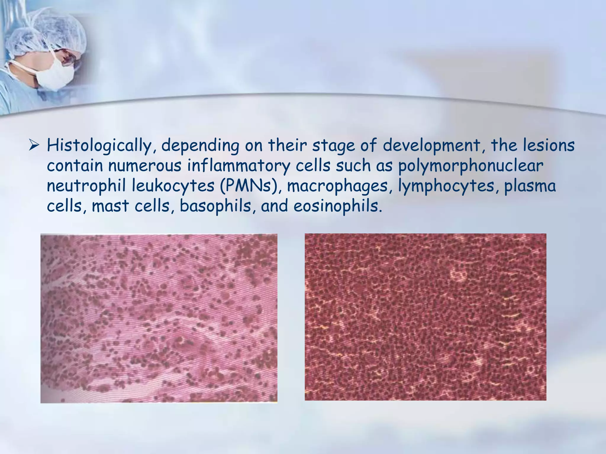
9. Histologically, depending on their stage of development, the lesions
contain numerous inflammatory cells such as polymorphonuclear
neutrophil leukocytes (PMNs), macrophages, lymphocytes, plasma
cells, mast cells, basophils, and eosinophils.
10. The interaction between the irritants
and the host defensive mechanisms
results in release of numerous
mediators that curtail progression of
infection and development of severe
local infection (osteomyelitis) and
systemic complication such as
septicemia.
11. IRRITANTS
Can b divided into-
• The LIVING irritants which include microorganisms and viruses.
• The NON-LIVING irritants which include mechanical, thermal &
chemical irritants.
Mild to moderate injuries of short duration cause REVERSIBLE tissue
damage and recovery of these tissues.
Persistent and/or severe injuries usually cause IRREVERSIBLE
changes in the pulp and development of periradicular lesions.
12. Microbial Irritants
Includes-
bacteria, bacterial toxins, bacterial fragments, and viruses
These irritants egress apically from the root canal system into the
periradicular tissues and initiate inflammation and tissue alterations.
A number of studies have shown that pulpal and/or periradicular
pathosis do not develop without the presence of bacterial
contamination.
13. In addition to bacterial irritation, the periradicular tissues can be
mechanically irritated and inflamed.
Physical irritation of periradicular tissues can also occur during root
canal therapy if the canals are instrumented or filled beyond their
anatomic boundaries.
Periradicular tissues can be irritated by impact trauma,
hyperocclusion, endodontic procedures and accidents, pulp
extirpation, overinstrumentation, root perforation, and
overextension of filling materials.
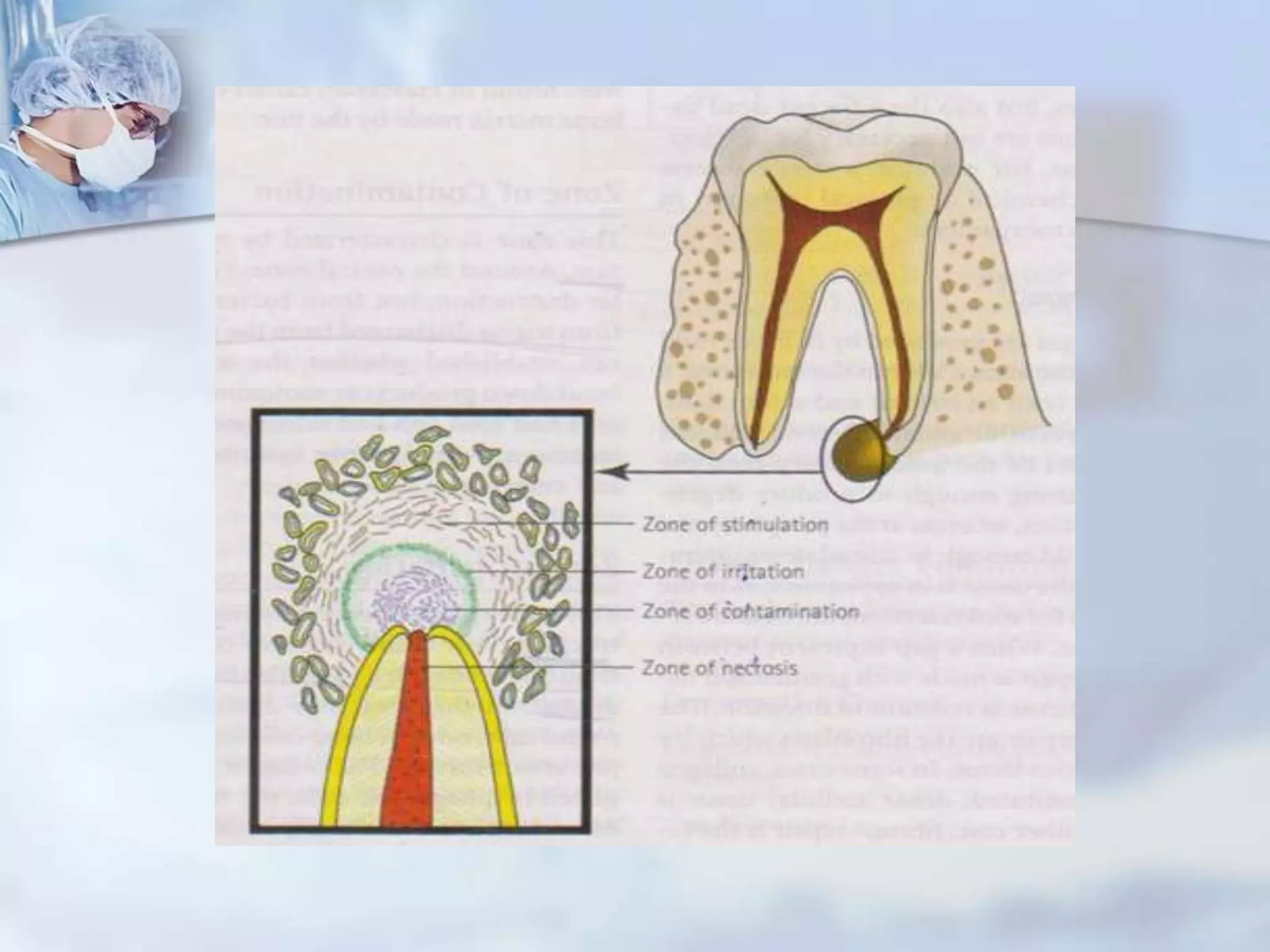
14. The reaction of the periradicular tissues to noxious products of
tissue necrosis, bacterial products, and antigenic agents from the
root canal has been described by FISH.
Four well-defined zones of reaction found were-
• Zone of infection
• Zone of contamination
• Zone of irritation
• Zone of stimulation
16. CHIEF COMPLAINT: Pain on biting, pain wid swelling, pus discharge etc.
DENTAL HISTORY : Recurring episodes of pain, swelling wid discharge,
swelling which reduces on its own.
OBJECTIVE EXAMINATION
• Xtraoral xamination- general appearance, skin tone, facial asymmetry,
swelling, extraoral sinus, sinus tract, tender or enlarged cervical lymph
nodes.
• Intraoral xamination- examination of soft tissues nd teeth to look 4
discolouration, abrasion, caries, restoration etc.
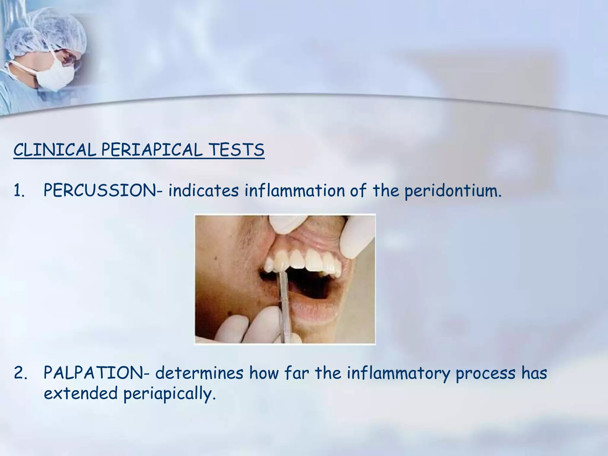
17. CLINICAL PERIAPICAL TESTS
1. PERCUSSION- indicates inflammation of the peridontium.
2. PALPATION- determines how far the inflammatory process has
extended periapically.
18. 3. PULP VITALITY-
• Thermal tests- includes heat and cold testing
• Anesthetic testing
• Test Cavity
• Electrical pulp testing
4. PERIODONTAL XAMINATION-
• Probing- determines the level of connective tissue attachment.
• Mobility- determines the status of pdl.
19. 5. RADIOGRAPHIC XAMINATION-
• Loss of lamina dura apically
• Radiolucency at apex regardless of cone angle nd usually resembles
a hanging drop.
• Cause of pulp necrosis is usually evident.
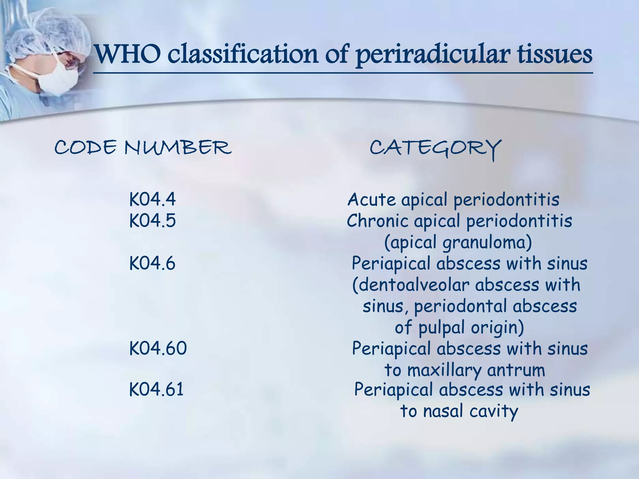
21. WHO classification of periradicular tissues
CODE NUMBER CATEGORY
K04.4 Acute apical periodontitis
K04.5 Chronic apical periodontitis
(apical granuloma)
K04.6 Periapical abscess with sinus
(dentoalveolar abscess with
sinus, periodontal abscess
of pulpal origin)
K04.60 Periapical abscess with sinus
to maxillary antrum
K04.61 Periapical abscess with sinus
to nasal cavity
22. CODE NUMBER CATEGORY
K04.62 Periapical abscess with sinus to
oral cavity
Ko4.63 Periapical abscess with sinus
to skin
Ko4.7 Periapical abscess with out
sinus
Ko4.8 Radicular cyst(apical periodontal
cyst, periapical cyst)
Ko4.80 Apical & Lateral cyst
Ko4.81 Residual cyst
Ko4.82 Inflammatory Paradental cyst
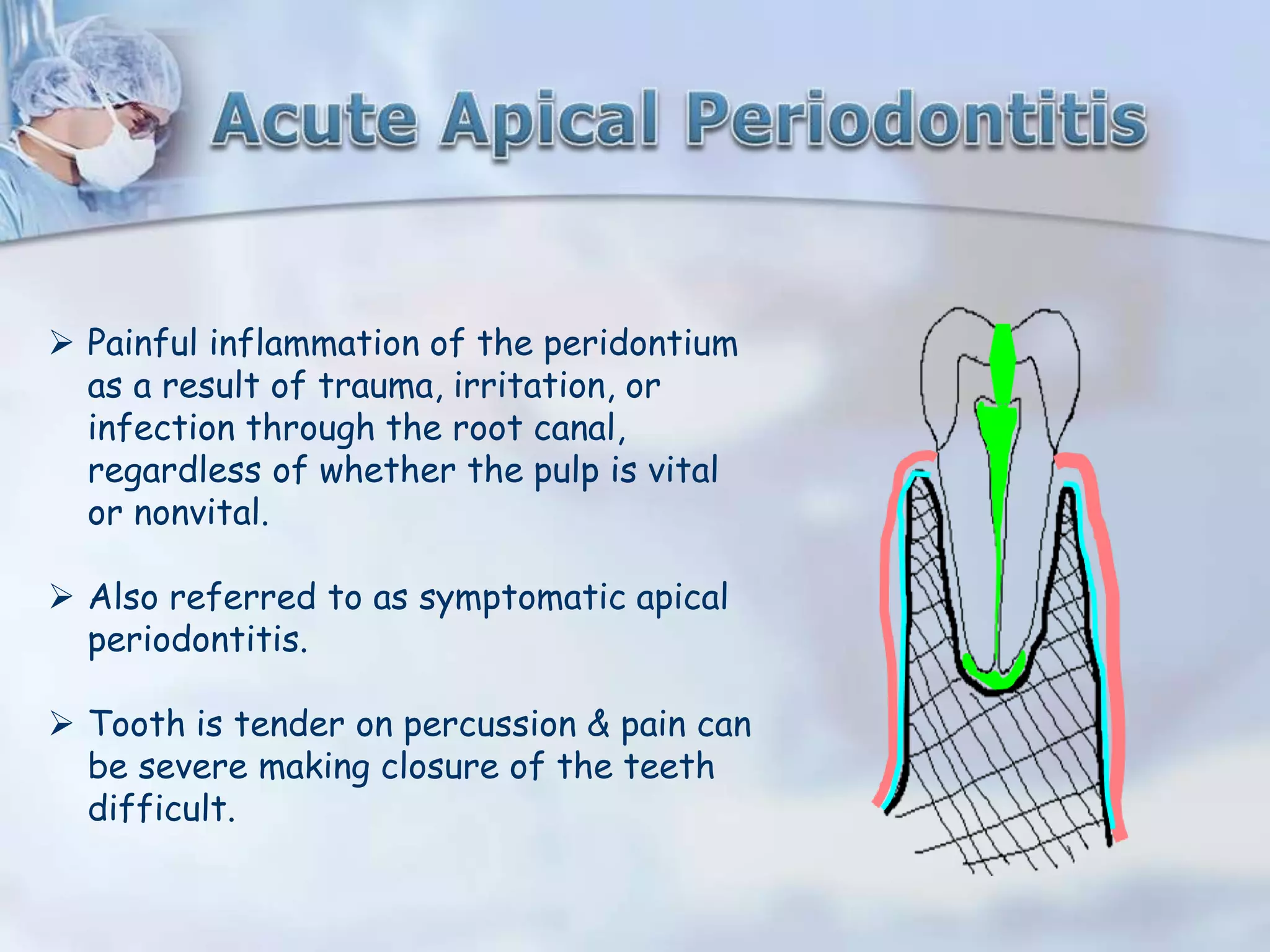
26. Painful inflammation of the peridontium
as a result of trauma, irritation, or
infection through the root canal,
regardless of whether the pulp is vital
or nonvital.
Also referred to as symptomatic apical
periodontitis.
Tooth is tender on percussion & pain can
be severe making closure of the teeth
difficult.
27. Etiology
IN VITAL TEETH-
Abnormal occlusal contacts
Recently inserted restoration extending beyond the occlusal plane
Wedging of a foreign object between the teeth such as a toothpick
or food
Traumatic blow to the teeth
28. IN NONVITAL TEETH-
a) Sequelae of pulpal diseases, i.e., the diffusion of bacteria & noxious
products from an inflamed or necrotic pulp.
b) Iatrogenic
• Root canal instrumentation forcing bacteria or debris
inadvertently through the apical foramen
• Forcing of irrigating irrigants or medicaments through the
apical foramen
• Extension of obturating material through the apical foramen
to impinge on periapical tissue
• Perforation of the root
• Overinstrumentation during cleaning & shaping of root canals.
29. Signs & Symptoms
Tooth is tender on percussion & may b slightly sore
Dull, throbbing & constant pain
Tooth may feel extruded & patient would have pain on closure &
mastication.
Negative or delayed vitality test
Radiographically, widening of the pdl space or a small area of
rarefaction if a pulpless tooth is involved & may show normal
periradicular structures if a vital pulp is present in the tooth.
30. HISTOPATHOLOGY
Inflammatory rxn in pdl
Dilation of blood vessels
Initiation of inflammatory response due to presence of
polymorphonuclear leukocytes & round cells
Accumulation of serous exudate
Distension of Pdl & extrusion of tooth, slight tenderness
If continuous irritation occurs, loss of alveolar bone
31. Periodontitis caused
due to trauma.
Widening of pdl space caused
due to abnormal occlusal contacts
Widening caused due to
orthodontic treatment
33. TREATMENT
Determining the cause & relieving the symptoms.
Adjustment of high points (in hyperocclusion cases).
Removal of irritatants (in case of nonvital infected pulp)
When the acute phase has subsided, the tooth is treated by
conservative means.
34. OUTCOME OF SYMPTOMATIC APICAL PERIODONTITIS
Spontaneous healing
Acute alveolar abscess
“Point” and open to the exterior (fistulation and sinus tract formation)
Lesion becomes asymptomatic and enters chronic phase.
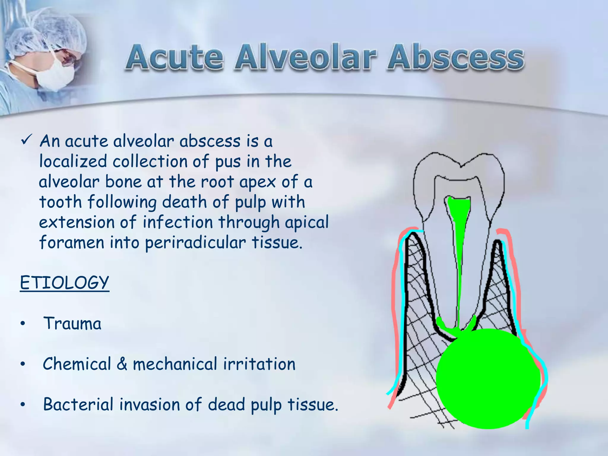
35. An acute alveolar abscess is a
localized collection of pus in the
alveolar bone at the root apex of a
tooth following death of pulp with
extension of infection through apical
foramen into periradicular tissue.
ETIOLOGY
• Trauma
• Chemical & mechanical irritation
• Bacterial invasion of dead pulp tissue.
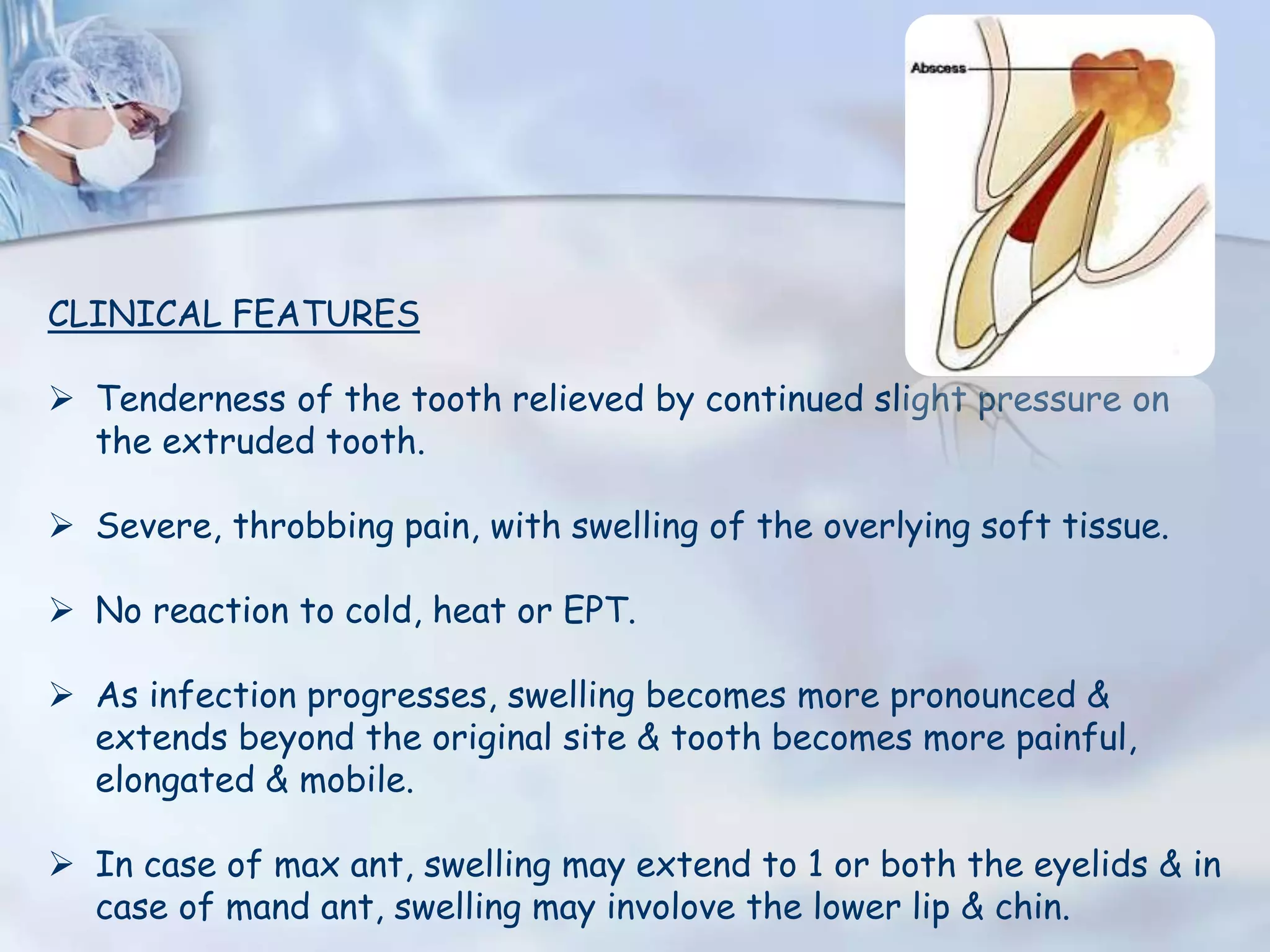
37. CLINICAL FEATURES
Tenderness of the tooth relieved by continued slight pressure on
the extruded tooth.
Severe, throbbing pain, with swelling of the overlying soft tissue.
No reaction to cold, heat or EPT.
As infection progresses, swelling becomes more pronounced &
extends beyond the original site & tooth becomes more painful,
elongated & mobile.
In case of max ant, swelling may extend to 1 or both the eyelids & in
case of mand ant, swelling may involove the lower lip & chin.
38. In case of max post, cheek may swell 2 an immense size, distorting
the facial structures & in case of mand post, swelling may xtend 2 ear
or round the border of the jaw into the submaxillary region.
SYSTEMIC REACTIONS include-
• Pt may appear pale, irritable & weakened due to pain & loss of
sleep.
• Mild cases- slight rise in temp (99-100˚C)
• Severe cases- temp above normal (102-103˚C)
• Fever often preceded or accompanies by chills.
• Intestinal stasis, manifesting orally by a coated tongue & foul
breath.
• Headache & malaise
43. LA is ineffective when injected into acutely inflamed tissue.
Conduction anesthesia may b administered to reduce the pain.
Hot saline rinses should b prescribed to assist drainage.
If the swelling is extensive, soft nd fluctuant, an incision through the
soft tissue to the bone may b necessary.
If it is hard, can b converted to soft, fluctuant state by rinsing with
hot saline solution 3-5 min at a time repeated every hr.
Antibiotics & analgesics can b prescribed as needed.
Finally, tooth shud b disoccluded slightly if extruded 4m its socket.
46. An acute inflammatory reaction superimposed on an existing chronic
lesion, such as cyst or granuloma.
Acute exacerbation of a chronic lesion.
ETIOLOGY
1.When state of equillibrium in granuloma /cyst is upset by:
• Influx of bacteria/necrotic products of high virulence and
antigenicity
• Lowering of host defenses
2. Mechanical irritation during RCT.
47. CLINICAL FEATURES
Clinically often indistinguishable from periapical abscess.
Initially, tooth may b tender on palpation.
As inflammation progresses, tooth gets elevated from the socket &
becomes sensitive.
Mucosa over the radicular area may appear red & swollen & sensitive
to palpation.
Most commonly associated with initiation of root canal therapy.
Do not respond to vitality testing.
48. Radiographically, well defined periradicular lesion may b present.
Histopathologically, shows areas of liquefaction necrosis with
disintegrated polymorphonuclear leukocytes & cellular debris
surrounded by macrophages, lymphocytes, plasma cells in periradicular
tissues.
Shud b differentiated from acute alveolar abscess through pt’s
history, symptoms & clinical tests results.
TREATMENT
Establishment of drainage
Once symptoms subside, complete root canal treatment.
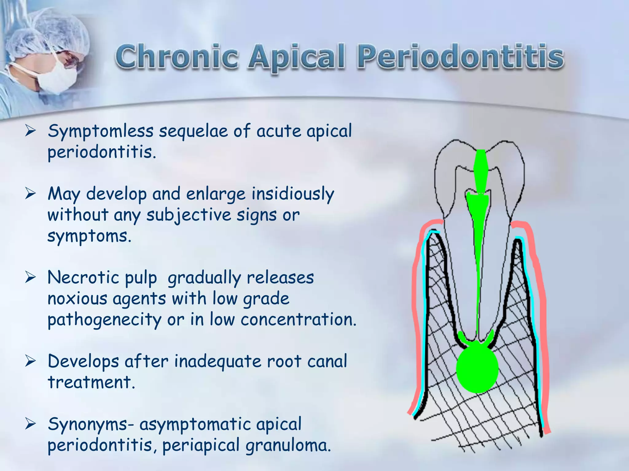
49. Symptomless sequelae of acute apical
periodontitis.
May develop and enlarge insidiously
without any subjective signs or
symptoms.
Necrotic pulp gradually releases
noxious agents with low grade
pathogenecity or in low concentration.
Develops after inadequate root canal
treatment.
Synonyms- asymptomatic apical
periodontitis, periapical granuloma.
51. ETIOLOGY
• Death of the pulp followed by mild irritation of periapical tissue that
stimulates a productive cellular reaction.
• Some cases preceded by chronic alveolar abscess.
CLINICAL FEATURES
Asymptomatic, discovered on routine radiographic xamination.
No pain on percussion
Associated tooth has a necrotic pulp therefore shud not respond to
the electrical or thermal stimuli.
53. A growth of
granulomatous tissue
continuous with the
periodontal ligament
resulting from death of
the pulp and the
diffusion of bacteria and
bacterial toxins from
the root canal into the
surrounding periradicular
tissues through the
apical and lateral
foramina.
55. Histologically, the periradicular granuloma consists predominantly of
granulation inflammatory tissue with many small capillaries,
fibroblasts, numerous connective tissue fibers, inflammatory
infiltrate, and usually a connective tissue capsule
57. Occasionally, needle-like spaces (the remnants of cholesterol
crystals), foam cells, and multinucleated foreign body giant cells are
seen in these lesions.
59. TREATMENT
Root canal treatment is recommended.
Removal of cause of inflammation is usually followed by
resorption of Granulomatous tissue & repair with
trabeculated bone.
60. Pathological cavity containing fluid, semi fluid or gaseous material
not created by accumulation of pus, frequently but not always lined
by epithelium(Kramer 1974).
Radicular cyst are generally considered to be a direct sequelae of
chronic apical periodontitis.
Not every lesion develops into cyst.
According to the studies 6-55% lesions are cyst.
There are two distinct categories of radicular cyst:-
• Periapical true cyst
• Periapical pocket cyst
61. ETIOLOGY
Acc to NAIR, cyst develops from dormant epithelial cell rests that
proliferate probably under the influence of inflammatory cytokines
& growth factors released by various cells residing in the lesion.
When proliferation occurs within the body of the granuloma, it plugs
the body of the apical foramen which limits the egress of the
bacteria.
Sometimes, epithelial plugs protrude out of the apical foramen
resulting in a pouch connected to the root & continuous with the
root canal.
63. Hypothesis related wid this growth-
a. Nutritional Deficient Theory-
• Periradicular inflammatory changes cause the epithelium 2
proliferate.
• As the epithelium grows into a mass of cells, the center loses
the source of nutrition from the peripheral tissues.
• This leads to necrosis in the center & a cavity is formed which
is lined by stratified squamous epithelium.
b. Abscess Theory-
• An abscess cavity is formed within the connective tissue & is
then surrounded with proliferating epithelial tissue, thereby
producing a cyst.
64. PRIAPICAL POCKET CYST
Originally designated as bay cyst.
Cyst contains an epithelial lined cavity that is
open towards the root canal of the affected
tooth.
initiated by the accumulation of neutrophils
around the apical foramen in response to the
bacterial presence in the apical root canal.
This forms a microabscess, that gets
enclosed by the proliferating epithelium,
forming a collar with epithelial attchment on
contacting the root tip.
65. PERIAPICAL TRUE CYST
Characterized by cavities
that are completely
enclosed in epithelial lining
and are totally
independent of the root
canal of the affected
tooth.
66. Clinical features
• No symptoms associated with
development of a cyst except
incidental to necrosis of the pulp.
• May become large enough, however,
to become obvious as a swelling.
• Pressure of the cyst may b sufficient
to cause movement of the teeth,
owing to accumulation of cystic fluid.
• If left untreated, may continue to
grow at the expense of the max or
mand.
67. HISTOPATHOLOGY
• Cavity is lined by stratified
squamous epithelium.
• Surrounded by connective
tissue that is infiltrated by
lymphocytes, plasma cells, and
polymorphonuclear neutrophils.
• Contains debris and
eosinophilic material.
• Cholestrol clefts,
macrophages, & giant cells also
present in CT.
68. RADIOLOGICAL FEATURES:
Classically presents as round
/ ovoid radiolucency with
sclerotic borders and
associated with pulpally
affected tooth / teeth.
If infection supervenes, the
margins become indistinct,
making it impossible to
distinguish it from a
periapical granuloma.
69. TREATMENT
• The treatment of choice is root canal therapy, followed by periodic
observation.
• Surgery required when lesion fails to resolve or symptoms develop.
• Extraction in case of severe bone loss.
70. Periapical cyst
Radiographically, shows a
well-circumscribed radiolucent
periapical lesion with a partial
sclerotic border, measuring
more than 1 cm in diameter.
Histologically, shows the
presence of an epithelial lining
with underlying dense
fibrocellular connective tissue
stroma
Granuloma
Radiographically, shows a well-
circumscribed radiolucent periapical
lesion without a sclerotic border,
measuring less than 1 cm in size.
Histologically, shows fibrocellular
connective tissue stroma consisting of
chronic inflammatory cell infiltrate
(mainly lymphocytes and plasma cells)
and endothelium-lined blood capillaries
with red blood cells, fibroblasts, and
collagen fibers.
72. Long standing, low grade
infection of the periradicular
alveolar bone.
Characterized by presence of
an abscess draining through a
sinus tract.
Synonyms- chronic suppurative
apical periodontitis,
asyptomatic apical abscess.
73. ETIOLOGY
• Source of infection is in
the root canal.
• It is a natural sequelae of
death of the pulp with
extension of the infective
process periapically, or
may result from a pre-
existing acute abscess.
74. CLINICAL FEATURES
• Tooth is asymptomatic or mildly
painful.
• Detected only during radiographic
examination or because of the
presence of a fistulous tract, which
can b either intraoral or extraoral.
• Exudate can also drain through
the gingival sulcus of the involved
tooth mimicking a periodontal
lesion with a pocket.
• Vitality test is negative.
76. RADIOGRAPHICAL EXAMINATION
A radiograph is taken after the insertion of GP cone into the sinus
tract which often shows the involved tooth by tracing the sinus tract
to its origin.
At times, the sinus tract is several teeth away from the cause.
Radiograph shows diffuse area of rarefaction which fade indistinctly
into normal bone.
The pdl is thickened.
78. HISTOPATHOLOGY
Some of the periodontal fibers at the root apex are detached
or lost followed by destruction of apical periodontal ligament.
lymphocytes & plasma cells are generally found toward the
periphery of abscessed area, with variable numbers of
polymorphonuclear leukocytes at the center.
Fibroblast may start to form a capsule at the periphery.
79. TREATMENT
Elimination of infection in root canal.
Once this is accomplished & root canal is filled, repair
of the periradicular tissues generally take place.
In case of smaller area of rarefaction, treatment is
similar to that of a tooth with a necrotic pulp.
80. It is a rapidly spreading inflammation of the soft tissues
characterized by diffuse pus formation.
This happens if an abscess is not able to establish drainage
through the skin surface or into oral cavity.
Cellulitis arising from dental infection and spreading through soft
tissues of head and neck can take various forms.
Mostly, infection spreads through tissue spaces like canine space,
infratemporal space, pharyngeal space, buccal space, submental and
submandibular space etc.
Two dangerous forms of cellulitis are –
- Ludwig’s angina
- Cavernous sinus thrombosis
82. TREATMENT
• Removal of the necrotic pulp.
• Extraction of the infected tooth.
• Incision & drainage of the swelling.
• In severe cases hospitalization required.
• Antibiotics & analgesics
83. Post treatment apical
periodontitis in an
endodontically treated
tooth.
ETIOLOGY
• Anatomical complexity
• Apical biofilms
• Cholestrol clefts
• Foreign body reaction to
gp
• Cellulose granuloma
• Periapical scar tissue
84. BACTERIOLOGY
E. Faecalis is the most consistently reported organism that can
survive prolonged starvation & can grow as a monoinfection in
endodontically treated teeth.
Studies have shown presence of yeast & candida albicans.
Gram+ cocci, rods & filaments, & Propioniobacterium have also
been implicated.
87. Apical scar is an area at the apex of a
tooth that fails to fill in with osseous
tissue after endodontic treatment.
88. CHARACTERISTIC FEATURES
Bone structures are recognized within the rarefaction.
The periphery of the rarefaction may be irregular and
may be demarcated by a compact bone border.
The rarefaction is often located asymmetrically around
the apex.
The connection of the rarefaction with the periodontal
space may be angular.
89. A surgical defect is that portion of bone that fails to form osseous
tissue.
It is frequently seen periapically after root resection in which the
site is filled with dense fibrous (collagen) tissue instead of bone.
It is an asymptomatic persistent radiolucency.
An extraction site can also form a surgical defect.
Approximately 75% of all surgically treated periapical
radiolucencies require 1 to 10 years or longer for complete
resolution. In the remaining 25%, complete healing does not occur.
91. Condensing osteitis is a reaction of bone induced by inflammation.
It occurs mainly at the apex of a tooth from an infected pulp.
The infection from tooth caries reaches the pulp and progresses
to the apical tissues to produce a small periapical radiolucency
called RAREFYING OSTEITIS.
The small rarefying osteitis may be either a periapical granuloma, a
radicular cyst or an abscess.
The bone surrounding this rarefying osteitis becomes dense in
order to prevent further spread of the lesion. This dense
radiopacity surrounding the rarefying osteitis is called
CONDENSING OSTEITIS.
92. CLINICAL FEATURES
Usually Asymptomatic
Discovered during routine radiographic
examination.
Pulp of the involved tooth is nonvital.
93. RADIOGRAPHIC EXAMINATION
• The tooth involved may exhibit a
large carious lesion or a large
restoration close to the dental
pulp.
• The lesion shows a diffuse
radiopacity surrounding a
small central radiolucency at the
apex (or apices) of a tooth.
• It is the area of dense bone with
reduced trabecular pattern.
94. HISTOPATHOLOGY
Appears as an area of dense bone with reduced trabecular pattern
lined with osteoblasts.
Chronic inflammatory cells, plasma cells, & lymphocytes are seen in the
scant bone marrow.
TREATMENT
Treatment consists of removing the infection either through tooth
extraction or root canal therapy.
96. CLASSIFICATION
Types of tooth resorption includes-
• External
a. External surface resorption
b. External inflammatory root resorption
c. External replacement resorption or ankylosis
• Internal
97. ETIOLOGY
• Trauma
• Excessive forces
• Granuloma
• Cyst
• Central jaw tumors
• Impaction of teeth
• Bleaching
• Systemic diseases
• If no cause is evident, the disorder is called as idiopathic
resorption.
98. CLINICAL FEATURES
Asymptomatic
On complete resorption, tooth may become mobile.
If extends into the crown, gives appearance of “pink tooth” as seen
in internal resorption.
In case of replacement resorption or ankylosis, root is gradually
replaced by bone, renders the tooth immobile, in infraocclusion, &
with a high mettalic percussion sound.
99. RADIOGRAPHICAL EXAMINATION
• Concave or ragged areas on the
root surface or blunting of the
apex.
• Inflammatory root resorption
caused by the pressure of a
growing granuloma, cyst or tumor
adjacent 2 the area of
radiolucency.
• Areas of ankylosis hav a resorbed
root with no pdl space & with bone
replacing the defects.
100. TREATMENT
• Varies with etiology
• If extended by pulpal
disease, root canal therapy.
• If due to excessive
orthodontic forces,
treatment of choice would b
reducing those forces.
• In case of cervical root
resorption, surgical xposure
of the defect & restoration
with a suitable rest material.
105. • Central giant cell granuloma
• Nasopalatine duct cyst
• Globulomaxillary cyst
• Simple bone cyst
• Enostosis
106. Establishment of proper diagnosis is of utmost importance
to carry out the effective clinical procedure for the
benefit of patient .
Review after the treatment is also to be given importance .
Researches done and ongoing research has thrown light on
various aspects untangling the puzzle which persisted for
long.
So this knowledge can be used in the practice to make
endodontic treatment a success when the controversy is
going on about to pull out or salvage the tooth with implant
coming strongly on the field of dentistry.
Editor's Notes
#4: Acellular afibrillar cementum- covers the teeth at and along the cej
Acellular extrinsic fiber cementum- is confined to the coronal half of the root.
Cellular intrinsic fiber cementum- is present on the apical half of the root where no extrinsic fiber cementum has been laid down.