Approach to chronic diarrhoea
148 likes29,488 views
This document discusses the evaluation and causes of chronic diarrhea. It begins by defining chronic diarrhea and outlining the normal stool production process. It then describes the main mechanisms that can cause diarrhea - osmotic, secretory, inflammatory, and dysmotility. Specific causes are discussed under each mechanism, including diseases, medications, toxins, and dietary factors. The document outlines the evaluation of a patient with chronic diarrhea, including history, physical exam, stool tests, imaging, and other lab tests. It provides guidance on testing for malabsorption and evaluating postsurgical causes of chronic diarrhea.


More Related Content
What's hot (20)
Similar to Approach to chronic diarrhoea (20)
More from Abhinav Srivastava (20)
Recently uploaded (20)
Approach to chronic diarrhoea
- 1. Approach to Chronic Diarrhea PRESENTER:DR.ABHINAV KUMAR
- 2. CHRONIC DIARRHOEA • AS A SYMPTOM – Frequency of bowel action – Looseness of stools – Increase in stool volume – AS A SIGN Stools weight more than 250 gm/24 hours
- 3. – Stools weight more than 250 gm/24 hours • Patients conception as regards symptoms is variable • Stools weighting is tedious. • A consensus statement by the AGA • Chronic Diarrhea = Decrease in fecal consistency for 4 or more weeks
- 4. Normal stool fluids processing • 8-9 L/d enter GI system – Ingest 1-2 L/d – Create approx 7 L/d • saliva, gastric, biliary, pancreatic secretions • Small bowel reabsorbs 6-7 L/d • Large bowel absorbs 1-2 L/d • 100-200 gm/d stool created • Reduction of water absorption, due to decrease in absorption or increase in secretion, by as little as 1% can lead to diarrhea
- 5. Causes of Diarrhea In almost all cases; it is a manifestation of 1 of 4 Mechanisms: – Osmotic diarrhea – Secretory diarrhea – Inflammatory and infectious diarrhea – Diarrhea associated with deranged motility • More than one of these mechanisms may be involved in the pathogenesis of a given case.
- 6. Classification of causes • Secretory • Osmotic • Steatorrheal • Inflammatory • Dysmotile Causes • Factitial Causes
- 7. Characterstics of Factitial diarrhea • accounts for up to 15% of unexplained diarrheas referred to tertiary care centers. • self-administer laxatives alone or in combination with other medications (e.g., diuretics) • surreptitously add water or urine to stool sent for analysis. • typially women with histories of psychiatric illness • disproportionately from careers in health care. • Hypotension and hypokalemia are common co-presenting features. • they benefit from psychiatric counseling
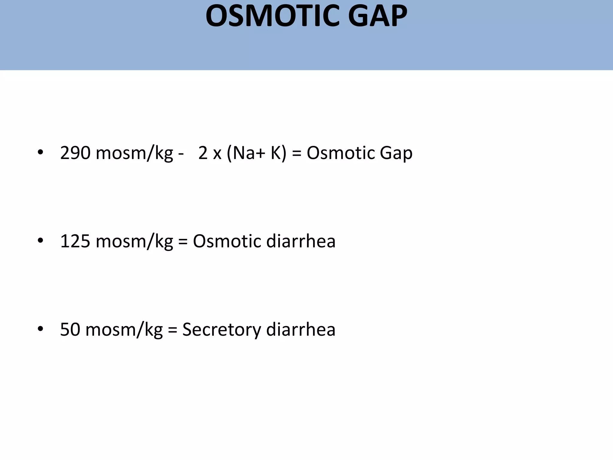
- 8. OSMOTIC GAP • 290 mosm/kg - 2 x (Na+ K) = Osmotic Gap • 125 mosm/kg = Osmotic diarrhea • 50 mosm/kg = Secretory diarrhea
- 9. Osmotic Diarrhea IN THE SMALL INTESTINE Ingestion of non-absorbable solutes Fluid entry into the small bowel Intraluminal solutions become iso-osmotic with the plasma Intraluminal Na+ concentration drop below 80 ml osmol Steep lumen to plasma gradient
- 10. Osmotic Diarrhea IN THE COLON Carbohydrate Metabolized by Bacteria Non metabolizable substrates Na+ and H2O may be absorbed by colon Short Chain fatty acids (Organic anions) A linear relation between ingested osmotic load & stool water output Quadrupling the Osmolality
- 11. Osmotic Diarrhea Short-Chain Fatty Acids (Organic Anions) Promote more fluid in the colon Obligate retention of inorganic cations Further increasing the osmotic load More fluid in the colon
- 12. Causes of Osmotic Diarrhea Exogenous • Osmotic Laxatives • Antacids containing MgO or Mg(OH)2 • Dietetic foods, candies and elixirs • Drugs e.g.: – Colchicine – Cholestyramine
- 13. Drugs Causing Diarrhea • Antibiotics – Clindamycin – Ampicillin – Cephalosporins • Antacids + Mg++ • Anti-HTN Agents – Propranolol – Methyldopa – Hydralazine • Antimetabolites • CV Agents – Digitalis • Alcohol • Nutritional Supplements • Potent Diuretics – Furosemide – Bumetanide
- 14. Causes of Osmotic Diarrhea Endogenous • Congenital – Specific Malabsorptive Disorders e.g Disaccharidase deficiencies – Generalized Malabsorptive Diseases e.g Abetalipoproteinemia – Pancreatic insufficiency e.g cystic fibrosis • Acquired – Specific Malabsorptive Diseases – Generalized Malabsorptive Diseases – Pancreatic insufficiency – Celiac disease – Infections
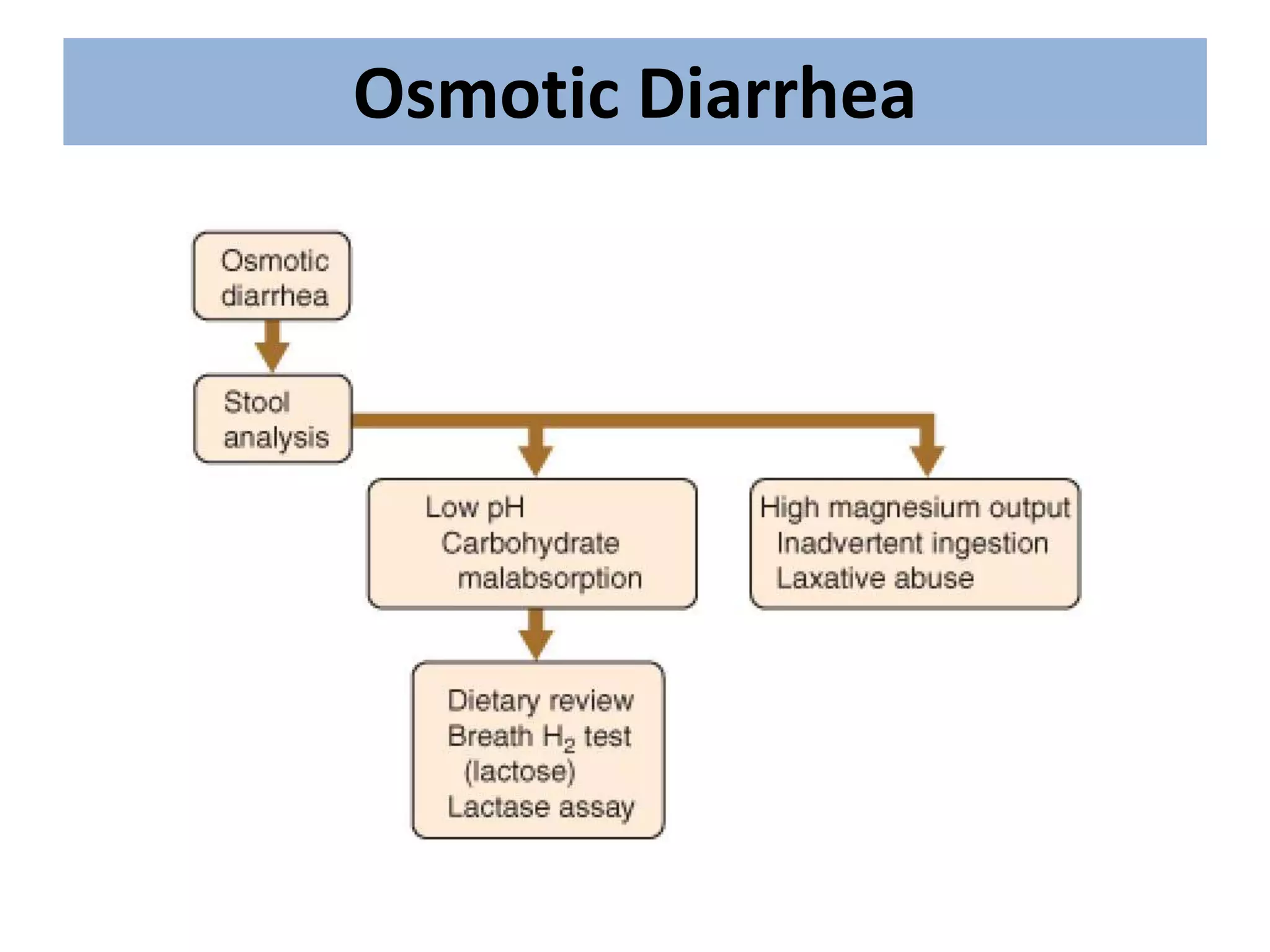
- 15. Osmotic Diarrhea
- 16. Osmotic Diarrhea • Mg can be measured directly in stool water by spectrophotometry. Excretion >30 mEq of Mg daily or concentrations in stool water > 90 mEq/L strongly suggests Mg-induced diarrhea • Ingestion of poorly absorbed carbohydrates ( fecal pH < 6 is highly suggestive, which is confirmed by Breath H2 test): • Lactose • Sorbitol or manitol • fructose • Acarbose (may lead to carbohydrate malabsorption)
- 17. • Electrolyte transport diarrhea • The intestine is able to – Secrete Fluids & electrolytes – Absorb • Secretion originates in the crypts • Absorption is mainly a villous function • Intracellular cyclic-AMP & -GMP are a corner stone in initiating Intestinal secretion
- 18. Mechanism of Secretory Diarrhea Neurotransmitters Hormones Bacterial Enterotoxins Cathartics Stimulate receptors on the enterochromaffin cells stimulate Cyclic AMP – Cyclic GMP Ca ions stimulate Cl-, H2O and CHO3 Secretion by the enterocytes
- 19. causes of Secretory Diarrhea • Exogenous • Stimulant Laxatives e.g. Anthraquinones, senna • Medications – Diuretics – Asthma medication – Eye drops – Bladder stimulants – Cardiac drug – Prostaglandins • Toxins – Metals – Organophosphorous – Seafood toxins – Bacterial toxins
- 20. Endogenous • Congenital e.g. Chloridorrhea • Bacterial enterotoxins • Hormone-producing tumors
- 21. Secretory Diarrhea peptide-secreting tumors : • Carcinoid syndrome (5HIAA, Metanephrine) • ZE syndrome (Gastrin) • Mastocytosis (Histamine) • VIPoma (VIP)
- 22. Secretory Diarrhea • Common endocrinologic diseases that cause diarrhea: • Diabetes mellitus • Hyperthyroidism (TSH) • Addison's disease (ACTH) • • Other blood tests for evaluating secretory diarrhea : • Serum protein electrophoresis and immunoglobulin electrophoresis (IgA deficiency recurrent intestinal infections such as giardiasis, CVID may mimic sprue , Testing for HIV and HIV2 ) • Cholestyramine trial is recommended for bile acid diarrhea.
- 23. Secretory Diarrhea small bowel Diseases that may be detected by small intestinal biopsy : • Crohn's disease, • Celiac sprue, • Intestinal lymphoma, • Eosinophilic gastroenteritis, • Tropical sprue, • Whipple's disease, • Lymphangiectasia, • Abetalipoproteinemia, • Amyloidosis, • Mastocytosis, • (Many of these disorders usually, but not always, present with steatorrhea)
- 24. Colon • Colonic causes of chronic secretory diarrhea tend to produce diffuse changes sigmoidoscopy usually is adequate • Colonoscopy is preferable : • older blood in the stool the clinical suspicion of right colonic or ileal disease is strong the patient has AIDS Diseases with colonic mucosa appears normal endoscopically, but that can be diagnosed histologically: • microscopic colitis (lymphocytic and collagenous colitis) amyloidosis • granulomatous infections schistosomiasis
- 25. • Cholera and enterotoxigenic E. coli • Bile acids and long chain fatty acids (postileal resection, Crohn’s disease, malabsorption syndromes) • Gastrointestinal hormones (VIPoma, gastrinoma, carcinoid) • Anthraquinone laxatives • Mechanism: Agents ↑ intracellular cAMP→ ↑secretion (Na+K+ ATPase is also inhibited)
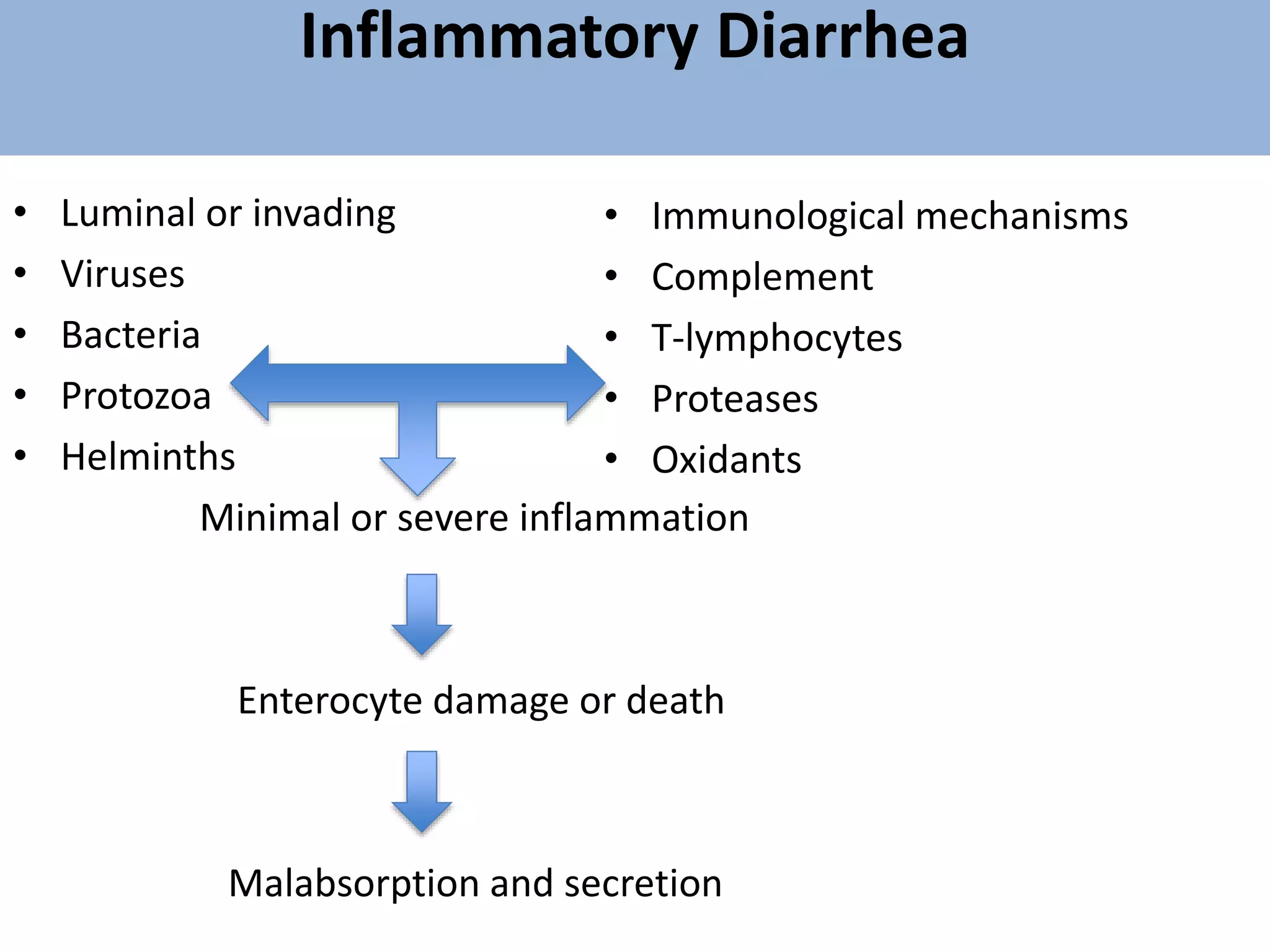
- 26. Inflammatory Diarrhea • Luminal or invading • Viruses • Bacteria • Protozoa • Helminths Minimal or severe inflammation Enterocyte damage or death Malabsorption and secretion • Immunological mechanisms • Complement • T-lymphocytes • Proteases • Oxidants
- 27. Chronic Inflammatory Diarrhea Diagnostic considerations include • IBD, • Infections, • Pseudomembranous enterocolitis, • Ischemia, • Radiation enteritis, • Neoplasia.
- 28. Chronic Inflammatory Diarrhea • Infections: – Bacteria – Viruses – Parasites – Helminthes • Anticancer agents • Radiation therapy • Hypersensitivity e.g. celiac disease • Autoimmune • Ulcerative colitis • Crohn's disease • Collagenous colitis • Lymphocytic colitis
- 29. Chronic Inflammatory Diarrhea • The pathogens most likely to cause chronic inflammatory diarrhea : • C. difficile • Cytomegalovirus • Entamoeba histolytica • Yersinia spp • Mycobacterium tuberculosis
- 31. Diarrhea Associated with Deranged Mobility • Adequate absorption requires • Adequate and long enough exposure to intestinal epithelium • Accelerated Transit time – Decreased absorption – Large fluid load to the colon – Colonic irritability – → Diarrhea • Diminished peristalsis – Bacterial overgrowth • → Secretory diarrhea
- 32. Diarrhea Associated with Deranged Mobility • IBS-D • Functional Diarrhea • Diabetic neuropathy • Scleroderma • Thyrotoxicosis
- 33. Evaluation of Chronic Diarrhea Systemic symptoms – Fever – Joint pains – Mouth ulcers – Eye redness Physical examination – Skin Rash – Mouth ulcers. – Eye manifestation and exophthalmos – Physical signs of anemia – Dehydration and cachexia – Abdominal masses – Anal sphincter tone – Presence of blood on PR examination
- 34. Diarrhea and Abdominal Pain • Suggests IBS if pain is in the left lower quadrant or suprapubic region • Suggests a disease of the small bowel (e.g., Crohn’s disease) if the pain is periumbilical or in right lower quadrant • Gastrinoma (Zollinger-Ellison Syndrome) – peptic ulcers responsible for upper abdominal pain
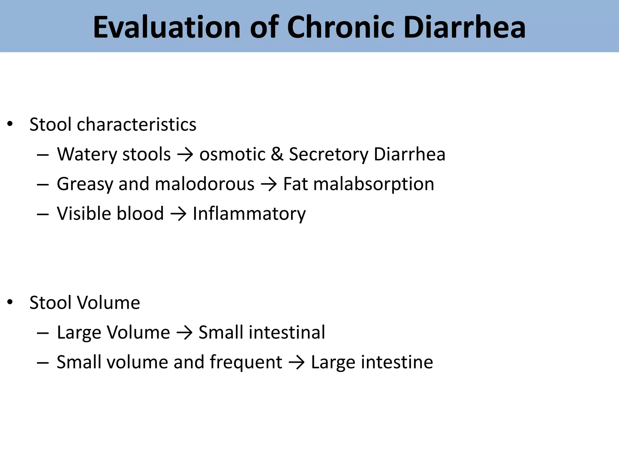
- 35. Evaluation of Chronic Diarrhea • Stool characteristics – Watery stools → osmotic & Secretory Diarrhea – Greasy and malodorous → Fat malabsorption – Visible blood → Inflammatory • Stool Volume – Large Volume → Small intestinal – Small volume and frequent → Large intestine
- 36. Evaluation of Chronic Diarrhea • Relation to food – After meals ?? → IBS – Stops with fasting → Osmotic diarrhea – Occurs in spite of fasting and during sleep → Secretory • Measure stools electrolytes • Calculate the osmolality
- 37. Evaluation of Chronic Diarrhea • Specific Testing • A large number of tests are available • No firm Rule as to what and to whom testing should be done – The minimum laboratory evaluation Complete blood picture and ESR – Total proteins and albumin – Serum electrolytes Thyroid functions – Stools examination and fecal occult blood are Mandatory. – Some patients may need endoscopic evaluation
- 38. Evaluation of Chronic Diarrhea • The Osmotic Gap • 290 mosm/kg - 2 x (Na+ K) = Osmotic Gap • 125 mosm/kg = Osmotic diarrhea • 50 mosm/kg = Secretory diarrhea – Further studies Stools culture – Selective testing for secretagogues • Gastrin • Vasoactive Intestinal Peptide (VIP) • Testing for bile acid malabsorption • Imaging the small and large intestine
- 39. • History • Physical Exam • Endoscopy • Laboratory studies • Radiological studies • Other studies
- 43. History of Organic Disease • Floating stools and undigested food are not helpful as they may be seen in functional and organic diarrhea • The association of diarrhea with milk or hyperosmolar solutions may not be recognized by the patient • Detailed drug history is important • Arthritis and arthralgias may suggest inflammatory bowel disease, Whipple’s disease, Reiter’s syndrome • Weight loss suggests malabsorption, hyperthyroidism or a malignant tumor
- 44. Physical Exam • General: Weight loss • Vital signs: Postural hypotension in diabetes with autonomic dysfunction • Neurology: – Peripheral neuropathy due to vitamin B deficiency – Carpopedal spasm due to hypocalcemia • Skin: – Erythema nodosum and pyoderma gangrenosum in inflammatory bowel disease – Hyperpigmentation in Whipple’s and Addison’s disease
- 45. • Musculoskeletal: – Non-deforming arthritis - Whipple’s disease/IBD • Abdominal: – Hepatosplenomegaly and lymphadenopathy suggest lymphoma or Whipple’s disease – Arterial bruit or an aortic aneurysm suggests ischemic bowel disease • Rectal: – Perianal disease (e.g., abscess, fistula Crohn’s disease) – Rectal tumor – Fecal impaction
- 46. Radiological Studies • Abdominal plain film – Pancreatic calcifications (chronic pancreatitis) – Dilated small bowel – Abnormal bowel contour suggesting lymphoma or inflammatory bowel disease • Small bowel series: – Distinguishes mucosal disease (celiac sprue), inflammatory conditions (Crohn’s disease) and infiltrative processes (Whipple’s disease or amyloidosis – Clarifies anatomy in post-surgical patients – Detects localized areas of dilation and stasis
- 47. Radiological Studies • Barium enema or Colonoscopy: – Patient >40 years with recurrent/chronic diarrhea – Not as helpful in the younger patient – If IBD is suspected, contrast material should be refluxed into the terminal ileum to rule out Crohn’s disease of the terminal ileum – A barium enema will interfere with the collection of stool for measurement of volume and fat and with the detection of parasites
- 48. Evaluation of Malabsorption When Steatorrhea is Present • <7% of fat is secreted into the stool per day (4-7g on average diet containing 60-100g fat) • Causes of steatorrhea: – Small intestinal disease – Pancreatic disease – Hepatobiliary disease – Gastric disease
- 49. Evaluation of Malabsorption D-Xylose Test • Measures absorptive capacity of proximal small bowel • Distinguishes malabsorption from maldigestion • Does not require pancreatic enzymes to be absorbed by an intact small bowel mucosa • Enters the blood and is excreted in urine • Give 25g D-Xylose; collect urine for 5 hours • Collect blood one hour after ingestion
- 50. D-Xylose Test • If test abnormal – small bowel biopsy • Celiac or Whipple’s disease – xylose is poorly absorbed with low levels in blood and urine • Massive bacterial overgrowth – abnormal xylose test may revert to normal after antibiotics • Dehydration, renal insufficiency, third spacing of fluid, vomiting, hypothyroidism all ↓urine but not serum levels of D-xylose
- 51. • If xylose test normal, maldigestion likely due to pancreatic insufficiency • Pancreatic insufficiency can be confirmed by intubation of the duodenum and collection of intraluminal contents by gastroenterology laboratory • Bentiramide test – utilizes a non-absorbable synthetic peptide, cleaved by pancreatic enzymes and measured in the urine
- 52. Postsurgical Diarrhea • Short bowel syndrome – diarrhea after extensive small bowel resections • Limited ileal resection (<100cm) – Sufficient bile acid pool - no steatorrhea – Diarrhea from bile acids delivered to the colon – Bind bile acids with Cholestyramine 4g QID • Extensive ileal resection (>100cm) – Steatorrhea – Do not use cholestyamine – depletes bile acids – Treat: medium chain triglycerides (Portagen)
- 53. • Gastric surgery with vagotomy – Altered intestinal motility – Increases concentration of fecal bile acids – May unmask latent lactase deficiency – May unmask celiac disease (rare) – Blind loop syndrome with bacterial overgrowth – Gastrocolic fistula
- 54. • Cholecystectomy – Increased fecal bile acids – Cholestyramine effective • Subtotal colectomy with ileal-rectal anastomosis (for multiple polyposis) – Diminishes with time – Treated with antidiarrheal medications
- 55. Osmotic diarrhea • For practical purposes osmotic diarrhea is caused by one of three conditions: • Ingestion of exogenous magnesium • Consumption of poorly absorbable carbohydrates • Carbohydrate malabsorption
- 58. Chronic Fatty Diarrhea • Major causes of maldigestion : • pancreatic exocrine insufficiency (e.g., chronic pancreatitis) lack of bile (e.g., advanced PBC) • Common causes of malabsorption : • Mucosal diseases (e.g., celiac sprue)
- 59. • The first step is to look for structural problems involving the small bowel. • This evaluation may include small bowel radiography, CT scan of the abdomen, and small bowel biopsy. • If no structural problems are discovered pancreatic exocrine function is evaluated by stool chymotrypsin activity or Secretin test.
- 60. EMPIRICAL THERAPY OF CHRONIC DIARRHEA • Empirical therapy is used in patients with chronic diarrhea in three situations: • as temporizing or initial treatment before diagnostic testing • after diagnostic testing has failed to confirm a specific diagnosis • when a diagnosis has been made but no specific treatment is available or specific treatment has failed to produce a cure.
- 61. • Therapeutic trials of pancreatic enzyme replacement and conjugated bile acid supplementation in patients with unexplained steatorrhea may be both diagnostic and therapeutic. • Symptomatic treatment with opiates often is necessary in patients with chronic diarrhea because specific treatment may not be available.
- 62. Other agents that sometimes are used to treat non-specific diarrhea : • Octreotide : (carcinoid syndrome and other endocrinopathies, dumping syndrome, and AIDS) • Clonidine (special role in diabetic diarrhea)