


The basics of retinal oct ophso.net
- 1. OCT Bootcamp: The Basics of Retinal OCT Optometry Symposium Hilary Wilson, M.D. November 2, 2008
- 2. Question • How many ophthalmic imaging tests can claim the following? – Non-invasive – Non-contact – No radiation – Painless – Fast – Reliable and sensitive (to 10 microns)
- 3. Optical Coherence Tomography • Diagnostic test that allows for imaging and measurement of various ocular structures
- 6. OCT: Retina
- 7. Goals • Quick overview of OCT function • Interpretation of macular OCT scan • Define indications for macular OCT • Practical examples
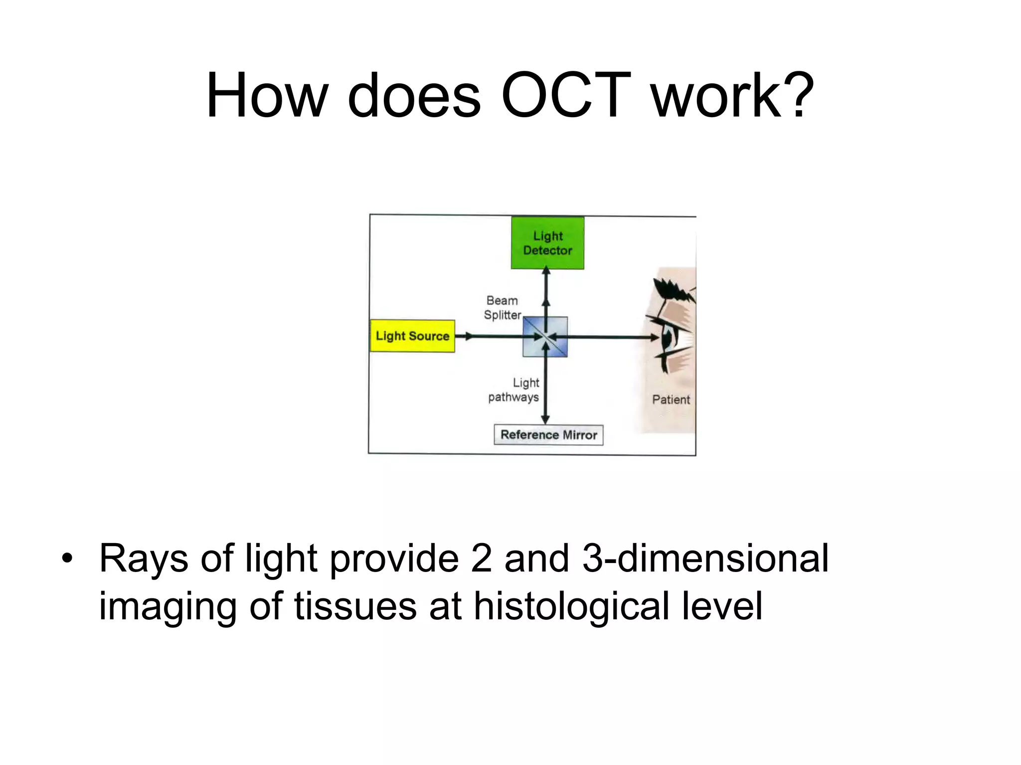
- 8. How does OCT work? • Rays of light provide 2 and 3-dimensional imaging of tissues at histological level
- 9. Optical Biopsy of Retinal Layers
- 10. Limitations of Retinal OCT • Mydriasis may sometimes be necessary • Dioptric media must be somewhat transparent • Exploration typically limited to posterior pole • Good lacrimal film necessary
- 11. Obtaining A Macular Scan
- 13. Interpretation of Macular OCT Printout • Assessment of reliability – Scan placement – Signal strength – Algorithm performance
- 14. Scan Placement
- 15. Signal Strength • Signal strength 6 = adequate • Signal strength 8 = very good
- 16. Algorithm Performance • For macular scan, the borders of algorithm should fit to ILM and PR inner and outer segment • If algorithm has failed, then the quantitative data should be disregarded
- 17. Interpretation of Macular OCT Printout • Color-coded qualitative thickness map
- 18. Interpretation of Macular OCT Printout • Color-coded quantitative thickness map – Macula 150 to 250 µ – Foveola ≤ 200 µ
- 19. Interpretation of Macular OCT Printout • Table of thickness and volume parameters
- 20. Indications for Retinal OCT • To examine the retina and its sub-layers – Atrophy, Edema, Traction, Subretinal fluid, RPE irregularity – ARMD, CME, CSME, CSR • To monitor progression • To aid in treatment planning • To monitor response to therapy
- 21. Indications for Retinal OCT • To examine the retina and its sub-layers – Extent of retinal defects or abnormalities – Detailed measurements
- 22. Indications for Retinal OCT • To monitor progression
- 23. Indications for Retinal OCT • To aid in treatment planning • To monitor response to therapy
- 24. Case Studies: Vitreoretinal Interface Disorders
- 25. Case 1 • A 67 year-old man notes progressive decrease in vision OS x 6 mos • VA 20/20 OD, 20/200 OS
- 26. Case 1 Fundus Photo
- 27. Case 1 OCT of Macula • Diagnosis?
- 28. Case 1 OCT Macular Scan • Diagnosis: Vitreomacular traction • Epiretinal membrane • Cystoid macular edema
- 29. OCT Macular Scan: 3 Months Post-op • No remaining ERM • Macular edema resolved • VA 20/40
- 30. Comparison OCT: Preop & Postop
- 31. OCT Advantage • Enhanced visualization of pathological process • Aided in determining optimal treatment • Postoperative OCT showed resolution
- 32. Case Studies: Retinal Vascular Diseases
- 33. Case 2 • 66-yo woman with severe NPDR OS treated with focal laser photocoagulation complains of subsequent worsening vision OS x several months • Her visual acuity 20/60 OD, 20/200 OS
- 34. Case 2 Fundus Photo
- 35. Case 2: FA Early and Late
- 36. Case 2: Initial OCT - CSME
- 37. Case 2: OCT 6 wks post-IVK
- 38. Case 2: Pre- and Post-Treatment
- 39. Case 2 OCT Advantage • Quantified morphological abnormality • Showed failure to respond to original laser treatment • Showed improvement with adjunctive intravitreal therapy
- 40. Case Studies: Other Retinal Entities
- 41. Case 3 • A 75-year-old woman complains of slowly deteriorating vision OS over 6 months • VA 20/30 OD, 20/60 OS
- 42. Case 3: Fundus Photo
- 43. Case 3: FA Early and Late
- 44. Case 3: OCT • Diagnosis
- 45. Case 3: OCT • Diagnosis: Wet ARMD with occult CNVM
- 46. Case 3 OCT Advantage • Effectively demonstrates the layers involved in the pathological process
- 47. Case 4 • A 70-year-old male was referred for evaluation of persistently decreased central visual acuity OD after retinal detachment repair 3 months earlier • VA remained 20/200 OD
- 48. Case 4 Fundus Photo
- 49. Case 4 OCT
- 50. Case 9 OCT Advantage • Diagnosis?
- 51. Case 9 OCT Advantage • Persistent shallow RD
- 52. Case 4 OCT Advantage • Reveals structural defect that is difficult to identify ophthalmoscopically
- 53. Unexpected Uses
- 55. Angioid Streaks
- 56. Summary • Retinal OCT as useful diagnostic tool for: – Evaluating structural integrity of posterior pole – Decision making – Following sequential change
- 57. References • Schuman, J, Puliafito, C. and Fujimoto, James. Everyday OCT. Slack. 2006. • Nussenblat, RB, Kaufman, SC, Palestine, AG, Davis, MD, Ferris, FL. (1987) Macular thickening and visual acuity Ophthalmology 94,1134-1139 • Hee, MR, Puliafito, CA, Duker, JS, et al (1998) Topography of diabetic macular edema with optical coherence tomography Ophthalmology 105,360-370 • Chauhan, DS, Marshall, J. (1999) The interpretation of optical coherence tomography images of the retina Invest Ophthalmol Vis Sci 40,2332-2342 • Koozekanani, D, Roberts, C, Katz, SE, Herderick, ED. (2000) Intersession repeatability of macular thickness measurements with the Humphrey 2000 OCT Invest Ophthalmol Vis Sci 41,1486-1491 • Munuera, JM, García-Layana, A, Maldonado, MJ, Aliseda, D, Moreno-Montañés, J. (1998) Optical coherence tomography in successful surgery of vitreomacular traction syndrome Arch Ophthalmol 116,1388-1389 • Hee, MR, Puliafito, CA, Wong, C, et al (1995) Quantitative assessment of macular edema with optical coherence tomography Arch Ophthalmol 113,1019-1029 • Otani, T, Kishi, S, Maruyama, Y. (1999) Patterns of diabetic macular edema with optical coherence tomography Am J Ophthalmol 127,688-693 • . Early Treatment Diabetic Retinopathy Study Research Group (1991) ETDRS report number 7: Early Treatment Diabetic Retinopathy Study design and baseline patient characteristics Ophthalmology 98,741-756 • Puliafito, CA, Hee, MR, Lin, CP, et al (1995) Imaging of macular diseases with optical coherence tomography Ophthalmology 102,217-229